. 2022 Mar 21:10:837295.
doi: 10.3389/fbioe.2022.837295. eCollection 2022.

Concentrated Growth Factors Promote hBMSCs Osteogenic Differentiation in a Co-Culture System With HUVECs

Affiliations

Concentrated Growth Factors Promote hBMSCs Osteogenic Differentiation in a Co-Culture System With HUVECs

Yunyang Liao et al. Front Bioeng Biotechnol. .

Abstract

Osteogenesis is a complex physiologic process that occurs during bone regeneration. This process requires several growth factors that act on bone marrow-derived mesenchymal stem cells (BMSCs). Concentrated growth factor (CGF) is a new-generation platelet-rich derivative that is an appealing autologous material for application in tissue repair and bone regenerative medicine because it contains a variety of fibrin and growth factors. In this study, the effects of CGF on the proliferation and osteogenic differentiation of hBMSCs and human umbilical vein endothelial cells (HUVECs) were explored with in vitro cell co-culture experiments. HBMSCs and HUVECs were directly co-cultured at the ratio of 1:2 under different concentrations (0, 2, 5, 10, 20%) of CGF for 7 days. Alkaline phosphatase (ALP) staining and quantitative reverse transcription polymerase chain reaction were used to detect the effects of CGF on the expression of osteogenic genes (ALP, osteocalcin [OCN], type I collagen [COL-1], Runt-related transcription factor 2 [RUNX2]) and connexin 43 (CX43). RNA sequencing was used to explore potential regulated genes and signaling pathways that affect the osteogenesis of co-cultured hBMSCs exposed to CGF. The results showed higher expressions of ALP, COL-1, RUNX2, OCN, and CX43 in the direct co-culture group containing 10% CGF compared to the direct co-culture group without CGF and the indirect co-culture group. In summary, 10% CGF can significantly promote osteogenesis in hBMSCs directly co-cultured with HUVECs. Intercellular communication between the direct co-culture of hBMSCs and HUVECs through CX43 may be one of the main regulatory mechanisms.

Keywords: Concentrated Growth Factors (CGF); cell co-culture; connexin 43 (cx43); hBMSCs; osteogenic differentiation.


Conflict of interest statement

The authors declare that the research was conducted in the absence of any commercial or financial relationships that could be construed as a potential conflict of interest.

Figures
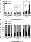
FIGURE 1
FIGURE 1
The number of hBMSC and HUVEC directly co-cultured at a ratio of 1:2 on the day 1, day 4 and day 7 under different concentrations of CGF (hBMSC: * p < 0.05, ** p < 0.01, HUVEC: # p < 0.05, ### p < 0.001, 0%: co-cultured control group). (A): numbers of the 2 cells on the 1st, 4th, and 7th day. (B): percentages of the 2 cells on the 1st, 4th, and 7th day.
FIGURE 2
FIGURE 2
Alkaline phosphatase (ALP) staining and its quantitative analysis.ALP staining and activity was detected in single cultured hBMSCs group (BMSC) and five directly co-cultured group with different concentrations of CGF (0, 2, 5, 10, and 20%), for 7 days. (A): General observation. (B): ALP staining under fluorescence microscope (ruler: 200 μm). (C): ALP quantitative analysis (*p < 0.05, **p < 0.01, ***p < 0.001, ****p < 0.0001 versus 0%CGF).
FIGURE 3
FIGURE 3
Under different CGF concentrations, the relative expressions of osteogenic-related genes of hBMSCs in direct co-culture (*p < 0.05, **p < 0.01, ***p < 0.001, ****p < 0.0001). (M: the group of single-cultured hBMSCs without CGF; E: the group of single-cultured HUVECs without CGF).
FIGURE 4
FIGURE 4
Immunofluorescence staining of CX43 between hBMSCs and HUVECs in direct co-culture. CX43 (orange) to label the linker protein, CD31 (green) to label HUVECS, DAPI (blue) to show nuclei.White arrows indicate the CX43 expression between hBMSCs and HUVECs.
FIGURE 5
FIGURE 5
Under 10% CGF, the relative expression of hBMSCs osteogenic-related genes in direct and indirect co-culture with HUVECs (*p < 0.05, **p < 0.01, ***p < 0.001, ****p < 0.0001).
FIGURE 6
FIGURE 6
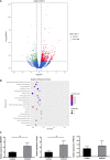
The results of RNA-seq, (A): Volcano plot of differentially expressed genes (DEGs) between 10%CGF and control group in directly co-cultured hBMSCs, (B): Representative up-regulated KEGG pathway categories, (C): mRNA expression level of MAP2K1, ULK1, PIK3CA (*p < 0.05).

References

    1. AlMuraikhi N., Alaskar H., Binhamdan S., Alotaibi A., Kassem M., Alfayez M. (2021). JAK2 Inhibition by Fedratinib Reduces Osteoblast Differentiation and Mineralisation of Human Mesenchymal Stem Cells. Molecules 26 (3), 606. 10.3390/molecules26030606 - DOI - PMC - PubMed
    1. Alsberg E., von Recum H. A., Mahoney M. J. (2006). Environmental Cues to Guide Stem Cell Fate Decision for Tissue Engineering Applications. Expert Opin. Biol. Ther. 6 (9), 847–866. 10.1517/14712598.6.9.847 - DOI - PubMed
    1. Bajada S., Mazakova I., Richardson J. B., Ashammakhi N. (2008). Updates on Stem Cells and Their Applications in Regenerative Medicine. J. Tissue Eng. Regen. Med. 2 (4), 169–183. 10.1002/term.83 - DOI - PubMed
    1. Bajaj P., Pradeep A. R., Agarwal E., Rao N. S., Naik S. B., Priyanka N., et al. (2013). Comparative Evaluation of Autologous Platelet-Rich Fibrin and Platelet-Rich Plasma in the Treatment of Mandibular Degree II Furcation Defects: a Randomized Controlled Clinical Trial[J]. J. Periodontal Res. 48 (5), 573–581. 10.1111/jre.12040 - DOI - PubMed
    1. Ball S. G., Shuttleworth A. C., Kielty C. M. (2004). Direct Cell Contact Influences Bone Marrow Mesenchymal Stem Cell Fate. Int. J. Biochem. Cel Biol. 36 (4), 714–727. 10.1016/j.biocel.2003.10.015 - DOI - PubMed

LinkOut - more resources