Review
. 2022 May;53(5):1473-1486.
doi: 10.1161/STROKEAHA.122.036946. Epub 2022 Apr 7.

Neuroinflammation, Stroke, Blood-Brain Barrier Dysfunction, and Imaging Modalities

Affiliations
Review

Neuroinflammation, Stroke, Blood-Brain Barrier Dysfunction, and Imaging Modalities

Eduardo Candelario-Jalil et al. Stroke. 2022 May.

Abstract

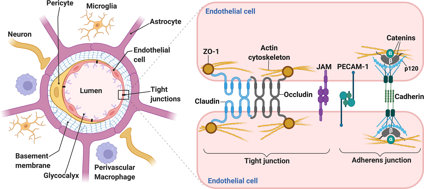
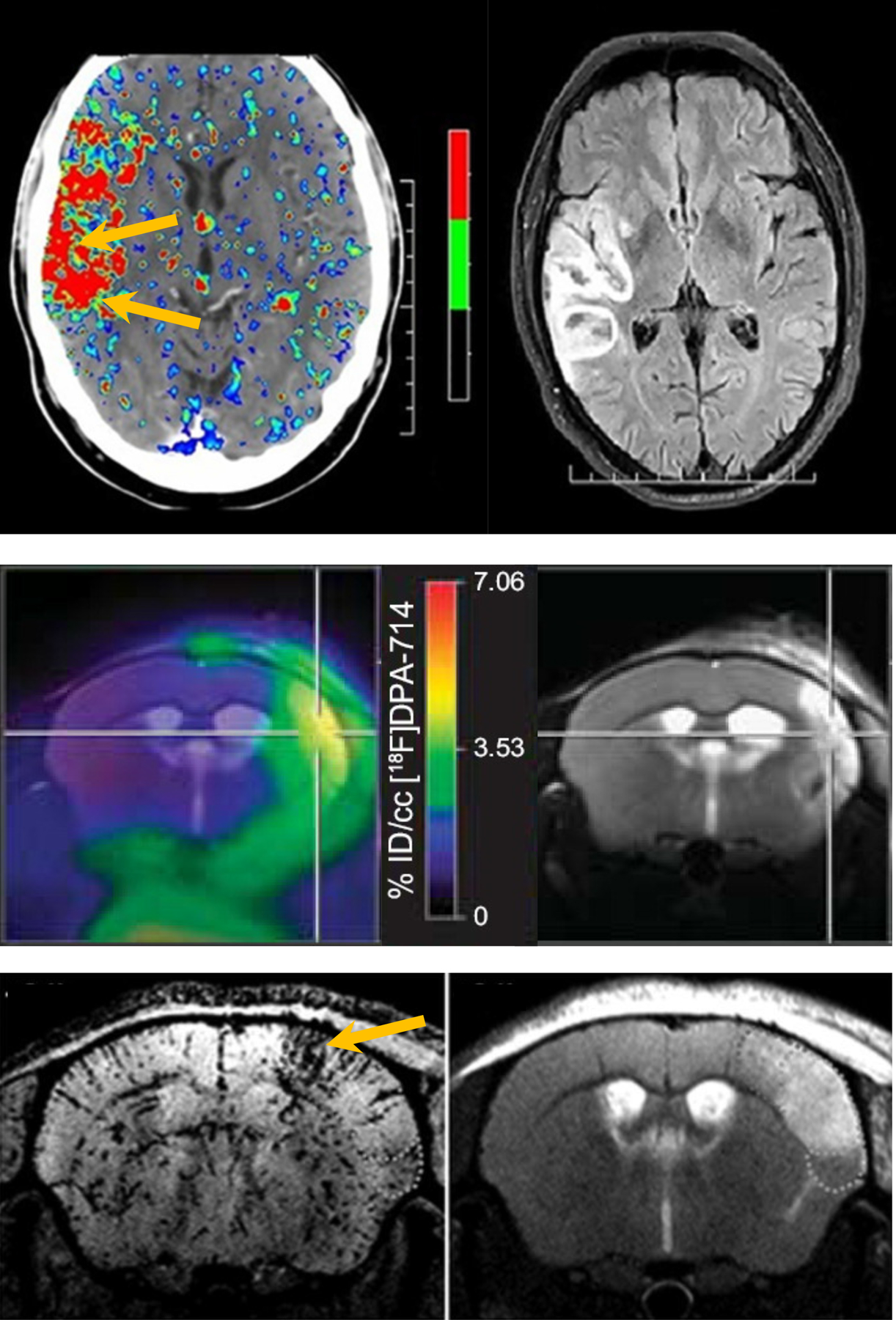
Maintaining blood-brain barrier (BBB) integrity is crucial for the homeostasis of the central nervous system. Structurally comprising the BBB, brain endothelial cells interact with pericytes, astrocytes, neurons, microglia, and perivascular macrophages in the neurovascular unit. Brain ischemia unleashes a profound neuroinflammatory response to remove the damaged tissue and prepare the brain for repair. However, the intense neuroinflammation occurring during the acute phase of stroke is associated with BBB breakdown, neuronal injury, and worse neurological outcomes. Here, we critically discuss the role of neuroinflammation in ischemic stroke pathology, focusing on the BBB and the interactions between central nervous system and peripheral immune responses. We highlight inflammation-driven injury mechanisms in stroke, including oxidative stress, increased MMP (matrix metalloproteinase) production, microglial activation, and infiltration of peripheral immune cells into the ischemic tissue. We provide an updated overview of imaging techniques for in vivo detection of BBB permeability, leukocyte infiltration, microglial activation, and upregulation of cell adhesion molecules following ischemic brain injury. We discuss the possibility of clinical implementation of imaging modalities to assess stroke-associated neuroinflammation with the potential to provide image-guided diagnosis and treatment. We summarize the results from several clinical studies evaluating the efficacy of anti-inflammatory interventions in stroke. Although convincing preclinical evidence suggests that neuroinflammation is a promising target for ischemic stroke, thus far, translating these results into the clinical setting has proved difficult. Due to the dual role of inflammation in the progression of ischemic damage, more research is needed to mechanistically understand when the neuroinflammatory response begins the transition from injury to repair. This could have important implications for ischemic stroke treatment by informing time- and context-specific therapeutic interventions.

Keywords: blood-brain barrier; immunity; ischemic stroke; microglia; neuroinflammatory diseases.


Conflict of interest statement

Disclosures

The authors report no conflicts.

Figures

Figure 1.
Figure 1.. Schematic of the neurovascular unit (NVU).
Anatomically comprising the blood-brain barrier (BBB), the endothelial cells are surrounded by pericytes, astrocytic end-feet processes, neuronal terminals, and the basement membrane, comprising extracellular matrix (ECM) proteins that provide mechanical support and facilitate cell-ECM and cell-cell interactions at the NVU. Perivascular microglial cells contact the brain vasculature and modulate BBB permeability. Perivascular macrophages are key mediators in immune surveillance and are located in penetrating arterioles and post-capillary venules. Situated on the luminal side of the endothelial cells, the glycocalyx is composed of proteoglycans and their linked glycosaminoglycan chains, including heparan sulfate and hyaluronan. Glycocalyx degradation and shedding dramatically increase BBB permeability. An intricate complex of tight junction and adherens junction proteins “zip” together adjacent endothelial cells, conferring the low permeability of the BBB under physiological conditions. Claudins and occludin form the seal between endothelial cells, and they associate with the actin cytoskeleton via accessory proteins, such as ZO-1. Junctional adhesion molecules (JAMs) form part of the tight junctions and facilitate the attachment of endothelial cell membranes. The brain vasculature expresses high levels of platelet endothelial cell adhesion molecule-1 (PECAM-1), also known as CD31, which contributes to BBB function. Vascular endothelial cadherin (VE-cadherin) and catenins are the main structural proteins comprising the adherens junctions. They are necessary to form tight junctions and provide physical integrity to the BBB.
Figure 2.
Figure 2.. Imaging neuroinflammation after stroke.
Multimodal brain images of different aspects of neuroinflammation (left images) and corresponding MR images of tissue injury (right images) after clinical or experimental stroke. Top: BBB permeability map, calculated from perfusion CT <4.5 h after ischemic stroke (before intravenous thrombolysis), and follow-up FLAIR MRI displaying hemorrhagic transformation after 24 h (after intravenous thrombolysis) (modified from Bivard et al., licensed under CC BY-NC-ND 4.0). Middle: Map of [18F]DPA-714 uptake, a marker of activated microglia, calculated from PET, overlaid on a T2-weighted MR image of the ischemic tissue lesion, at 7 days after transient MCAO in mice (from Zinnhardt et al., licensed under CC BY-NC-ND). Bottom: T2*-weighted MRI after injection of anti-VCAM-1 antibody functionalized MPIO, revealing upregulation of VCAM-1 in and around the lesion, shown on a T2-weighted MR image, at 2 days after transient MCAO in mouse (from Gauberti et al., reproduced with permission, Copyright Clearance Center, license# 5233371245585). Arrows or crosshair point at elevated BBB permeability preceding hemorrhagic transformation (top), maximal microglial activation in the lesion core (middle), and high VCAM-1 expression in the lesion borderzone (bottom).

References

    1. Hawkins BT, Davis TP. The blood-brain barrier/neurovascular unit in health and disease. Pharmacological reviews 2005;57:173–185. doi: 10.1124/pr.57.2.4 - DOI - PubMed
    1. Schaeffer S, Iadecola C. Revisiting the neurovascular unit. Nature neuroscience 2021;24:1198–1209. doi: 10.1038/s41593-021-00904-7 - DOI - PMC - PubMed
    1. Iadecola C The Neurovascular Unit Coming of Age: A Journey through Neurovascular Coupling in Health and Disease. Neuron 2017;96:17–42. doi: 10.1016/j.neuron.2017.07.030 - DOI - PMC - PubMed
    1. Heo JH, Han SW, Lee SK. Free radicals as triggers of brain edema formation after stroke. Free Radic Biol Med 2005;39:51–70. doi: 10.1016/j.freeradbiomed.2005.03.035 - DOI - PubMed
    1. Ishibashi N, Prokopenko O, Weisbrot-Lefkowitz M, Reuhl KR, Mirochnitchenko O. Glutathione peroxidase inhibits cell death and glial activation following experimental stroke. Brain Res Mol Brain Res 2002;109:34–44. doi: 10.1016/s0169-328x(02)00459-x - DOI - PubMed

Publication types