. 2022 Apr;16(S1):S17-S25.
doi: 10.14444/8232.

Single-Position Anterior Column Lateral Lumbar Interbody Fusion

Affiliations

Single-Position Anterior Column Lateral Lumbar Interbody Fusion

Gregory Lopez et al. Int J Spine Surg. 2022 Apr.

Abstract

Lateral lumbar fusion is a commonly used spinal fusion technique that allows for indirect neural decompression while correcting sagittal malalignment. The lateral position has evolved to include placement of percutaneous pedicle screw fixation, anterior longitudinal ligament release, and approach the L5-S1 segment. This review article focuses on the anatomy and technique of the single-position anterior column spinal fusion and highlights the recent trends, outcomes, and future directions for the approach.

Keywords: LLIF; lumbar fusion; spine surgery.


Conflict of interest statement

Declaration of Conflicting Interests: The authors report no conflicts of interest related to this work.

Figures
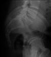
Figure 1
Figure 1
Radiograph demonstrating a patient with a recessed L5-S1 segment. The patient’s pubic symphysis is blocking easy entry into the L5-S1 segment, making anterior exposure difficult and especially not recommended for the lateral position.
Figure 2
Figure 2
Images depicting a 55-year-old man with a Grade 1 isthmic spondylolisthesis with severe foraminal stenosis (A), (B). The common iliac vessels are on either side of the midline of L5-S1 disc space (A), allowing for adequate lateral anterior lumbar interbody fusion exposure. The postoperative fluoroscopic images (C) demonstrate adequate reduction of the spondylolisthesis.
Figure 3
Figure 3
Axial cut T2-magnetic resonance imaging demonstrates anteriorly-migrated psoas, similarly migrating the lumbar plexus and increasing the risk for injury with a direct lateral approach in this patient.
Figure 4
Figure 4
A patient in the lateral decubitus position for single-position lateral surgery is depicted prior to draping. The patient is taped to the bed with the posterior holders in place on the inferior portion of the sacrum and thoracic spine.
Figure 5
Figure 5
Illustration of the lateral anterior lumbar interbody fusion retroperitoneal exposure.
Figure 6
Figure 6
Intraoperative fluoroscopic image of the L5-S1 segment that allows for analysis of the vertebral body rotation, retractor placement, and assessment of the midline.
Figure 7
Figure 7
An intraoperative picture depicting simultaneous anterior and posterior manipulation of the spine.

References

    1. Ozgur BM, Aryan HE, Pimenta L, Taylor WR, et al. . Extreme lateral interbody fusion (XLIF): a novel surgical technique for anterior lumbar interbody fusion. Spine J. 2006;6(4):435–443. 10.1016/j.spinee.2005.08.012 - DOI - PubMed
    1. Talia AJ, Wong ML, Lau HC, Kaye AH, et al. . Comparison of the different surgical approaches for lumbar interbody fusion. J Clin Neurosci. 2015;22(2):243–251. 10.1016/j.jocn.2014.08.008 - DOI - PubMed
    1. Heth JA, Hitchon PW, Goel VK, Rogge TN, Drake JS, Torner JC, et al. . A biomechanical comparison between anterior and transverse interbody fusion cages. Spine (Phila Pa 1976). 2001;26(12):E261-7. 10.1097/00007632-200106150-00012 - DOI - PubMed
    1. Turner JD, Akbarnia BA, Eastlack RK, et al. . Radiographic outcomes of anterior column realignment for adult sagittal plane deformity: a multicenter analysis. Eur Spine J. 2015;24 Suppl 3:427–432. 10.1007/s00586-015-3842-0 - DOI - PubMed
    1. Saigal R, Mundis GM, Eastlack R, Uribe J, Phillips F, Akbarnia BA, et al. . Anterior column realignment (ACR) in adult sagittal deformity correction. SPINE. 2016:1. 10.1097/BRS.0000000000001483 - DOI - PubMed

LinkOut - more resources