. 2022 Apr;604(7906):541-545.
doi: 10.1038/s41586-022-04545-z. Epub 2022 Apr 6.

Basis of narrow-spectrum activity of fidaxomicin on Clostridioides difficile

Affiliations

Basis of narrow-spectrum activity of fidaxomicin on Clostridioides difficile

Xinyun Cao et al. Nature. 2022 Apr.

Abstract

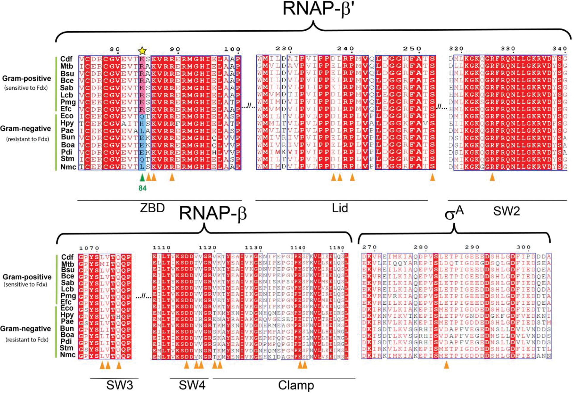
Fidaxomicin (Fdx) is widely used to treat Clostridioides difficile (Cdiff) infections, but the molecular basis of its narrow-spectrum activity in the human gut microbiome remains unknown. Cdiff infections are a leading cause of nosocomial deaths1. Fidaxomicin, which inhibits RNA polymerase, targets Cdiff with minimal effects on gut commensals, reducing recurrence of Cdiff infection2,3. Here we present the cryo-electron microscopy structure of Cdiff RNA polymerase in complex with fidaxomicin and identify a crucial fidaxomicin-binding determinant of Cdiff RNA polymerase that is absent in most gut microbiota such as Proteobacteria and Bacteroidetes. By combining structural, biochemical, genetic and bioinformatic analyses, we establish that a single residue in Cdiff RNA polymerase is a sensitizing element for fidaxomicin narrow-spectrum activity. Our results provide a blueprint for targeted drug design against an important human pathogen.


Conflict of interest statement

Competing interests: Authors declare that they have no competing interests.

Figures

Extended Data Fig. 1.
Extended Data Fig. 1.. Overexpression and purification of Cdiff RNAP.
a, pXC026, overexpression plasmid for the Cdiff rpoA, rpoZ, rpoB, and rpoC genes (encoding the α, ω, β, and β′ subunits of Cdiff RNAP, respectively). The β and β′ subunits were fused with an inter-subunit 10-amino-acid (aa) linker (LARHVGGSGA) and a C-terminal Rhinovirus 3C protease-cleavable His10 tag. b, (Top) Size-exclusion chromatography profile for the assembled Cdiff RNAP EσA. (Bottom) Coomassie-stained SDS-PAGE of individual fractions from major peaks. RNAP subunits are labeled on the right of the gel. Cdiff RNAP for biochemistry and structural biology was taken from pooled fractions of the second peak. c, Abortive transcription assay with Cdiff core and EσA using the Cdiff rrnC promoter as DNA template. The transcriptional activity of CdiffA was inhibited with increasing concentrations of Fdx. Lane 1, Cdiff RNAP core; lane 2, CdiffA; lane 3, CdiffA with 0.2 μM Fdx added; lane 4, CdiffA with 2 μM Fdx added.
Extended Data Fig. 2.
Extended Data Fig. 2.. Cryo-EM processing pipeline.
Flow chart showing the image-processing pipeline for the cryo-EM data of CdiffA/Fdx complexes, starting with 6,930 dose-fractionated movies collected on a 300-keV Titan Krios (FEI) equipped with a K3 Summit direct electron detector (Gatan). Movies were frame-aligned and summed using MotionCor2. CTF estimation for each micrograph was calculated with cryoSPARC2. A representative micrograph is shown following processing by MotionCor2. Particles were auto-picked from each micrograph with cryoSPARC2 Blob Picker and then sorted by 2D classification using cryoSPARC2 to assess quality. The selected classes from the 2D classification are shown. After picking and cleaning by 2D classification, the dataset contained 2,415,902 particles. A subset of particles was used to generate an ab initio templates in cryoSPARC2 and 3D heterogeneous refinement was performed with these templates using cryoSPARC2. One major, high-resolution class emerged, which was polished using RELION and further cleaned with two more 3D heterogenous refinements. The final 182,390 particles were refined using cryoSPARC Non-Uniform refinement.
Extended Data Fig. 3.
Extended Data Fig. 3.. Cryo-EM analysis.
a, Top left, the 3.26 Å-resolution cryo-EM density map of CdiffA/Fdx. Top right, a cross-section of the structure, showing the Fdx. Bottom, same views as above, but colored by local resolution, The boxed region is magnified and displayed as an inset. Density for Fdx is outlined in red. b, Gold-standard FSC plots of the CdiffA/Fdx complex from cryoSPARC. The dotted line represents the gold-standard 0.143 FSC cutoff which indicates a nominal resolution of 3.26 Å. c, Angular distribution calculated in cryoSPARC for CdiffA /Fdx particle projections. Heat map shows number of particles for each viewing angle (less = blue, more = red). d, Cross-validation FSC plots for map-to-model fitting were calculated between the refined structure of CdiffA/Fdx and the half-map used for refinement (work, red), the other half-map (free, blue), and the full map (black). The dotted black line represents the 0.5 FSC cutoff determined for the full map.
Extended Data Fig. 4.
Extended Data Fig. 4.. Differences between Cdiff and other bacterial RNAPs.
The lineage-specific β inserts are shown for Cdiff RNAP in dark blue, Eco RNAP in red (PDB ID:4LK1), Bsub RNAP in green (PDB ID:6ZCA). The Fdx is shown in green spheres, and the active site Mg2+ is shown as a yellow sphere. Superimposition of the RNAPs from each organism was performed in PyMOL. Only the CdiffA is shown.
Extended Data Fig. 5.
Extended Data Fig. 5.. Differences in σA–Fdx contacts between Cdiff and Mtb and σA sequence alignment.
a, Conserved regions of Cdiff σA compared to Mtb σA and Eco σ70. Mtb σA has a much shorter σA NCR than Eco σ70, but the residues in the short Mtb NCR that contact RbpA are not present in either Cdiff or Eco. Mtb RbpA contacts Fdx whereas Cdiff σA makes more contacts to Fdx than does Mtb σA. Black arrows indicate RpbA- σA contacts whereas colored arrows indicate Fdx contacts to σA and RpbA, which includes one shared contact between Mtb and Cdiff σA (red arrow). b, Amino acid-sequence alignment of σA for diverse representatives of bacteria species. Identical residues are highlighted in yellow. Gaps are indicated by dashed lines. Conserved σ regions are labeled underneath the alignment. The three letter species code is as follows: Cdf, Clostridioides difficile; Bsu, Bacillus subtilis; Bun, Bacteroides uniformis; Eco, Escherichia coli; Mtb, Mycobacterium tuberculosis.
Extended Data Fig. 6
Extended Data Fig. 6. Fdx binding residues in Mtb RbpA-EσA and CdiffA.
Ligplot was used to determine contacts between Fdx and Mtb RbpA-EσA (left) and Cdiff EσA (right). Cyan sphere, H2O; green dashed line, hydrogen bond or salt bridge; red arc, van der Waals interactions; red dashed line, cation-π interactions. Note that in ligplot of the CdiffA/Fdx interactions, V1143 (discussed in the text as one of the residues when mutated cause Fdx-resistance) did not make the distance cutoff (4.5 Å) as it was located 4.7 Å away from Fdx. The RNAP β, β’ and σA residues are in cyan, pink, and orange respectively. The two Mtb RbpA residues (E17, R10) that interact with Fdx are colored in purple and indicated in the text. The Fdx-interacting residues that do not have corresponding interactions between Cdiff and Mtb are highlighted in red circles.
Extended Data Fig. 7.
Extended Data Fig. 7.. The cryo-EM density map of residues interacting with Fdx.
Coloring of the residues is consistent with RNAP subunits coloring in Fig. 3, the stick model and cryo-EM densities are color-coded as follows: Pink: β-subunit, cyan: β′- subunit, and orange: σA. Water molecules are shown as red spheres. The residues that form hydrogen bonds (black dotted line) with Fdx are labeled.
Extended Data Fig. 8.
Extended Data Fig. 8.. In vitro abortive transcription assays used to determine Fdx IC50 of Cdiff and MtbAand Eco70 related to Fig. 2d.
Abortive 32P-RNA products (GpUpG) synthesized on Cdiff rrnC promoter were quantified in the presence of increasing concentrations of Fdx. For each EσA (or Eσ70), three independent experiments were performed and analyzed on the same gel.
Extended Data Fig. 9.
Extended Data Fig. 9.. Comparative sequence alignment of key structural components of RNAPs that interact with Fdx between Fdx-resistant and sensitive bacteria.
The Fdx interacting regions are labeled on the top of sequence alignment. Locations of residues contacting Fdx in both Cdiff and Mtb are labeled by triangles underneath sequences. For gram-positive bacteria that are sensitive to Fdx, the corresponding residue at Cdiff β′K84 is either K or R, which is highlighted in pink background. For gram-negative bacteria that are resistant to Fdx, the residue at β′K84 is neutral Q, L, or negative E, which is highlighted in blue background. Conserved residues are shown as white letters on a red background, and similar residues are shown as red letters in blue boxes. Cdf, Clostridioides difficile; Mtb, Mycobacterium tuberculosis; Bsu, Bacillus subtilis; Bce, Bacillus cereus, Sab, Staphylococcus aureus; Lcb, Lactobacillus casei; Pmg, Peptococcus magna; Efc, Enterococcus faecium; Eco, Escherichia coli; Hpy, Helicobacter pylori; Pae, Pseudomonas aeruginosa; Bun, Bacteroides uniformis; Boa, Bacteroides ovatus; Pdi, Parabacteroides distasonis; Stm, Salmonella Choleraesuis; Nmc, Neisseria meningitidis.
Extended Data Fig. 10.
Extended Data Fig. 10.. Phylogenetic tree demonstrating the clade-specific distribution of the identity of the Fdx-sensitizer.
The tree displays the identity of the amino acid corresponding to position β′K84 of Cdiff in the most common species from human gut microbiota. Bacterial species were largely picked from and . The tree was built from 66 small subunit ribosomal RNA sequences by using RaxML and iTol. Species with experimentally confirmed resistance (MIC > 32 μg/mL) and sensitivity (MIC < 0.125 μg/mL) to Fdx are marked with solid and open orange circle respectively. The amino acid sequence at β′K84 position for corresponding bacteria phyla is denoted by capital letters. The detailed bacterial species are listed in Extended Dada Table 4.
Extended Data Fig. 11.
Extended Data Fig. 11.. In vitro abortive transcription assays measuring Fdx IC50 on Cdiff and Eco WT and mutant holo enzymes. a, Transcription assays for Cdiff WT, β′K94E and β′K84Q EσAs related to Fig. 4b.
The Cdiff rrnC promoter (Fig. 2c) was used as a template. b, Transcription assays for Eco WT and β’Q94K Eσ70s related to Fig. 4c. The same Cdiff rrnC promoter was used. For each RNAP, three independent experiments were performed and analyzed on the same gel.
Extended Data Fig.12.
Extended Data Fig.12.. In vivo assays on agar plates for E. coli WT and Q94K mutant strains.
a. Temperature-sensitive strain RL602 was transformed with control plasmid pRL662 encoding no rpoC, WT rpoC and mutant rpoC-Q94K. Strains were grown overnight at 40 °C. Bacteria containing plasmids expressing rpoC WT and Q94K grew well while the empty plasmid does not cell support growth. b. Antibiotic inhibition assays using E. coli rpoC WT and mutant strains from panel (a). Antibiotics in 3 μL DMSO (Fdx, SPR741, and Rif) or water (Kan) were pipetted onto overlay soft agar containing the bacteria (see Methods). SPR741 did not inhibit cell growth but increased the potency of Rif and Fdx, suggesting that it increased antibiotic diffusion into the cells. Rif (± SPR741) and Kan, an antibiotic targeting ribosomes rather than RNAP and not affected by SPR741, equally inhibited the WT and mutant Q94K strains. In contrast, Fdx potently inhibited only the mutant strain, establishing that the Q94E mutation conferred specific sensitivity to Fdx.
Fig. 1.
Fig. 1.. Fidaxomicin is a narrow-spectrum antimicrobial that inhibits RNAP.
a, Diagram illustrating how fidaxomicin specifically targets Cdiff without affecting gut commensals and thus reduces recurrence (upper circles). For CDI patients treated with broad-spectrum antibiotics (lower circles), the abundance of gut commensals drops simultaneously with Cdiff resulting in high rates of Cdiff recurrence. b, Chemical structure of Fdx. c, Cryo-EM structure of CdiffA in complex with Fdx. The EσA model is colored by subunits according to the key, and the 3.26 Å cryo-EM map is represented in a white transparent surface. The cryo-EM density for Fdx is shown in the inset as a green transparent surface.
Fig. 2.
Fig. 2.. Fdx binding and inhibition of the CdiffA
a, Clamp differences between Cdiff and Mtb RNAP. The Cdiff RNAP clamp (pink) is twisted (5.8°) towards Fdx compared with the Mtb RNAP–Fdx clamp (yellow) (PDB ID: 6BZO). The actinobacterial specific insert in the clamp is partially cropped. The clamp residues, β′K314 and β′M319, that interact with Fdx (shown in green spheres) in Cdiff but not Mtb RNAP are shown as red spheres. The zinc in the ZBD is shown as a blue sphere. b, The interactions between Fdx and Cdiff RNAP are shown. Hydrogen-bonding interactions are shown as black dashed lines. The cation-π interaction of β′R89 is shown with a red dashed line. Arches represent hydrophobic interactions. RNAP residues are colored corresponding to subunits: cyan (β) and pink (β′). The Fdx-contacting residues that are not present in the MtbA–Fdx structure (pdb 6BZO) are marked with red circles. c, The sequence of the native Cdiff ribosomal RNAP rrnC promoter used in the in vitro transcription assay in d. The –10 and –35 promoter elements are shaded in purple and green, respectively. The abortive transcription reaction used to test Fdx effects is indicated below the sequence (*, [α-32P]GTP used to label the abortive product GpUpG). d, Fdx inhibits CdiffA and Mtb RbpA-EσA similarly and ~100 times more effectively than Eco70. Error bars are standard deviations (SD) of three independent replicates (for some points, SD was smaller than the data symbols).
Fig. 3.
Fig. 3.. Analysis of Fdx-interacting residues across bacterial lineages.
Cdiff RNAP EσA–Fdx is shown as a molecular surface for orientation (left inset). The boxed region is magnified on the right with RNAP subunits shown as α-carbon backbone worms, Fdx-interacting residues conserved between Mtb and Cdiff shown as side-chain sticks, and Fdx (green) shown as sticks. Non-carbon atoms are red (oxygen) and blue (nitrogen). The MtbCdiff-conserved, Fdx-interacting residues shown in the cartoon structure are labeled under the sequence logos. Amino acids that make hydrophilic interactions with Fdx are labeled on the structure. Representative bacterial species with published Fdx MICs were used to make the logos. See Extended Data Fig. 9 for detailed sequence alignments. Most Fdx-interacting residues are conserved except residues corresponding to Cdiff β′S85 and the sensitizer (yellow star corresponding to Cdiff β’K84).
Fig. 4.
Fig. 4.. The sensitizer position (β′K84 in Cdiff RNAP) explains Fdx narrow-spectrum activity in the gut microbiota.
a, Sequence logos for the Fdx-interaction region of the β′ZBD is highly conserved among the four most common bacterial phyla in the human microbiota: Bacteroidetes, Firmicutes, Actinobacteria, and Proteobacteria, except for the sensitizer position and C-adjacent residue (β′K84 and S85 in Cdiff RNAP). The logos were derived using 66 representative species in the human microbiota, (Extended Data Fig. 10, Extended Data Table 4). b, Fdx effects on abortive transcription reveal that the β′K84E substitution increases resistance 10-fold, whereas β′K84Q and β′K84R have much lesser effects. c, Transcription assays with Eco70 show the β′Q94K substitution in Eco RNAP reduces Fdx IC50 by a factor of ~20 relative to the WT enzyme. d, Zone-of-inhibition assays with WT and β′R84E B. subtilis demonstrate that an R84-to-E change increases Fdx-resistance and establish this residue as a sensitizing determinant in vivo. The mutant (right) displayed reduced zones of inhibition relative to the WT (left) bacteria for Fdx but not for a control (spectinomycin; Spec). e, Zone-of-inhibition assays show that the β′Q94K substitution sensitizes E. coli to Fdx in the presence of the outer-membrane-weakening compound SPR741 (45 μM). E. coli WT (left) was not inhibited by high concentrations of Fdx (3mM), whereas the β′Q94K cells (right) gave inhibited zones at 250 μM.

Comment in

References

    1. (https://www.cdc.gov/drugresistance/pdf/threats-report/2019-ar-threats-re..., 2019).
    1. Louie TJ et al. Fidaxomicin versus vancomycin for Clostridium difficile infection. N Engl J Med 364, 422–431, doi: 10.1056/NEJMoa0910812 (2011). - DOI - PubMed
    1. Crawford T, Huesgen E & Danziger L Fidaxomicin: a novel macrocyclic antibiotic for the treatment of Clostridium difficile infection. Am J Health Syst Pharm 69, 933–943, doi: 10.2146/ajhp110371 (2012). - DOI - PubMed
    1. Louie TJ, Emery J, Krulicki W, Byrne B & Mah M OPT-80 eliminates Clostridium difficile and is sparing of bacteroides species during treatment of C. difficile infection. Antimicrob Agents Chemother 53, 261–263, doi: 10.1128/AAC.01443-07 (2009). - DOI - PMC - PubMed
    1. Lee YJ et al. Protective Factors in the Intestinal Microbiome Against Clostridium difficile Infection in Recipients of Allogeneic Hematopoietic Stem Cell Transplantation. J Infect Dis 215, 1117–1123, doi: 10.1093/infdis/jix011 (2017). - DOI - PMC - PubMed

Methods References

    1. Welch M et al. Design parameters to control synthetic gene expression in Escherichia coli. PLoS One 4, e7002, doi: 10.1371/journal.pone.0007002 (2009). - DOI - PMC - PubMed
    1. Salis HM, Mirsky EA & Voigt CA Automated design of synthetic ribosome binding sites to control protein expression. Nat Biotechnol 27, 946–950, doi: 10.1038/nbt.1568 (2009). - DOI - PMC - PubMed
    1. Czyz A, Mooney RA, Iaconi A & Landick R Mycobacterial RNA polymerase requires a U-tract at intrinsic terminators and is aided by NusG at suboptimal terminators. Mbio 5, e00931, doi: 10.1128/mBio.00931-14 (2014). - DOI - PMC - PubMed
    1. Yang X & Lewis PJ Overproduction and purification of recombinant Bacillus subtilis RNA polymerase. Protein Expr Purif 59, 86–93, doi: 10.1016/j.pep.2008.01.006 (2008). - DOI - PubMed
    1. Davis E, Chen J, Leon K, Darst SA & Campbell EA Mycobacterial RNA polymerase forms unstable open promoter complexes that are stabilized by CarD. Nucleic Acids Res 43, 433–445, doi: 10.1093/nar/gku1231 (2015). - DOI - PMC - PubMed

Publication types

MeSH terms

LinkOut - more resources