

Tractional retinal detachment and juxtapapillary retinal capillary hemangioma in a 6-year-old girl: A case report
- PMID: 35388237
- PMCID: PMC8979396
- DOI: 10.4103/ojo.ojo_348_20
Tractional retinal detachment and juxtapapillary retinal capillary hemangioma in a 6-year-old girl: A case report
Abstract
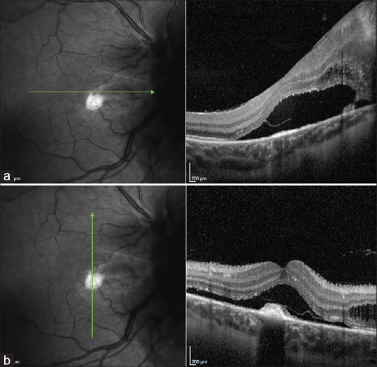
A 6-year-old girl with visual impairment in the right eye (OD) was referred for an eye evaluation. The fundus of the OD showed a fibrotic orange endophytic lesion located adjacent to the optic disc. In retinal optical coherence tomography, a local tractional retinal detachment and choroidal neovascular membrane were observed together also with the presence of subretinal fluid. Due to the vision of the OD evolved to nonlight perception in the following exam, enucleation was performed. The pathology report was correlated with hemangioblastoma. Herein, we describe a case of a young girl with a retinal hemangioblastoma with quick evolution and without prior systemic diagnosis.
Keywords: Hemangioblastoma; Von Hippel-Lindau; ocular oncology; oncology; pediatric retina.
Copyright: © 2022 Oman Ophthalmic Society.
Conflict of interest statement
There are no conflicts of interest.
Figures


References
-
- Gass JD. Developmental tumors of the retinal pigment epithelium (RPE) and retina. In: Gass JD, editor. Stereoscopic Atlas of Macular Diseases, Diagnosis and Treatment. 2nd ed. St Louis, Missouri: Mosby; 1997. p. 1061.
-
- Singh AD, Rundle PA, Rennie I. Retinal vascular tumors. Ophthalmol Clin North Am. 2005;18:167–76. - PubMed
-
- Kase S, Ishida S. Retinal capillary hemangioma in von Hippel-Lindau disease: Current concept, diagnosis and managements. J Transl Med Epidemiol. 2014;2:1010.
-
- McCabe CM, Flynn HW, Jr, Shields CL, Shields JA, Regillo CD, McDonald HR, et al. Juxtapapillary capillary hemangiomas. Clinical features and visual acuity outcomes. Ophthalmology. 2000;107:2240–8. - PubMed
-
- Magee MA, Kroll AJ, Lou PL, Ryan EA. Retinal capillary hemangiomas and von Hippel-Lindau disease. Semin Ophthalmol. 2006;21:143–50. - PubMed
Publication types
LinkOut - more resources
Full Text Sources