. 2022 Aug 1;71(4):206-213.
doi: 10.1093/jmicro/dfac018.

Improved chemical fixation of lipid-secreting plant cells for transmission electron microscopy

Affiliations

Improved chemical fixation of lipid-secreting plant cells for transmission electron microscopy

Shingo Kiyoto et al. Microscopy (Oxf). .

Abstract

Cultured Lithospermum erythrorhizon cells were fixed with a new fixation method to visualize the metabolism of shikonin derivatives, the lipophilic naphthoquinone pigments in Boraginaceae. The new fixation method combined glutaraldehyde containing malachite green, imidazole-osmium and p-phenylenediamine treatments, and cells were then observed with a transmission electron microscope. The method prevented the extraction of lipids, including shikonin derivatives, and improved the visualization of subcellular structures, especially the membrane system, when compared with that of conventional fixation. The improved quality of the transmission electron micrographs is because malachite green ionically binds to the plasma membrane, organelles and lipids and acts as a mordant for electron staining with osmium tetroxide. Imidazole promotes the reaction of osmium tetroxide, leading to enhanced electron staining. p-Phenylenediamine reduces osmium tetroxide bound to cellular materials and increases the electron density. This protocol requires only three additional reagents over conventional chemical fixation using glutaraldehyde and osmium tetroxide.

Keywords: Lithospermum erythrorhizon; p-phenylenediamine; imidazole; malachite green; shikonin; transmission electron microscopy.


Figures

Fig. 1.
Fig. 1.
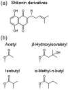
Shikonin derivatives produced by L. erythrorhizon. (a) Structures of shikonin derivatives. (b) Chemical prefixes of the shikonin derivatives and corresponding structures of R in (a).
Fig. 2.
Fig. 2.
Light micrographs of L. erythrorhizon cultured cells fixed with the MGIP method. The sections were observed without post-embedded staining. (a) Cells cultured in M9 medium producing shikonin derivatives. (b) Cells cultured in LS medium as the nonproducing control. (c) Magnified image of the boxed region in (a). Solid and open arrowheads indicate lipid droplets in cytoplasm and materials in vacuole, respectively. Arrows indicate shikonin granules secreted to the extracellular spaces. (d) Magnified image of the boxed region in (b). Arrowheads indicate lipid droplets.
Fig. 3.
Fig. 3.
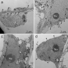
TEM images of L. erythrorhizon cultured cells producing shikonin derivatives. (a) Fixed with conventional fixation. (b) Fixed with the malachite green method. (c) Fixed with the imidazole–osmium method. (d) Fixed with the p-phenylenediamine method. The sections were stained with uranyl acetate and lead citrate. Arrowheads indicate lipid droplets. M, mitochondria; Nu, nucleolus; P, plastid.
Fig. 4.
Fig. 4.
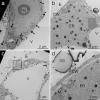
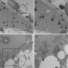
TEM images of L. erythrorhizon cultured cells producing shikonin derivatives fixed with the MGIP method. The sections were stained with uranyl acetate and lead citrate. (a) Nuclear membrane, organelles, lipid droplets and tonoplasts are clearly visible. Arrowheads indicate lipid droplets. (b) A highly developed ER and a lipid-storing plastid were observed. (c) A cell secreting shikonin derivatives. (d) Magnified image of the boxed region in (c). CW, cell wall; E, extracellular spaces; ER, endoplasmic reticulum; M, mitochondria; Nu, nucleolus; P, plastid; SG, shikonin granules secreted to extracellular spaces; T, tonoplast; V, vacuole.
Fig. 5.
Fig. 5.
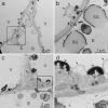
TEM images of L. erythrorhizon cultured cells producing shikonin derivatives fixed using the MGIP method. The sections were stained with uranyl acetate and lead citrate. (a, c) Cells secreting shikonin derivatives. (b) Magnified image of the boxed region in (a). Arrows indicate the folded plasma membrane and tonoplast. An arrowhead indicates a lipid droplet on the outer side of the folded plasma membrane. (d) Magnified image of the boxed region in (c). Arrows indicate a folded tonoplast. Asterisks indicate high-electron-dense materials attached to tonoplasts. CW, cell wall; E, extracellular spaces; SG, shikonin granules secreted to extracellular spaces; V, vacuole.
Fig. 6.
Fig. 6.
TEM images of L. erythrorhizon cultured cells not producing shikonin derivatives fixed with the MGIP method. The sections were stained with uranyl acetate and lead citrate. (a) A cell adjacent to the extracellular spaces. (b) A cell that is not adjacent to the extracellular spaces. Arrowheads indicate lipid droplets. CW, cell wall; E, extracellular spaces; V, vacuole.
Fig. 7.
Fig. 7.
TEM images of L. erythrorhizon cultured cells producing shikonin derivatives fixed with the MGIP method. The sections were observed without post-embedded staining. (a) A cell adjacent to the extracellular spaces. (b) Magnified image of the boxed region in (a). Mitochondria, ER, lipid droplets and tonoplasts are visible. (c) The cytoplasm containing the nucleus and several organelles. (d) Magnified image of the boxed region in (c). A nuclear membrane and a lipid-storing plastid are visible. Arrowheads indicate lipid droplets. CW, cell wall; E, extracellular spaces; ER, endoplasmic reticulum; M, mitochondria; Nu, nucleolus; P, plastid; V, vacuole.
Fig. 8.
Fig. 8.
TEM images of L. erythrorhizon cultured cells producing shikonin derivatives fixed with the MGIP method. The sections were stained with potassium permanganate. (a) A cell adjacent to the extracellular spaces. (b) Magnified image of the boxed region in (a). Mitochondria, ER, lipid droplets and tonoplasts are visible. (c) The cytoplasm containing the nucleus and several organelles. (d) Magnified image of the boxed region in (c). A nuclear membrane and a lipid-storing plastid are clearly visible. Arrowheads indicate lipid droplets. CW, cell wall; E, extracellular spaces; ER, endoplasmic reticulum; M, mitochondria; Nu, nucleolus; P, plastid; V, vacuole.
Fig. 9.
Fig. 9.
Spot tests on specific lipid standards. Stereoscopic micrographs of filter paper fixed with (a) the imidazole–osmium method, (b) a combination of the imidazole–osmium and p-phenylenediamine methods and (c) the MGIP method. Light micrographs of filter paper where (d) triolein, (e) oleic acid, (f) palmitic acid and (g) ethanol only, as a negative control, were spotted and fixed with the MGIP method. Scale bars indicate 10 μm. E, ethanol only, as a negative control; O, oleic acid; P, palmitic acid; TO, triolein.

References

    1. Hussein R A, El-Anssary A A (2019) Plants secondary metabolites: the key drivers of the pharmacological actions of medicinal plants. In: Builders P (ed.), Herbal Medicine, pp 11–30 (IntechOpen, Rijeka; ).doi: 10.5772/intechopen.76139. - DOI
    1. Guo C, He J, Song X, Tan L, Wang M, Jiang P, Li Y, Cao Z, and Peng C (2019) Pharmacological properties and derivatives of shikonin—a review in recent years. Pharmacol. Res. 149: 104463. - PubMed
    1. Yazaki K (2017) Lithospermum erythrorhizon cell cultures: present and future aspects. Plant Biotech. 34: 131–142. - PMC - PubMed
    1. Tabata M, Mizukami H, Hiraoka N, and Konoshima M (1974) Pigment formation in callus cultures of Lithospermum erythrorhizon. Phytochem 13: 927–932.
    1. Shimomura K, Sudo H, Saga H, and Kamada H (1991) Shikonin production and secretion by hairy root cultures of Lithospermum erythrorhizon. Plant Cell Rep. 10: 282–285. - PubMed