Review
. 2023 May 2;119(3):668-690.
doi: 10.1093/cvr/cvac049.

Cellular interplay between cardiomyocytes and non-myocytes in diabetic cardiomyopathy

Affiliations
Review

Cellular interplay between cardiomyocytes and non-myocytes in diabetic cardiomyopathy

Ren Jie Phang et al. Cardiovasc Res. .

Abstract

Patients with Type 2 diabetes mellitus (T2DM) frequently exhibit a distinctive cardiac phenotype known as diabetic cardiomyopathy. Cardiac complications associated with T2DM include cardiac inflammation, hypertrophy, fibrosis, and diastolic dysfunction in the early stages of the disease, which can progress to systolic dysfunction and heart failure. Effective therapeutic options for diabetic cardiomyopathy are limited and often have conflicting results. The lack of effective treatments for diabetic cardiomyopathy is due in part, to our poor understanding of the disease development and progression, as well as a lack of robust and valid preclinical human models that can accurately recapitulate the pathophysiology of the human heart. In addition to cardiomyocytes, the heart contains a heterogeneous population of non-myocytes including fibroblasts, vascular cells, autonomic neurons, and immune cells. These cardiac non-myocytes play important roles in cardiac homeostasis and disease, yet the effect of hyperglycaemia and hyperlipidaemia on these cell types is often overlooked in preclinical models of diabetic cardiomyopathy. The advent of human-induced pluripotent stem cells provides a new paradigm in which to model diabetic cardiomyopathy as they can be differentiated into all cell types in the human heart. This review will discuss the roles of cardiac non-myocytes and their dynamic intercellular interactions in the pathogenesis of diabetic cardiomyopathy. We will also discuss the use of sodium-glucose cotransporter 2 inhibitors as a therapy for diabetic cardiomyopathy and their known impacts on non-myocytes. These developments will no doubt facilitate the discovery of novel treatment targets for preventing the onset and progression of diabetic cardiomyopathy.

Keywords: Autonomic neurons; Cardiac fibroblasts; Cardiomyocytes; Diabetic cardiomyopathy; Endothelial cells; Immune cells.


Conflict of interest statement

Conflict of interest: none declared.

Figures

Figure 1
Figure 1
The contribution of non-cardiac tissues to the pathogenesis of diabetic cardiomyopathy. In diabetes, pancreatic beta-cell dysfunction and insulin resistance lead to impaired insulin-mediated glucose uptake and increased ectopic lipid accumulation in various non-cardiac tissues, including liver, adipose tissue, and skeletal muscle. Impaired insulin signalling in the liver increases hepatic glucose production, while glucose disposal in skeletal muscle is reduced, both of which contribute to systemic hyperglycaemia. To compensate for hyperglycaemia, the pancreas produces more insulin leading to hyperinsulinaemia. Furthermore, increased lipolysis of adipose tissue increases the levels of circulating free fatty acids leading to lipotoxicity and hyperlipidaemia. These pathological events result in overproduction of pro-inflammatory adipokines, dysregulation of hepatokines, and a reduction in protective myokines, all of which contributes to the pathogenesis of diabetic cardiomyopathy.
Figure 2
Figure 2
Cellular interactions among non-myocytes in the heart and their pathological phenotypes in T2DM. Non-myocytes populations play important roles in maintaining healthy cardiac function and promoting cardiomyopathy under diabetic conditions. Cardiac fibroblasts receive pro-fibrotic factors from immune and vascular cells to regulate the dynamics of the extracellular matrix. Conversely, cardiac fibroblasts can induce angiogenesis and infiltration of immune cells to evoke an inflammatory response via secretion of angiogenic growth factors and chemoattractants, respectively. The function of autonomic nervous system is tightly linked with the activity of the immune cells, where the modulation of neuronal activity can be achieved by pro-inflammatory cytokines. Under diabetic conditions, these intercellular interactions can be exacerbated resulting in maladaptive cellular phenotype (highlighted in boxes) and contribute to the pathogenesis of diabetic cardiomyopathy. Signalling molecules include proteins, EVs, messenger RNA (mRNA), microRNA (miRNA), long non-coding RNA (lncRNA), and circular RNA (circRNA).
Figure 3
Figure 3
An overview of current preclinical models of Type 2 diabetes. ob/ob, leptin deficient mouse (obese); db/db, leptin resistant mouse (obese); KK, Kuo-Kondo mouse (obese); KK/Ay, yellow KK mouse (obese); NZO, New Zealand obese mouse (obese); TSOD, Tsumara Suzuki obese diabetes mouse (obese); M16 mouse (obese); ZDF, Zucker diabetic fatty rat; GK, Goto-Kakizaki rat; OLETF, Otsuka Long-Evans Tokushima Fat rat; SHR/N-cp, spontaneously hypertensive rat/NIH-corpulent rat (obese); Cohen, Cohen diabetic rat (non-obese); Torri, Torri rat (non-obese); T2DM, Type 2 diabetes mellitus.
Figure 4
Figure 4
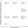
The physiological and human relevance of preclinical disease models. The development of effective therapies for patients is largely impeded by the lack of reliable models with strong biological relevance to human disease. In vivo (blue circles) animal models, especially small animal models, have predominantly been employed for their physiological complexity at the expense of biological relevance to the human. Although primary cells from human cardiac tissues represent the most ideal cell type to model human disease, their availability and accessibility are limited. The establishment of 3D cardiac organoids from human PSCs has revolutionized human disease modelling, providing a mimicry of human heart physiology in vitro (red circles). Cardiac organoids provide enhanced biological complexity such as a 3D microenvironment allowing interaction between cardiomyocytes and non-myocytes compared with the conventional in vitro culture, which is generally monocellular 2D culture.

References

    1. DeFronzo RA, Ferrannini E, Groop L, Henry RR, Herman WH, Holst JJ, Hu FB, Kahn CR, Raz I, Shulman GI, Simonson DC, Testa MA, Weiss R. Type 2 diabetes mellitus. Nat Rev Dis Primers 2015;1:15019. - PubMed
    1. Htay T, Soe K, Lopez-Perez A, Doan AH, Romagosa MA, Aung K. Mortality and cardiovascular disease in type 1 and type 2 diabetes. Curr Cardiol Rep 2019;21:45. - PubMed
    1. Saeedi P, Petersohn I, Salpea P, Malanda B, Karuranga S, Unwin N, Colagiuri S, Guariguata L, Motala AA, Ogurtsova K, Shaw JE, Bright D, Williams R. Global and regional diabetes prevalence estimates for 2019 and projections for 2030 and 2045: results from the International Diabetes Federation Diabetes Atlas, 9th edition. Diabetes Res Clin Pract 2019;157:107843. - PubMed
    1. DiMeglio LA, Evans-Molina C, Oram RA. Type 1 diabetes. Lancet 2018;391:2449–2462. - PMC - PubMed
    1. Tan Y, Zhang Z, Zheng C, Wintergerst KA, Keller BB, Cai L. Mechanisms of diabetic cardiomyopathy and potential therapeutic strategies: preclinical and clinical evidence. Nat Rev Cardiol 2020;17:585–607. - PMC - PubMed

Publication types

MeSH terms