. 2022 Aug;49(10):3557-3570.
doi: 10.1007/s00259-022-05763-3. Epub 2022 Apr 7.

Intraoperative MET-receptor targeted fluorescent imaging and spectroscopy for lymph node detection in papillary thyroid cancer: novel diagnostic tools for more selective central lymph node compartment dissection

Affiliations

Intraoperative MET-receptor targeted fluorescent imaging and spectroscopy for lymph node detection in papillary thyroid cancer: novel diagnostic tools for more selective central lymph node compartment dissection

Pascal K C Jonker et al. Eur J Nucl Med Mol Imaging. 2022 Aug.

Abstract

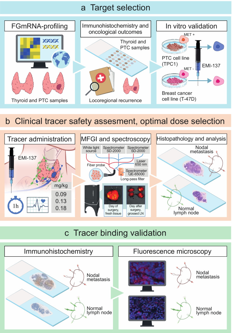
Purpose: Patients undergoing prophylactic central compartment dissection (PCLND) for papillary thyroid cancer (PTC) are often overtreated. This study aimed to determine if molecular fluorescence-guided imaging (MFGI) and spectroscopy can be useful for detecting PTC nodal metastases (NM) and to identify negative central compartments intraoperatively.

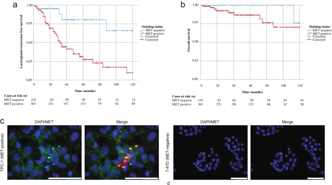
Methods: We used a data-driven prioritization strategy based on transcriptomic profiles of 97 primary PTCs and 80 normal thyroid tissues (NTT) to identify tumor-specific antigens for a clinically available near-infrared fluorescent tracer. Protein expression of the top prioritized antigen was immunohistochemically validated with a tissue microarray containing primary PTC (n = 741) and NTT (n = 108). Staining intensity was correlated with 10-year locoregional recurrence-free survival (LRFS). A phase 1 study (NCT03470259) with EMI-137, targeting MET, was conducted to evaluate safety, optimal dosage for detecting PTC NM with MFGI, feasibility of NM detection with quantitative fiber-optic spectroscopy, and selective binding of EMI-137 for MET.

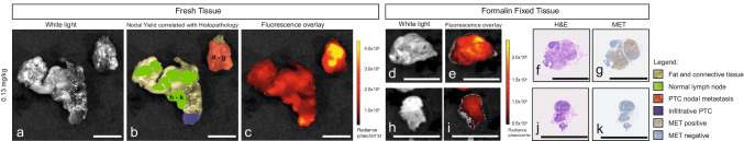
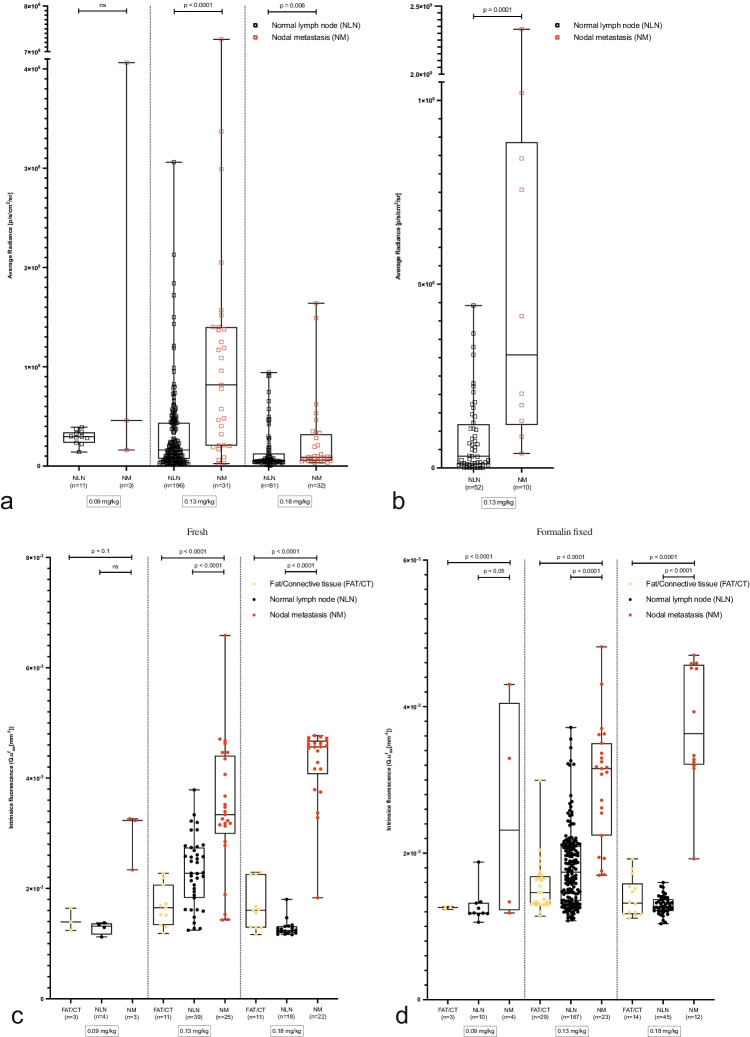
Results: MET was selected as the most promising antigen. A worse LRFS was observed in patients with positive versus negative MET staining (81.9% versus 93.2%; p = 0.02). In 19 patients, no adverse events related to EMI-137 occurred. 0.13 mg/kg EMI-137 was selected as optimal dosage for differentiating NM from normal lymph nodes using MFGI (p < 0.0001) and spectroscopy (p < 0.0001). MFGI identified 5/19 levels (26.3%) without NM. EMI-137 binds selectively to MET.

Conclusion: MET is overexpressed in PTC and associated with increased locoregional recurrence rates. Perioperative administration of EMI-137 is safe and facilitates NM detection using MFGI and spectroscopy, potentially reducing the number of negative PCLNDs with more than 25%.

Clinical trial registration: NCT03470259.

Keywords: Lymph node imaging; Molecular fluorescence-guided imaging; Papillary thyroid cancer; Spectroscopy.


Conflict of interest statement

GMvD is a member of the scientific board of SurgVision BV, founder, shareholder, and CEO of TRACER Europe BV (Groningen, the Netherlands). None of the other authors reported any potential conflicts of interest.

Figures

Fig. 1
Fig. 1
Study workflow illustrating each of the steps for target selection (a), clinical tracer safety assessment and optimal dosage selection (b), and validation of in vivo tracer binding following intravenous administration (c). Abbreviations: EMI-137, investigational medicinal product; FGmRNA-profling, functional genomic mRNA profiling; h, hour; IHC, immunohistochemistry; mg, milligram; MFGI, molecular fluorescence-guided imaging; kg, kilogram; LN, lymph nodes; NLN, normal lymph nodes; NM, nodal metastases; PTC, papillary thyroid cancer
Fig. 2
Fig. 2
In vitro specific binding analysis of EMI-137 and association of MET immunohistochemical staining intensities with oncological outcome parameters in PTC. (a) The 10-year locoregional recurrence-free survival for patients with positive (red line) and negative (blue line) MET staining status is depicted. Patients with a positive staining status have worse 10-year locoregional recurrence-free survival rates compared to patients with negative staining (p = 0.017). (b) The similar 10-year overall survival between both groups (p = 0.21). Immunofluorescent staining of MET-positive (TPC-1, c) and MET-negative (T-47D, d) cell lines, with nuclei (blue), MET (green), and EMI-137 (red); scale bar is 50 µm; abbreviation: PTC, papillary thyroid cancer
Fig. 3
Fig. 3
Representative images from PTC nodal metastases and normal lymph nodes imaged with the IVIS Spectrum in the 0.13 mg/kg dosage cohort. MFGI images from the fresh nodal dissection specimen and representative formalin-fixed PTC nodal metastasis (d–g) and normal lymph node (h–k) are presented. The exact location of the grossed lymph nodes is provided and correlated to final histopathology (b). Fluorescence intensities for the formalin-fixed PTC nodal metastases and normal lymph nodes are scaled. The scale is provided in radiance. Corresponding H&E (f and j) and MET stained slides (g and k) are provided per presented lymph node. Scale bars represent 10 mm
Fig. 4
Fig. 4
(a) An overview of fluorescent intensities per dosage cohort of grossed formalin-fixed PTC nodal metastases and normal lymph nodes imaged with the IVIS Spectrum. (b) The fluorescence intensities of PTC nodal metastases and normal lymph nodes of patients in the 0.13 mg/kg dosage cohort imaged using the IVIS Lumina. An overview of the intrinsic fluorescence measured with quantitative spectroscopy in fresh (c) and formalin-fixed (d) PTC nodal metastases and normal lymph nodes, respectively. Abbreviations: CT, connective tissue; NLN, normal lymph node; NM, nodal metastases; PTC, papillary thyroid cancer

References

    1. Haugen BR, et al. 2015 American Thyroid Association management guidelines for adult patients with thyroid nodules and differentiated thyroid cancer: the American Thyroid Association Guidelines Task Force on Thyroid Nodules and Differentiated Thyroid Cancer. Thyroid : official journal of the American Thyroid Association. 2016;26:1–133. doi: 10.1089/thy.2015.0020. - DOI - PMC - PubMed
    1. Hwang HS, Orloff LA. Efficacy of preoperative neck ultrasound in the detection of cervical lymph node metastasis from thyroid cancer. Laryngoscope. 2011;121:487–491. doi: 10.1002/lary.21227. - DOI - PubMed
    1. Choi JS, et al. Preoperative staging of papillary thyroid carcinoma: comparison of ultrasound imaging and CT. Am J Roentgenol. 2009;193:871–878. doi: 10.2214/AJR.09.2386. - DOI - PubMed
    1. Kim JW, et al. Extent of extrathyroidal extension as a significant predictor of nodal metastasis and extranodal extension in patients with papillary thyroid carcinoma. Ann Surg Oncol. 2016;24:460–468. doi: 10.1245/s10434-016-5594-4. - DOI - PubMed
    1. Viola D, et al. Prophylactic central compartment lymph node dissection in papillary thyroid carcinoma: clinical implications derived from the first prospective randomized controlled single institution study. J Clin Endocrinol Metab. 2015;100:1316–1324. doi: 10.1210/jc.2014-3825. - DOI - PubMed

MeSH terms

Associated data