. 2022 Apr 7;17(4):e0266111.
doi: 10.1371/journal.pone.0266111. eCollection 2022.

Comprehensive cancer-oriented biobanking resource of human samples for studies of post-zygotic genetic variation involved in cancer predisposition

Natalia Filipowicz  1 Kinga Drężek  1 Monika Horbacz  1 Agata Wojdak  1 Jakub Szymanowski  1   2 Edyta Rychlicka-Buniowska  1 Ulana Juhas  1 Katarzyna Duzowska  1 Tomasz Nowikiewicz  3   4 Wiktoria Stańkowska  1 Katarzyna Chojnowska  1 Maria Andreou  1 Urszula Ławrynowicz  1 Magdalena Wójcik  1 Hanna Davies  5 Ewa Śrutek  4   6 Michał Bieńkowski  7 Katarzyna Milian-Ciesielska  8 Marek Zdrenka  6 Aleksandra Ambicka  9 Marcin Przewoźnik  9 Agnieszka Harazin-Lechowska  9 Agnieszka Adamczyk  9 Jacek Kowalski  7 Dariusz Bała  4   10 Dorian Wiśniewski  10 Karol Tkaczyński  10 Krzysztof Kamecki  11 Marta Drzewiecka  3 Paweł Wroński  11 Jerzy Siekiera  11 Izabela Ratnicka  12 Jerzy Jankau  12 Karol Wierzba  13 Jarosław Skokowski  14   15 Karol Połom  14 Mikołaj Przydacz  16 Łukasz Bełch  16 Piotr Chłosta  16 Marcin Matuszewski  17 Krzysztof Okoń  8 Olga Rostkowska  18 Andrzej Hellmann  18 Karol Sasim  19 Piotr Remiszewski  18 Marek Sierżęga  20 Stanisław Hać  18 Jarosław Kobiela  18 Łukasz Kaska  18 Michał Jankowski  4   10 Diana Hodorowicz-Zaniewska  20 Janusz Jaszczyński  21 Wojciech Zegarski  4   10 Wojciech Makarewicz  14   22 Rafał Pęksa  7 Joanna Szpor  8 Janusz Ryś  9 Łukasz Szylberg  6   23 Arkadiusz Piotrowski  1   24 Jan P Dumanski  1   5   24
Affiliations

Comprehensive cancer-oriented biobanking resource of human samples for studies of post-zygotic genetic variation involved in cancer predisposition

Natalia Filipowicz et al. PLoS One. .

Abstract

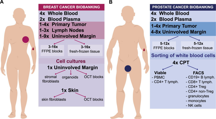
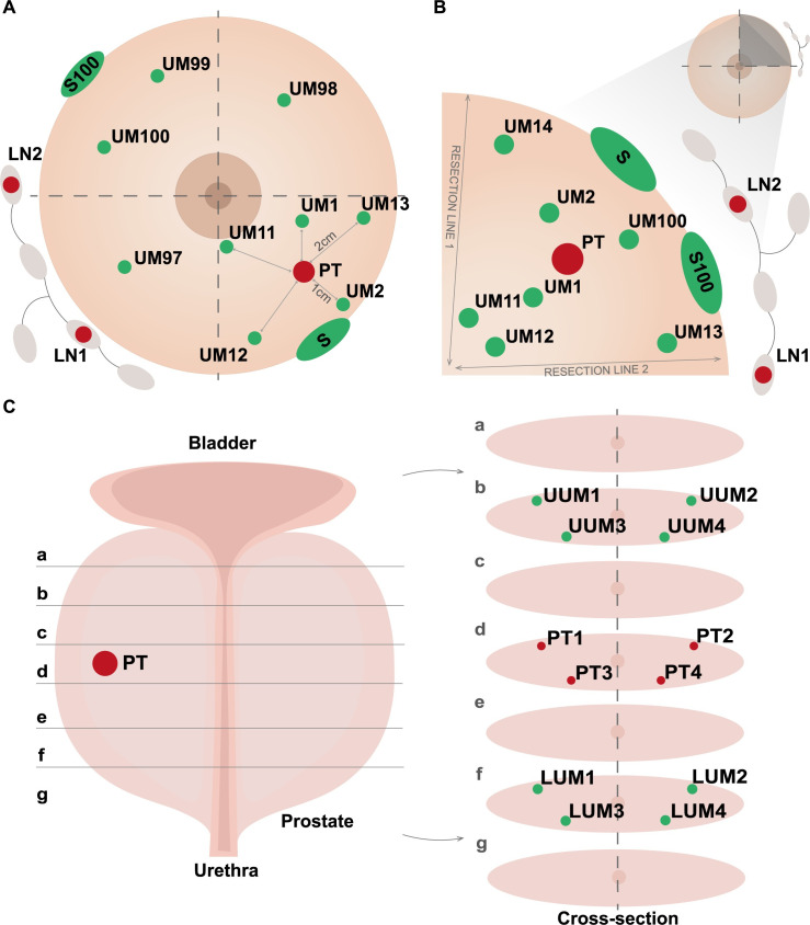
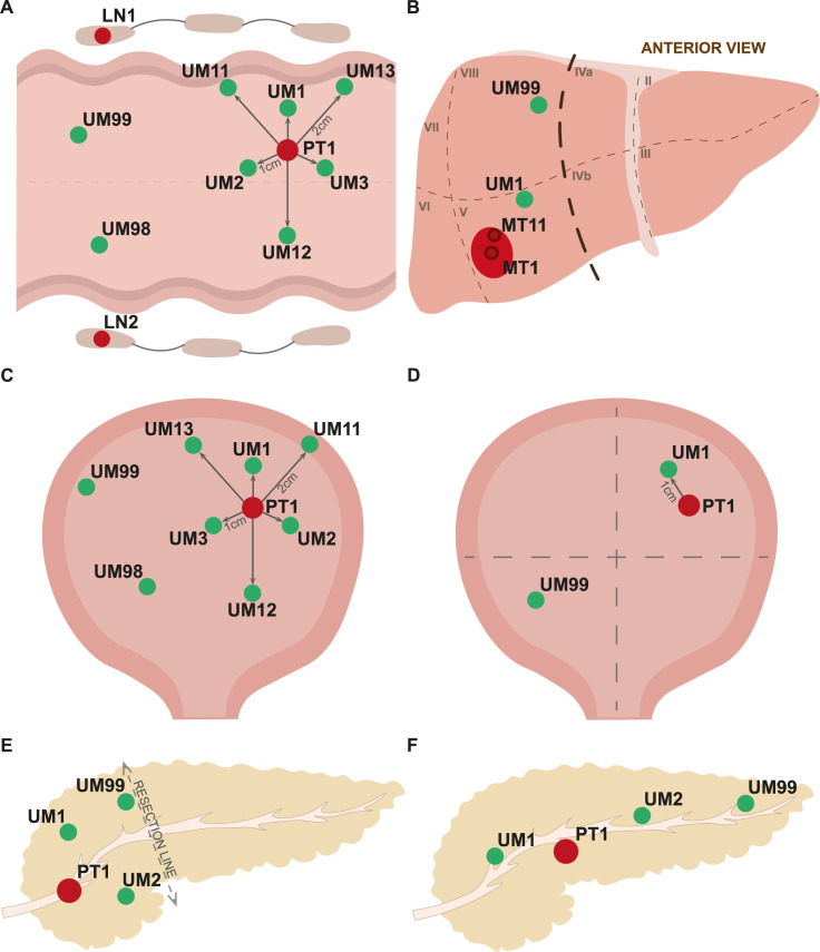
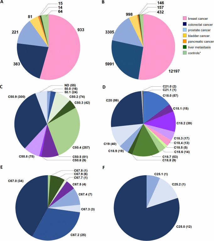
The progress in translational cancer research relies on access to well-characterized samples from a representative number of patients and controls. The rationale behind our biobanking are explorations of post-zygotic pathogenic gene variants, especially in non-tumoral tissue, which might predispose to cancers. The targeted diagnoses are carcinomas of the breast (via mastectomy or breast conserving surgery), colon and rectum, prostate, and urinary bladder (via cystectomy or transurethral resection), exocrine pancreatic carcinoma as well as metastases of colorectal cancer to the liver. The choice was based on the high incidence of these cancers and/or frequent fatal outcome. We also collect age-matched normal controls. Our still ongoing collection originates from five clinical centers and after nearly 2-year cooperation reached 1711 patients and controls, yielding a total of 23226 independent samples, with an average of 74 donors and 1010 samples collected per month. The predominant diagnosis is breast carcinoma, with 933 donors, followed by colorectal carcinoma (383 donors), prostate carcinoma (221 donors), bladder carcinoma (81 donors), exocrine pancreatic carcinoma (15 donors) and metachronous colorectal cancer metastases to liver (14 donors). Forty percent of the total sample count originates from macroscopically healthy cancer-neighboring tissue, while contribution from tumors is 12%, which adds to the uniqueness of our collection for cancer predisposition studies. Moreover, we developed two program packages, enabling registration of patients, clinical data and samples at the participating hospitals as well as the central system of sample/data management at coordinating center. The approach used by us may serve as a model for dispersed biobanking from multiple satellite hospitals. Our biobanking resource ought to stimulate research into genetic mechanisms underlying the development of common cancers. It will allow all available "-omics" approaches on DNA-, RNA-, protein- and tissue levels to be applied. The collected samples can be made available to other research groups.


Conflict of interest statement

J.P.D. is cofounder and shareholder in Cray Innovation AB. The remaining authors have declared that no competing interests exist.

Figures

Fig 1
Fig 1. A summary of samples collected for two common cancer diagnoses.
(A) Collection for breast carcinoma patients. (B) Collection for prostate carcinoma patients. FFPE, Formalin-Fixed Paraffin-Embedded blocks; OCT, Optimal Cutting Temperature compound for fresh-frozen tissue; PBMC, Peripheral Blood Mononuclear Cells; CPT, Cell Preparation Tube with Sodium Heparin (BD Bioscience) for separation of granulocyte- and PBMC-fraction of white blood cells; FACS, Fluorescent Activated Cell Sorting; lymph., lymphocytes; Treg, T-regulatory lymphocytes; NK, Natural Killer cells.
Fig 2
Fig 2. An illustration of sample collection protocols for breast- and prostate cancer.
(A) Procedure for breast carcinoma samples treated with mastectomy. (B) Procedure for breast carcinoma patients treated with Breast Conservative Therapy (BCT). The distances in centimeters between samples of primary tumor and normal tissue are illustrated in panel A with solid lines. (C) Protocol for prostatectomy with detailed scheme of sample collection in different cross-sections (a–g). Abbreviations: UM, uninvolved margin composed of macroscopically normal tissue; PT, primary tumor; S, skin; LUM, lower uninvolved margin; UUM, upper uninvolved margin; LN, regional lymph node. Detailed description of particular fragments for the protocols is given in the Materials and Method section.
Fig 3
Fig 3. An illustration of sample collection protocols for colorectal carcinoma and metastases of colorectal cancer to liver, urinary bladder- and exocrine pancreas carcinomas.
(A) Scheme of sample collection for colorectal carcinoma. (B) Protocol of samples collection for metastases of colorectal cancer to liver. (C) Protocol of sample collection for urinary bladder after cystectomy. (D) Collection of samples for transurethral resection of tumor (TURBT). (E) Scheme of sample collection for the surgical removal of pancreas head. (F) Scheme of sample collection for the total pancreatomy. Primary tumors in all panels are drawn in red and samples of normal tissues in green. Abbreviations: UM, uninvolved margin composed of macroscopically normal tissue; PT, primary tumor; LN, regional lymph node; the lines show distances in centimeters from primary tumor.
Fig 4
Fig 4. The statistics of donors and samples collected in five collaborating hospitals; status as of May 12, 2021.
(A) The total number of donors with compulsory set of samples (as described in Materials and Methods-section and shown in Figs 1 and 2). (B) The sum of all samples collected from recruited donors. The numbers for donors and samples are divided for six different cancer diagnoses and controls (*). The control (*) category represents a healthy male cohort ≥ 65-year-old recruited as controls for patients with prostate- and colorectal cancer, used in the Loss of Y Chromosome (LOY) project. (C–F) show distribution of diagnoses according to International Classification of Diseases (ICD-10, World Health Organization) for breast, colorectal, bladder and pancreatic cancer patients, respectively. Abbreviations: C50, Malignant neoplasm of breast; C50.0, Nipple and areola; C50.1, Central portion of breast; C50.2, Upper-inner quadrant of breast; C50.3, Lower-inner quadrant of breast; C50.4, Upper-outer quadrant of breast; C50.5, Lower-outer quadrant of breast; C50.6, Axillary tail of breast; C50.8, Overlapping lesion of breast; C50.9, Breast, unspecified; C18, Malignant neoplasm of colon; C18.0, Caecum, Ileocaecal valve; C18.1, Appendix; C18.2, Ascending colon; C18.3, Hepatic flexure; C18.4, Transverse colon; C18.5, Splenic flexure; C18.6, Descending colon; C18.7, Sigmoid colon, Sigmoid (flexure); C18.8, Overlapping lesion of colon;C18.9, Colon, unspecified, Large intestine, unspecified; C19, Malignant neoplasm of rectosigmoid junction, including colon with rectum, rectosigmoid colon; C20, Malignant neoplasm of rectum, Including rectal ampulla; C21, Malignant neoplasm of anus and anal canal; C21.0, Anus, unspecified, excluding anal margin and perianal skin; C21.1, Anal canal, Anal sphincter; C67, Malignant neoplasm of bladder; C67.0, Trigone of bladder; C67.2, Lateral wall of bladder; C67.3, Anterior wall of bladder; C67.4, Posterior wall of bladder; C67.5, Bladder neck, Internal urethral orifice; C67.7, Urachus; C67.8, Overlapping lesion of bladder; C67.9, Bladder, unspecified; C25, Malignant neoplasm of pancreas; C25.0, Head of pancreas; C25.1, Body of pancreas; C25.2, Tail of pancreas. ND–not yet defined due to temporary lack of medical documentation.
Fig 5
Fig 5. Smoking status declared by donors in the questionnaire.
(A) Female cohort. (B) Male cohort. (C) Control male group, as described in Materials and Methods.
Fig 6
Fig 6. DNA quality isolated from collected tissues.
(A) Concentration of DNA (ng/μl) obtained from blood and tissues from donors per diagnoses, measured with the fluorometric method (Qubit Fluorometric Quantification and/or Agilent TapeStation System). (B) DNA Integrity Number (DIN) for DNA obtained from blood and tissue in four diagnoses measured by Genomic DNA ScreenTape Analysis (Agilent). (C) DNA quality measured by UV-Vis spectroscopic method for DNA obtained from blood and tissues from donors with five diagnoses. The number of samples for each diagnosis that was used for calculations are: 88 for breast cancer; 202 for colorectal cancer; 3092 for prostate cancer; 189 for bladder cancer; 12 for pancreas cancer; and 62 for controls. The amount of frozen tissue used for DNA extractions range as follows: Breast cancer 15–60 mg; bladder cancer 2–19 mg; prostate cancer 6–43 mg; colorectal cancer 15–33 mg; pancreas cancer 12–21 mg; and controls (sorted leukocytes) 0.05x10⁶ - 1x10⁶ cells.

References

    1. Frank C, Fallah M, Sundquist J, Hemminki A, Hemminki K. Population Landscape of Familial Cancer. Sci Rep. 2015;5:12891. Epub 2015/08/11. doi: 10.1038/srep12891 ; PubMed Central PMCID: PMC4530455. - DOI - PMC - PubMed
    1. Hemminki K, Sundquist J, Bermejo JL. How common is familial cancer? Ann Oncol. 2008;19(1):163–7. Epub 2007/09/07. doi: 10.1093/annonc/mdm414 . - DOI - PubMed
    1. Scheuner MT, McNeel TS, Freedman AN. Population prevalence of familial cancer and common hereditary cancer syndromes. The 2005 California Health Interview Survey. Genet Med. 2010;12(11):726–35. Epub 2010/10/06. doi: 10.1097/GIM.0b013e3181f30e9e . - DOI - PubMed
    1. Forsberg LA, Gisselsson D, Dumanski JP. Mosaicism in health and disease—clones picking up speed. Nat Rev Genet. 2017;18(2):128–42. Epub Dec 12. doi: 10.1038/nrg.2016.145 . - DOI - PubMed
    1. Ronowicz A, Janaszak-Jasiecka A, Skokowski J, Madanecki P, Bartoszewski R, Balut M, et al.. Concurrent DNA Copy-Number Alterations and Mutations in Genes Related to Maintenance of Genome Stability in Uninvolved Mammary Glandular Tissue from Breast Cancer Patients. Hum Mutat. 2015;36(11):1088–99. Epub 2015/07/30. doi: 10.1002/humu.22845 . - DOI - PubMed

Publication types