. 2022 Aug;25(3):331-342.
doi: 10.1007/s10456-022-09836-7. Epub 2022 Apr 7.

NRASQ61R mutation in human endothelial cells causes vascular malformations

Affiliations

NRASQ61R mutation in human endothelial cells causes vascular malformations

Elisa Boscolo et al. Angiogenesis. 2022 Aug.

Abstract

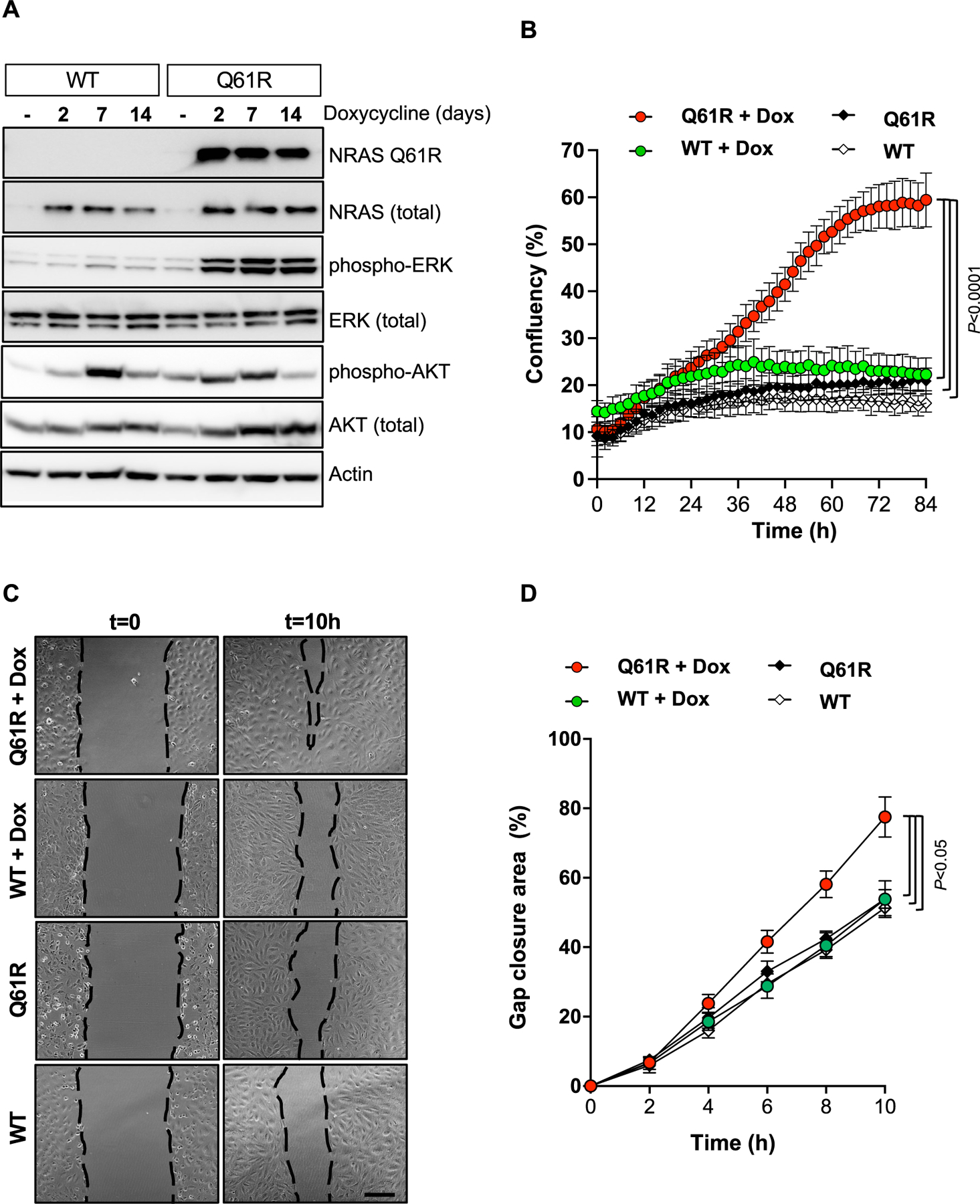
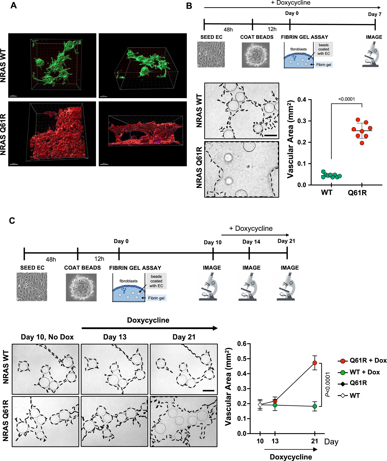
Somatic mutations in NRAS drive the pathogenesis of melanoma and other cancers but their role in vascular anomalies and specifically human endothelial cells is unclear. The goals of this study were to determine whether the somatic-activating NRASQ61R mutation in human endothelial cells induces abnormal angiogenesis and to develop in vitro and in vivo models to identify disease-causing pathways and test inhibitors. Here, we used mutant NRASQ61R and wild-type NRAS (NRASWT) expressing human endothelial cells in in vitro and in vivo angiogenesis models. These studies demonstrated that expression of NRASQ61R in human endothelial cells caused a shift to an abnormal spindle-shaped morphology, increased proliferation, and migration. NRASQ61R endothelial cells had increased phosphorylation of ERK compared to NRASWT cells indicating hyperactivation of MAPK/ERK pathways. NRASQ61R mutant endothelial cells generated abnormal enlarged vascular channels in a 3D fibrin gel model and in vivo, in xenografts in nude mice. These studies demonstrate that NRASQ61R can drive abnormal angiogenesis in human endothelial cells. Treatment with MAP kinase inhibitor U0126 prevented the change to a spindle-shaped morphology in NRASQ61R endothelial cells, whereas mTOR inhibitor rapamycin did not.

Keywords: Kaposiform lymphangiomatosis; Lymphatic anomaly; RASopathies; Vascular Anomaly; Vascular Malformation.


Figures

Fig. 1
Fig. 1
Morphology of human endothelial progenitor cells (EPC)-expressing NRASWT and NRASQ61R. Panel A: Representative images of EPC monolayer with no Doxycycline (No Dox) and 24 and 48 h after addition of Doxycycline to the cell culture media. n = 4 independent experiments. Scale bar = 200 μm Panel B and C: Circularity index measurements for EPC-expressing NRASWT and NRASQ61R in No Dox and Dox-containing media at 24 and 48 h. In B, measurements for each cell is shown. One-way Anova followed by Tukey’s test for multiple comparisons. In C, data points are mean ± SD; n = 4 independent experiments. Two-way Anova followed by Bonferroni test for multiple comparisons, p values are at 24 and 48 h
Fig. 2
Fig. 2
Signaling pathways and angiogenic properties of human endothelial progenitor cells (EPC) expressing NRASWT and NRASQ61R. Panel A: Cell lysates were analyzed 2, 7 and 14 days after doxycycline (Dox) was added to the cell culture media. Antibodies specific to the mutated form of NRAS (NRASQ61R), wild-type NRAS (total), phospho-ERK, phospho-AKT Ser473, total ERK, total AKT, and actin were used. Representative blot of n = 3 independent repeats. Panel B: Proliferation of endothelial cells was measured using an Incucyte® system in endothelial basal medium (EBM2) containing 20% fetal bovine serum (FBS). Data points are mean ± SD; n = 8 wells. Two-way Anova followed by Bonferroni test for multiple comparisons. P value < 0.0001 for times ≥ 36 h. Panel C: Representative images of migration of endothelial cells at t = 0 and t = 10 h after the wound was made. Scale bar: 200 μm. Panel D: Migration area of endothelial cells was measured every 2 h from 0 to 10 h after creating the gap. Percentage gap closure was determined. Data points are mean ± SD; shown is one representative of n = 2 independent experiments with 3–4 wells analyzed. Two-way Anova followed by Bonferroni test for multiple comparisons. P value < 0.05 at 6, 8, 10 h
Fig. 3
Fig. 3
Human endothelial progenitor cells (EPC)-expressing NRASQ61R form enlarged vascular structures in a 3D lumen formation assay. Panel A: Imaris-generated 3D structural models of vascular channels formed by human EPC-expressing NRASWT and NRASQ61R stained for Ulex europaeus agglutinin-I (UEA) on day Scale Bars = 200 μμ. Panel B: Experimental protocol schematic (top), Doxycycline was added 48 h prior starting the 3D lumen formation assay. Representative 2D images of vascular structures (bottom, left) and quantification of vascular area (normalized by bead number) at day 7 (bottom, right). Data points are mean ± SD; n = 3 independent experiments. Welch’s t test. Panel C: Experimental protocol schematic, Doxycycline was not added until day 10 (top). Representative 2D images of vascular structures (bottom, left) and quantification of vascular area at days 10, 13, and 21 (bottom, right). Data points are mean ± SD; n = 3 independent experiments. Two-way Anova followed by Bonferroni test for multiple comparisons. P value < 0.0001 at day 21. Scale Bars in Panel B and C = 100 μμ
Fig. 4
Fig. 4
Human endothelial progenitor cells (EPC)-expressing NRASQ61R form enlarged and perfused vascular structures in a murine xenograft model. Panel A: Experimental protocol showing time of cell injection and Doxycycline treatment period, and time of harvest of xenograft plugs. Panel B: Pictures and graph of weight measurements of the xenograft plugs after removal from mice at day 8. Data points are mean ± SD; n = 2–6 mice with 2 plugs. One-way Anova followed by Fisher test for multiple comparisons. Panel C: Histology of xenograft plugs after removal from mice at day 8. Tissue sections were stained with hematoxylin and eosin. Graph shows vascular area measured from histology. One-way Anova followed by Fisher test for multiple comparisons. Panel D: Immunostaining of xenograft plug sections for Ulex europaeus agglutinin-I (UEA) that is expressed by human endothelial cells and not mouse endothelial cells. Scale bars = 1 cm in B, 100 μm in C and D
Fig. 5
Fig. 5
Treatment of endothelial progenitor cells (EPC) expressing NRASQ61R with MAPK/ERK and mTOR inhibitors. Panel A: Western blot analysis of phospho-ERK, ERK, phospho-S6, S6, and actin 48 h hours after addition of doxycycline and either vehicle (DMSO control), MAPK/ERK inhibitor U0126 (U; 10 μM), or rapamycin (R; 15 nM). Panel B: Representative images of EPC monolayers with no Doxycycline (No Dox), plus Doxycycline, and either vehicle (DMSO control), MAPK/ERK inhibitor U0126 (10 μM), or rapamycin (15 nM). Scale bar = 200 μm. Panel C: Circularity index measurements for EPC-expressing NRASWT and NRASQ61R in Dox-containing media at 48 h and DMSO control (−), MAPK/ERK inhibitor U0126 (U; 10 μM), or rapamycin (R; 15 nM). Data are expressed in % change relative to No Dox control. One-way Anova followed by Tukey’s test for multiple comparisons. ns: p > 0.05

References

    1. Degirmenci U, Wang M, Hu J (2020) Targeting aberrant RAS/RAF/MEK/ERK signaling for cancer therapy. Cells 9(1):198 - PMC - PubMed
    1. Munoz-Couselo E et al. (2017) NRAS-mutant melanoma: current challenges and future prospect. Onco Targets Ther 10:3941–3947 - PMC - PubMed
    1. Queisser A, Boon LM, Vikkula M (2018) Etiology and genetics of congenital vascular lesions. Otolaryngol Clin North Am 51(1):41–53 - PubMed
    1. Groesser L et al. (2016) BRAF and RAS mutations in sporadic and secondary pyogenic granuloma. J Invest Dermatol 136(2):481–486 - PubMed
    1. Lim YH et al. (2015) Somatic activating RAS mutations cause vascular tumors including pyogenic granuloma. J Invest Dermatol 135(6):1698–1700 - PMC - PubMed

Publication types

LinkOut - more resources