Review
. 2022 Mar 22:13:860931.
doi: 10.3389/fmicb.2022.860931. eCollection 2022.

Intestinal Damage in COVID-19: SARS-CoV-2 Infection and Intestinal Thrombosis

Affiliations
Review

Intestinal Damage in COVID-19: SARS-CoV-2 Infection and Intestinal Thrombosis

Xiaoming Wu et al. Front Microbiol. .

Abstract

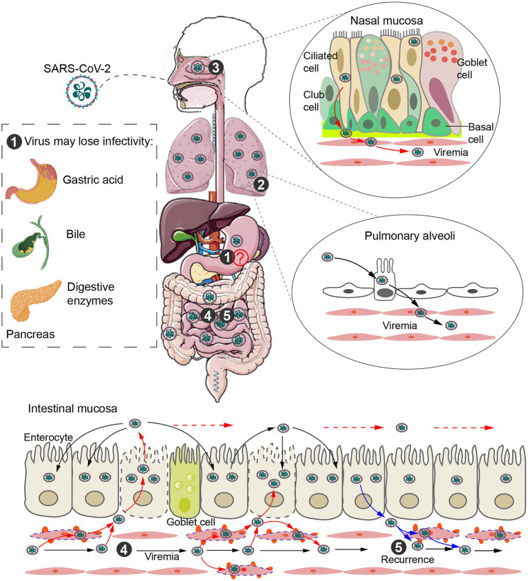
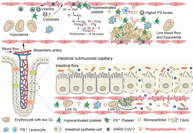
The intestinal tract, with high expression of angiotensin-converting enzyme 2 (ACE2), is a major site of extrapulmonary infection in COVID-19. During pulmonary infection, the virus enters the bloodstream forming viremia, which infects and damages extrapulmonary organs. Uncontrolled viral infection induces cytokine storm and promotes a hypercoagulable state, leading to systemic microthrombi. Both viral infection and microthrombi can damage the gut-blood barrier, resulting in malabsorption, malnutrition, and intestinal flora entering the blood, ultimately increasing disease severity and mortality. Early prophylactic antithrombotic therapy can prevent these damages, thereby reducing mortality. In this review, we discuss the effects of SARS-CoV-2 infection and intestinal thrombosis on intestinal injury and disease severity, as well as corresponding treatment strategies.

Keywords: COVID-19; antithrombotic therapy; blood transmission; intestinal infection; intestinal thrombosis.


Conflict of interest statement

The authors declare that the research was conducted in the absence of any commercial or financial relationships that could be construed as a potential conflict of interest. The reviewer (UDA) declared a shared affiliation with the authors (VN and JS) to the handling editor at the time of review.

Figures

Figure 1
Figure 1
Intestinal infection and transmission routes. ① Direct evidence for fecal–oral transmission is still lacking. SARS-CoV-2 may be unable to enter the small intestine from the stomach due to gastric acid, bile and digestive enzymes. ② SARS-CoV-2 released from type II alveolar cells infects alveolar capillary endothelial cells (ECs). The virus replicates in ECs and is released into the blood to form viremia. ③ SARS-CoV-2 is released from infected ciliary cells of the nasal cavity and breaks through the basement membrane, infecting the vascular ECs and eventually entering circulation. ④ Blood transmission after alveolar or nasal infection is a potential route of intestinal infection. Eventually, SARS-CoV-2 is released into the gut and infects surrounding intestinal epithelial cells along the intestinal tract. ⑤ SARS-CoV-2 in the gut can also enter the capillaries and cause viremia, leading to recurrence of disease.
Figure 2
Figure 2
Intestinal thrombosis leads to intestinal mucosal necrosis and dissemination of gut bacteria, endotoxins, and microbial metabolites in blood. (Top) Mesenteric vascular endotheliitis (initiated by viremia and accelerated by cytokines), hyperactivated platelets and high levels of phosphatidylserine (PS) promote a high rate of mesenteric thrombus in COVID-19 patients (mesenteric vein is shown in Supplementary Figure 1). (Bottom) Intestinal microthrombi and hypoxemia rapidly lead to intestinal mucosal ischemia and necrosis. The damaged gut–blood barrier leads to dissemination of gut bacteria, endotoxins, and microbial metabolites in blood.
Figure 3
Figure 3
Phosphatidylserine exposure on activated/apoptotic cells and microparticles (MPs) promotes fibrin formation. (A) Phosphatidylserine is usually confined to the inner leaflet of the cell membrane. This asymmetry is maintained through ATP-dependent inward transport of PS by flippases and outward transport of non-PS by floppases (left). Upon stimulation, calcium transients will inhibit ATP-dependent transport and stimulate the nonselective lipid transporter scramblase (ATP-independent), resulting in PS exposure (right). (B–D) Human umbilical vein ECs were treated with healthy human plasma and TNF-ɑ (our previous study; He et al., 2016). (B) ECs retracts the cell margins, extends PS positive filopods and releases endothelial-MPs. (C) The PS+ filopods and MPs can be co-stained by Xa and Va. (D) ECs (green) were incubated with MPs-depleted plasma (MDP) in the presence of calcium for 30 min and stained with Alexa Fluro 647-anti-fibrin for 30 min. Considerable fibrin stands among cultured ECs along with filopodia. (E) Confocal images showed PS expression on platelets of patients stained with Alexa 488 lactadherin (our previous study; Ma et al., 2017). MPs from the activated platelet (*) had formed at the margin area located between the distinct outlines. (F) MPs from plasma were co-stained by Xa and Va (or lactadherin and annexin V; our previous study; Gao et al., 2015). (G) MPs that were incubated with recalcified MDP for 30 min and stained with Alexa Fluro 647-anti-fibrin for 30 min. Converted fibrin networks were detected around MPs. The inset bars represent 5 μm in (B–D,G) and 2 μm in (E,F).

References

    1. Aiyegbusi O. L., Hughes S. E., Turner G., Rivera S. C., McMullan C., Chandan J. S., et al. . (2021). Symptoms, complications and management of long COVID: a review. J. R. Soc. Med. 114, 428–442. doi: 10.1177/01410768211032850, PMID: - DOI - PMC - PubMed
    1. Althaus K., Marini I., Zlamal J., Pelzl L., Singh A., Häberle H., et al. . (2021). Antibody-induced procoagulant platelets in severe COVID-19 infection. Blood 137, 1061–1071. doi: 10.1182/blood.2020008762, PMID: - DOI - PMC - PubMed
    1. Ananworanich J., Mogg R., Dunne M. W., Bassyouni M., David C. V., Gonzalez E., et al. . (2021). Randomized study of rivaroxaban vs. placebo on disease progression and symptoms resolution in high-risk adults with mild COVID-19. Clin. Infect. Dis. doi: 10.1093/cid/ciab813, PMID: [Epub ahead of print]. - DOI - PMC - PubMed
    1. Antonelli M., Penfold R. S., Merino J., Sudre C. H., Molteni E., Berry S., et al. . (2022). Risk factors and disease profile of post-vaccination SARS-CoV-2 infection in UK users of the COVID symptom study app: a prospective, community-based, nested, case-control study. Lancet Infect. Dis. 22, 43–55. doi: 10.1016/S1473-3099(21)00460-6 - DOI - PMC - PubMed
    1. Baden L. R., El Sahly H. M., Essink B., Kotloff K., Frey S., Novak R., et al. . (2021). Efficacy and safety of the mRNA-1273 SARS-CoV-2 vaccine. N. Engl. J. Med. 384, 403–416. doi: 10.1056/NEJMoa2035389 - DOI - PMC - PubMed