. 2021 Nov 10;4(2):e210160.
doi: 10.1148/ryai.2021210160. eCollection 2022 Mar.

Reader Perceptions and Impact of AI on CT Assessment of Air Trapping

Affiliations

Reader Perceptions and Impact of AI on CT Assessment of Air Trapping

Tara A Retson et al. Radiol Artif Intell. .

Abstract

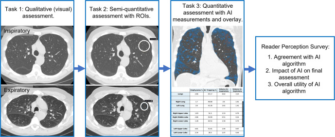
Quantitative imaging measurements can be facilitated by artificial intelligence (AI) algorithms, but how they might impact decision-making and be perceived by radiologists remains uncertain. After creation of a dedicated inspiratory-expiratory CT examination and concurrent deployment of a quantitative AI algorithm for assessing air trapping, five cardiothoracic radiologists retrospectively evaluated severity of air trapping on 17 examination studies. Air trapping severity of each lobe was evaluated in three stages: qualitatively (visually); semiquantitatively, allowing manual region-of-interest measurements; and quantitatively, using results from an AI algorithm. Readers were surveyed on each case for their perceptions of the AI algorithm. The algorithm improved interreader agreement (intraclass correlation coefficients: visual, 0.28; semiquantitative, 0.40; quantitative, 0.84; P < .001) and improved correlation with pulmonary function testing (forced expiratory volume in 1 second-to-forced vital capacity ratio) (visual r = -0.26, semiquantitative r = -0.32, quantitative r = -0.44). Readers perceived moderate agreement with the AI algorithm (Likert scale average, 3.7 of 5), a mild impact on their final assessment (average, 2.6), and a neutral perception of overall utility (average, 3.5). Though the AI algorithm objectively improved interreader consistency and correlation with pulmonary function testing, individual readers did not immediately perceive this benefit, revealing a potential barrier to clinical adoption. Keywords: Technology Assessment, Quantification © RSNA, 2021.

Keywords: Quantification; Technology Assessment.


Conflict of interest statement

Disclosures of Conflicts of Interest: T.A.R. RSNA Machine Learning Committee member, unrelated to this work. K.A.H. No relevant relationships. S.J.K. Deputy editor of Radiology: Cardiothoracic Imaging. K.E.J. No relevant relationships. A.C.Y. No relevant relationships. S.S.B. No relevant relationships. L.D.H. No relevant relationships. A.H. Grants from GE Healthcare and Bayer; cofounder and shareholder in Arterys.

Figures

Design of the reader study. Cardiothoracic radiologists performed
lobar-level assessment of air trapping severity in three stages. 1, Air
trapping severity was rated visually with inspiratory and expiratory images.
2, Air trapping was assessed semiquantitatively after placement of any
desired regions of interest (ROIs) to measure lung attenuation. 3, Air
trapping was rated after providing artificial intelligence
(AI)–generated quantitative measurements and color overlays showing
areas of air trapping (blue) and emphysema (red). Finally, readers were
surveyed for their perceptions of the AI algorithm.
Figure 1:
Design of the reader study. Cardiothoracic radiologists performed lobar-level assessment of air trapping severity in three stages. 1, Air trapping severity was rated visually with inspiratory and expiratory images. 2, Air trapping was assessed semiquantitatively after placement of any desired regions of interest (ROIs) to measure lung attenuation. 3, Air trapping was rated after providing artificial intelligence (AI)–generated quantitative measurements and color overlays showing areas of air trapping (blue) and emphysema (red). Finally, readers were surveyed for their perceptions of the AI algorithm.
Example case in a 45-year-old man with history of stem cell transplant and
graft-versus-host disease with bronchiolitis obliterans syndrome (BOS) causing
diffuse air trapping. At (A) inspiration and (B) expiration, lung attenuation in
the left upper lobe in the regions of interest (ROIs) were 896 HU and 797 HU,
respectively. (C) On the artificial intelligence–generated quantitative
overlay, there is extensive air trapping throughout the lungs (shown in blue,
with areas of emphysema shown in red). For this case, readers’ assessment
of air trapping increased by nearly one grade between placement of ROIs and
provision of quantitative maps. (D) Two months after the initial CT, the patient
went on to develop spontaneous pneumomediastinum (arrows), a rare complication
of severe BOS.
Figure 2:
Example case in a 45-year-old man with history of stem cell transplant and graft-versus-host disease with bronchiolitis obliterans syndrome (BOS) causing diffuse air trapping. At (A) inspiration and (B) expiration, lung attenuation in the left upper lobe in the regions of interest (ROIs) were 896 HU and 797 HU, respectively. (C) On the artificial intelligence–generated quantitative overlay, there is extensive air trapping throughout the lungs (shown in blue, with areas of emphysema shown in red). For this case, readers’ assessment of air trapping increased by nearly one grade between placement of ROIs and provision of quantitative maps. (D) Two months after the initial CT, the patient went on to develop spontaneous pneumomediastinum (arrows), a rare complication of severe BOS.

References

    1. Lowe KE, Regan EA, Anzueto A, et al. . COPDGene® 2019: Redefining the Diagnosis of Chronic Obstructive Pulmonary Disease. Chronic Obstr Pulm Dis (Miami) 2019;6(5):384–399. - PMC - PubMed
    1. Devaraj A, van Ginneken B, Nair A, Baldwin D. Use of Volumetry for Lung Nodule Management: Theory and Practice. Radiology 2017;284(3):630–644. - PubMed
    1. Miller WT Jr, Chatzkel J, Hewitt MG. Expiratory air trapping on thoracic computed tomography. A diagnostic subclassification. Ann Am Thorac Soc 2014;11(6):874–881. - PubMed
    1. Criado E, Sánchez M, Ramírez J, et al. . Pulmonary sarcoidosis: typical and atypical manifestations at high-resolution CT with pathologic correlation. RadioGraphics 2010;30(6):1567–1586. - PubMed
    1. Hall GL, Logie KM, Parsons F, et al. . Air trapping on chest CT is associated with worse ventilation distribution in infants with cystic fibrosis diagnosed following newborn screening. PLoS One 2011;6(8):e23932. - PMC - PubMed