. 2022 Mar 22:12:872012.
doi: 10.3389/fcimb.2022.872012. eCollection 2022.

Lactobacillus plantarum Disrupts S. mutans-C. albicans Cross-Kingdom Biofilms

Affiliations

Lactobacillus plantarum Disrupts S. mutans-C. albicans Cross-Kingdom Biofilms

Yan Zeng et al. Front Cell Infect Microbiol. .

Abstract

Dental caries, an ecological dysbiosis of oral microflora, initiates from the virulent biofilms formed on tooth surfaces where cariogenic microorganisms metabolize dietary carbohydrates, producing acid that demineralizes tooth enamel. Forming cariogenic biofilms, Streptococcus mutans and Candida albicans are well-recognized and emerging pathogens for dental caries. Recently, probiotics have demonstrated their potential in treating biofilm-related diseases, including caries. However, limited studies have assessed their effect on cariogenic bacteria-fungi cross-kingdom biofilm formation and their underlying interactions. Here, we assessed the effect of four probiotic Lactobacillus strains (Lactobacillus rhamnosus ATCC 2836, Lactobacillus plantarum ATCC 8014, Lactobacillus plantarum ATCC 14917, and Lactobacillus salivarius ATCC 11741) on S. mutans and C. albicans using a comprehensive multispecies biofilm model that mimicked high caries risk clinical conditions. Among the tested probiotic species, L. plantarum demonstrated superior inhibition on the growth of C. albicans and S. mutans, disruption of virulent biofilm formation with reduced bacteria and exopolysaccharide (EPS) components, and formation of virulent microcolonies structures. Transcriptome analysis (RNA sequencing) further revealed disruption of S. mutans and C. albicans cross-kingdom interactions with added L. plantarum. Genes of S. mutans and C. albicans involved in metabolic pathways (e.g., EPS formation, carbohydrate metabolism, glycan biosynthesis, and metabolism) were significantly downregulated. More significantly, genes related to C. albicans resistance to antifungal medication (ERG4), fungal cell wall chitin remodeling (CHT2), and resistance to oxidative stress (CAT1) were also significantly downregulated. In contrast, Lactobacillus genes plnD, plnG, and plnN that contribute to antimicrobial peptide plantaricin production were significantly upregulated. Our novel study findings support further assessment of the potential role of probiotic L. plantarum for cariogenic biofilm control.

Keywords: Candida albicans; Lactobacillus plantarum; Streptococcus mutans; cross-kingdom interactions; dental caries; multispecies biofilms.


Conflict of interest statement

The authors declare that the research was conducted in the absence of any commercial or financial relationships that could be construed as a potential conflict of interest.

Figures
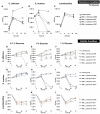
Figure 1
Figure 1
Inhibition of C. albicans and S. mutans by Lactobacilli in multispecies biofilms. The growth curves of C. albicans, S. mutans, and Lactobacilli in multispecies planktonic and biofilm conditions are plotted. The control group consists of C. albicans and S. mutans. The group with added Lactobacilli was marked as “with Lactobacillus”. (A) Lactobacilli significantly inhibited the growth of C. a lbicans by 1 log after 6 h and 1–2 logs after a 20-h incubation. (B) Lactobacilli significantly inhibited the growth of S. mutans at 6 and 20 h. S. mutans was inhibited to non-detectable level (<20 CFU/ml) after a 20-h incubation with L. plantarum 8014 and L. salivarius 11741. (C) Lactobacilli maintained a stable growth in all groups. (D–L) The growth curves of C. albicans, S. mutans, and Lactobacilli in multispecies biofilm conditions are plotted. (D–F) Lactobacilli (L. plantarum and L. salivarius) inhibited the growth of C. albicans in high-sucrose conditions (1%) by 72 h, a 3-log reduction compared to the control group. No difference of C. albicans growth was detected with the addition of L. rhamnosus in all sugar conditions. (G–I) Lactobacilli (L. plantarum and L. salivarius) inhibit the growth of S. mutans in high-sugar conditions (1% sucrose and 1% glucose). Significantly, L. plantarum 8014 and 14917 inhibited S. mutans in the biofilms to non-detectable level (<20 CFU/ml) as early as 48 h, and the treated biofilms remained non-detectable S. mutans (<20 CFU/ml) at 72 h. L. rhamnosus had poor performance on inhibiting the growth of S. mutans growth in all sugar conditions. (J–L) Lactobacilli maintained a stable growth in all groups. * Indicates that the CFU values of the multispecies biofilms were significantly less than the control group at all follow-up time points (p < 0.05). # Indicates that the CFU values of the multispecies biofilms were significantly less than the control group at specific marked time points (p < 0.05).
Figure 2
Figure 2
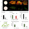
Morphogenesis, 3D architecture, and quantitative measurement of microcolonies in 72-h multispecies biofilms (1% sucrose condition). The 72-h biofilms of the control group (C. albicans and S. mutans) and experimental groups (with L. plantarum 14917) in 1% sucrose condition were visualized using a two-photon laser confocal microscope. The three-dimensional structure of the biofilms was rendered using Amira software. The green color indicates bacteria and the red color indicates the exopolysaccharides (EPS). L. plantarum 14917 dramatically reduced biofilm formation, compared to the control group (A, B). Biofilm dry weight was significantly reduced with added L. plantarum 14917 (C). *p < 0.05. The biomass of the two biofilm components, bacteria and exopolysaccharides (EPS), was calculated using image-processing software COMSTAT (Heydorn et al., 2000). Both L. plantarum 14917 significantly reduced the biomass of bacteria and EPS (D). The confocal images indicate the cross-sectional and sagittal views of microcolonies formed in the control group (S. mutans and C. albicans duo-species) and with added L. plantarum 14917. Well-formed mushroom-shaped microcolonies were seen in the control group, and the largest size microcolonies were seen in the S. mutans and C. albicans duo-species biofilm. Microcolonies formed with added L. plantarum 14917 were much less structured. Bacterial components were less encapsulated with EPS. The amount of co-localization between bacteria and EPS was calculated using DUOSTAT (E), which was consistent with the findings revealed in the images (*p < 0.05). The surface-attached and free-floating microcolonies were evaluated using COMSTAT and DUOSTAT software. Panel (F) illustrates that biofilms treated by L. plantarum 14917 had significantly reduced microcolony size (p > 0.05; ANOVA, comparison for all pairs using Tukey–Kramer HSD).
Figure 3
Figure 3
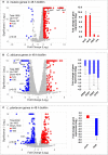
Comparison of transcriptome profiling between multispecies biofilms treated with L. plantarum 14917 and their controls. (A) Volcano plots from transcriptome analysis of S. mutans in multispecies (L. plantarum 14917 + S. mutans + C. albicans) biofilm (48 h, 1% sucrose) compared to S. mutans in duo-species (S. mutans + C. albicans) biofilm. (B) C. albicans in multispecies biofilm compared to C. albicans in duo-species biofilm. (C) L. plantarum 14917 in multispecies biofilm compared to L. plantarum 14917 single-species biofilms. Data represent three independent replicates of each condition. qRT-PCR validation results of selected genes are shown on the right side of each volcano plots. * Indicates that the expression of genes in the multispecies biofilms was significantly different from that in the control group (p < 0.05).
Figure 4
Figure 4
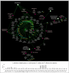
KEGG pathway network for S. mutans differentially expressed genes between the multispecies and duo-species biofilms. The genes of S. mutans differentially expressed genes between the comparison groups with FDR p-values < 0.05 and log2 fold changes > 2 were defined as DEGs and are listed in Supplementary Table 2 . Overall, 33 impacted pathways were found for 441 S. mutans DEGs. The fold change of the DEGs involved in the identified pathways is shown in the lower panel.
Figure 5
Figure 5
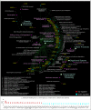
KEGG pathway network for C. albicans differentially expressed genes between the multispecies and duo-species biofilms. The genes of C. albicans that differentially expressed between the comparison groups with FDR p-values < 0.05 and log2 fold changes > 2 were defined as DEGs and are listed in Supplementary Table 3 . Overall, 66 impacted pathways were found for 232 C. albicans DEGs. The fold change of the DEGs involved in the identified pathways is shown in the lower panel.

References

    1. Alkhars N Z. Y., Alomeir N., Al Jallad N., Wu T. T., Aboelmagd S., Youssef M., et al. . (2021). Oral Candida Predicts Streptococcus Mutans Emergence in Underserved US Infants. J. Dental Res. 101 (1), 54–62. doi: 10.1177/00220345211012385 - DOI - PMC - PubMed
    1. Arasu M. V., Al-Dhabi N. A., Ilavenil S., Choi K. C., Srigopalram S. (2016). In Vitro Importance of Probiotic Lactobacillus Plantarum Related to Medical Field. Saudi J. Biol. Sci. 23 (1), S6–s10. doi: 10.1016/j.sjbs.2015.09.022 - DOI - PMC - PubMed
    1. Baker J. L., Faustoferri R. C., Quivey R. G. (2017). Acid-Adaptive Mechanisms of Streptococcus Mutans-the More We Know, the More We Don't. Mol. Oral. Microbiol. 32 (2), 107–117. doi: 10.1111/omi.12162 - DOI - PMC - PubMed
    1. Bowen W. H. (2016). Dental Caries - Not Just Holes in Teeth! A Perspective. Mol. Oral. Microbiol. 31 (3), 228–233. doi: 10.1111/omi.12132 - DOI - PubMed
    1. Bowen W. H., Burne R. A., Wu H., Koo H. (2018). Oral Biofilms: Pathogens, Matrix, and Polymicrobial Interactions in Microenvironments. Trends Microbiol. 26 (3), 229–242. doi: 10.1016/j.tim.2017.09.008 - DOI - PMC - PubMed

Publication types