

Are there differences in arthroscopic and histological features between traumatic and degenerative rotator cuff tears in elderly patients? A prospective dual-center analysis
- PMID: 35392942
- PMCID: PMC8991962
- DOI: 10.1186/s13018-022-03100-w
Are there differences in arthroscopic and histological features between traumatic and degenerative rotator cuff tears in elderly patients? A prospective dual-center analysis
Abstract
Background: Discriminating traumatic rotator cuff tears (RCTs) from degenerative RCTs is sometimes difficult in elderly patients because the prevalence of asymptomatic RCTs increases with age. Little intraoperative information is available on the characteristics of traumatic and degenerative RCTs in elderly patients. The purpose of this study was to compare the arthroscopic findings and histological changes of the coracoacromial ligament (CAL) between traumatic and degenerative RCTs in elderly patients.
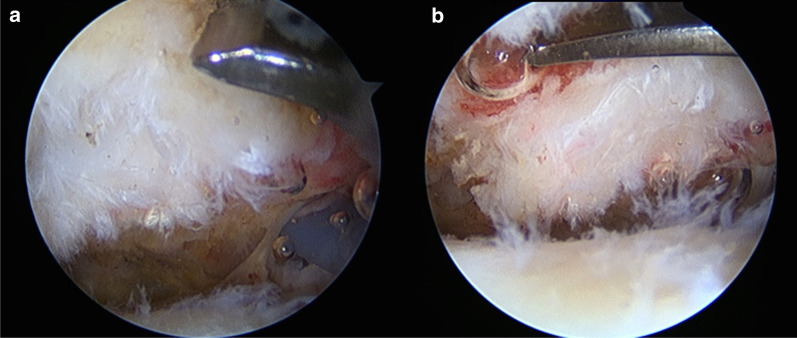
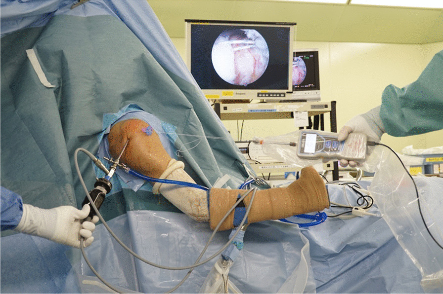
Methods: Forty-two shoulders of 42 patients aged ≥ 65 years underwent arthroscopic rotator cuff repair. Nineteen patients had traumatic full-thickness RCTs (Group T), and 23 had degenerative full-thickness RCTs (Group D). The quality of the rotator cuff tissue and the condition of the long head of the biceps were examined. The grade of CAL was evaluated both arthroscopically and histologically. The stiffness of the musculotendinous unit was calculated by measuring the force and displacement using a tensiometer. The arthroscopic and histological findings of the two groups were compared.
Results: Although the mean tendon displacement was comparable, the stiffness was different between Group T and Group D (0.56 ± 0.31 and 1.09 ± 0.67 N/mm, respectively; p < 0.001). Both arthroscopic and histological analysis of the CAL showed that the degenerative changes in the CAL were milder in Group T than in Group D (p < 0.001 and p < 0.001, respectively). There was a moderate positive correlation between the arthroscopic findings of CAL degeneration and the histopathological changes in this ligament (r = 0.47, p = 0.002).
Conclusions: Traumatic RCTs were characterized by preserved elasticity of the musculotendinous unit and milder CAL degeneration compared with degenerative RCTs even in elderly patients.
Keywords: Arthroscopy; Coracoacromial ligament; Elderly; Histology; Repair tension; Traumatic rotator cuff tears.
© 2022. The Author(s).
Conflict of interest statement
The authors declare that they have no competing interests.
Figures

Similar articles
-
No differences in histopathological degenerative changes found in acute, trauma-related rotator cuff tears compared with chronic, nontraumatic tears.Knee Surg Sports Traumatol Arthrosc. 2022 Jul;30(7):2521-2527. doi: 10.1007/s00167-022-06884-w. Epub 2022 Feb 8. Knee Surg Sports Traumatol Arthrosc. 2022. PMID: 35133449 Free PMC article.
-
WITHDRAWN: Higher coracoacromial ligament thickness, critical shoulder angle and acromion index are associated with rotator cuff tears in patients who undergo arthroscopic rotator cuff repair.Arthroscopy. 2021 Jun 11:S0749-8063(21)00570-3. doi: 10.1016/j.arthro.2021.05.057. Online ahead of print. Arthroscopy. 2021. PMID: 34126216
-
Histological analysis of the coracoacromial arch: correlation between age-related changes and rotator cuff tears.Arthroscopy. 1996 Oct;12(5):531-40. doi: 10.1016/s0749-8063(96)90190-5. Arthroscopy. 1996. PMID: 8902125
-
Does Distal Clavicle Resection Decrease Pain or Improve Shoulder Function in Patients With Acromioclavicular Joint Arthritis and Rotator Cuff Tears? A Meta-analysis.Clin Orthop Relat Res. 2018 Dec;476(12):2402-2414. doi: 10.1097/CORR.0000000000000424. Clin Orthop Relat Res. 2018. PMID: 30334833 Free PMC article.
-
All-arthroscopic versus mini-open rotator cuff repair: A long-term retrospective outcome comparison.Arthroscopy. 2003 Mar;19(3):234-8. doi: 10.1053/jars.2003.50036. Arthroscopy. 2003. PMID: 12627146 Review.
Cited by
-
Study on Shoulder Joint Parameters and Available Supraspinatus Outlet Area Using Three-Dimensional Computed Tomography Reconstruction.Tomography. 2024 Aug 29;10(9):1331-1341. doi: 10.3390/tomography10090100. Tomography. 2024. PMID: 39330746 Free PMC article.
-
Risk Factors for High Repair Tension During Rotator Cuff Repair.Orthop J Sports Med. 2024 Oct 9;12(10):23259671241276445. doi: 10.1177/23259671241276445. eCollection 2024 Oct. Orthop J Sports Med. 2024. PMID: 39399768 Free PMC article.
-
Association of Coracoacromial Ligament Degeneration With Rotator Cuff Tear Patterns and Retear Rate.Orthop J Sports Med. 2023 Jun 7;11(6):23259671231175873. doi: 10.1177/23259671231175873. eCollection 2023 Jun. Orthop J Sports Med. 2023. PMID: 37347016 Free PMC article.
-
Position Paper of the Italian Society of Orthopaedics and Traumatology (SIOT) on the Treatment of Rotator Cuff Tears.Orthop J Sports Med. 2025 Jul 1;13(7):23259671251351901. doi: 10.1177/23259671251351901. eCollection 2025 Jul. Orthop J Sports Med. 2025. PMID: 40612329 Free PMC article. Review.
References
-
- Fehringer EV, Sun J, VanOeveren LS, Keller BK, Matsen FA., 3rd Full-thickness rotator cuff tear prevalence and correlation with function and co-morbidities in patients sixty-five years and older. J Shoulder Elbow Surg. 2008;17(6):881–885. - PubMed
-
- Loew M, Magosch P, Lichtenberg S, Habermeyer P, Porschke F. How to discriminate between acute traumatic and chronic degenerative rotator cuff lesions: an analysis of specific criteria on radiography and magnetic resonance imaging. J Shoulder Elbow Surg. 2015;24(11):1685–1693. - PubMed
-
- Balke M, Liem D, Greshake O, Hoeher J, Bouillon B, Banerjee M. Differences in acromial morphology of shoulders in patients with degenerative and traumatic supraspinatus tendon tears. Knee Surg Sports Traumatol Arthrosc. 2016;24(7):2200–2205. - PubMed
-
- Bassett RW, Cofield RH. Acute tears of the rotator cuff. The timing of surgical repair. Clin Orthop Relat Res. 1983;175:18–24. - PubMed
-
- Hantes ME, Karidakis GK, Vlychou M, Varitimidis S, Dailiana Z, Malizos KN. A comparison of early versus delayed repair of traumatic rotator cuff tears. Knee Surg Sports Traumatol Arthrosc. 2011;19(10):1766–1770. - PubMed
MeSH terms
LinkOut - more resources
Full Text Sources
Medical
