Review
. 2022 Apr 7;41(1):127.
doi: 10.1186/s13046-022-02340-2.

The role of tumor-infiltrating lymphocytes in cholangiocarcinoma

Affiliations
Review

The role of tumor-infiltrating lymphocytes in cholangiocarcinoma

Dong Liu et al. J Exp Clin Cancer Res. .

Abstract

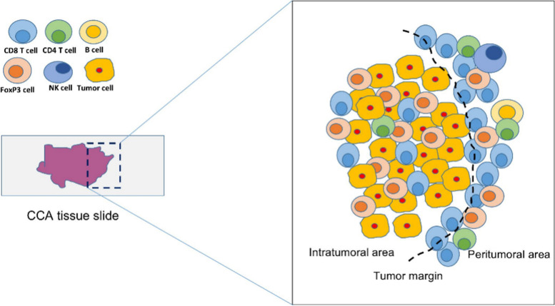
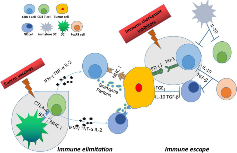
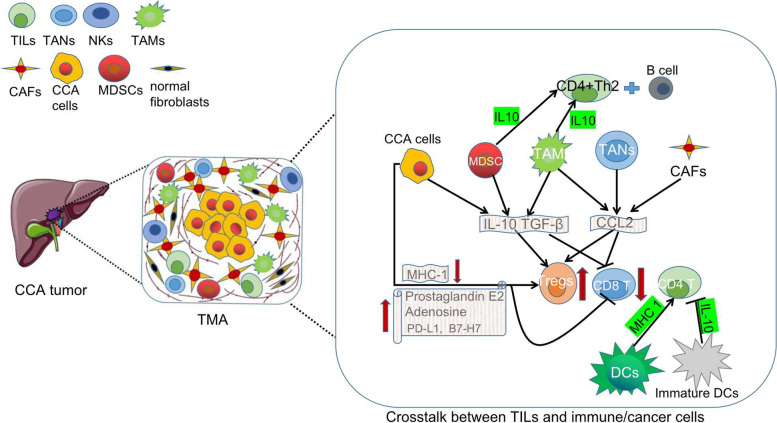
Cholangiocarcinoma (CCA) is the second most common primary liver cancer and associated with a dismal prognosis due to the lack of an efficient systemic therapy. In contrast to other cancers, new immunotherapies have demonstrated unsatisfactory results in clinical trials, underlining the importance of a deeper understanding of the special tumor microenvironment of CCA and the role of immune cells interacting with the tumor. Tumor-infiltrating lymphocytes (TILs) are an important component of the adaptive immune system and the foundation of current immunotherapy. Therefore, the aim of this systemic review is to summarize the current literature focusing on the proportions and distribution, molecular pathogenesis, prognostic significance of TILs and their role in immunotherapy for CCA patients.In CCA, CD8+ and CD4+ T lymphocytes represent the majority of TILs and are mostly sequestered around the cancer cells. CD20+ B lymphocytes and Natural Killer (NK) cells are less frequent. In contrast, Foxp3+ cells (regulatory T cells, Tregs) are observed to infiltrate into the tumor. In the immune microenvironment of CCA, cancer cells and stromal cells such as TAMs, TANs, MSDCs and CAFs inhibit the immune protection function of TILs by secreting factors like IL-10 and TGF-β. With respect to molecular pathogenesis, the Wnt/-catenin, TGF-signaling routes, aPKC-i/P-Sp1/Snail Signaling, B7-H1/PD-1Pathway and Fas/FasL signaling pathways are connected to the malignant potential and contributed to tumor immune evasion by increasing TIL apoptosis. Distinct subtypes of TILs show different prognostic implications for the long-term outcome in CCA. Although there are occasionally conflicting results, CD8+ and CD4+ T cells, and CD20+ B cells are positively correlated with the oncological prognosis of CCA, while a high number of Tregs is very likely associated with worse overall survival. TILs also play a major role in immunotherapy for CCA.In summary, the presence of TILs may represent an important marker for the prognosis and a potential target for novel therapy, but more clinical and translationaldata is needed to fully unravel the importance of TILs in the treatment of CCA.

Keywords: Cholangiocarcinoma, Tumor-infiltrating lymphocytes (TIL); Immunotherapy; Molecular pathogenesis; Oncological prognosis; Systematic review.


Conflict of interest statement

The authors declare no competing interests.

Figures

Fig. 1
Fig. 1
PRISMA flowchart of study selection for this systematic review
Fig. 2
Fig. 2
Spatial distribution of tumor-infiltrating lymphocytes in CCA. In CCA, CD8+ T lymphocytes represent the majority of T lymphocytes, whereas CD4+ T lymphocytes were also common. B lymphocytes are only seen occasionally. The total number of NK cells is also modest, though higher than B cells. While CD8+ and CD4+ cells are mainly distributed around the cancer, while Foxp3 cells infiltrate into the tumor. CCA, cholangiocarcinoma; CD; cluster of differentiation; Foxp3, forkhead box p3; NK, natural killer; TGF, transforming growth factor
Fig. 3
Fig. 3
Overview of different types of tumor-infiltrating lymphocytes in CCA. TILs are a highly heterogeneous group of lymphocytes. Distinct cell subsets play different roles in the tumor microenvironment. CD4+ cells are activated by reaction with peptide antigens delivered by major histocompatibility complex II (MHC II) and secrete cytokines such as IFN-γ, TNF-α and IL-2, which mediate cellular immunity and enhance the killing ability of NK cells and cytotoxic T cells. CD8+ cytotoxic T cells destroy tumor cells directly by releasing chemicals like perforin and granzyme and indirectly by inducing apoptosis by expressing FasL or secreting TNF-α attaching to target cell surface receptors. NK cells kill tumor cells by the same mechanisms as CD8+ cytotoxic T cells. Tregs suppress CD8+ cytotoxic T cells and NK cells by secreting soluble anti-inflammatory chemicals such as IL-10 and TGF-β. CD, cluster of differenciation; FasL, Fas ligand; IFN; interferon; IL, interleukin; MHC; major histocompatibility complex; NK, natural killer; TGF, transforming growth factor; TILs, tumor-infiltrating lymphocytes; TNF, tumor necrosis factor; Tregs; regulatory T cells
Fig. 4
Fig. 4
Overview of the crosstalk between TILs and immune/cancer cells in the tumor microenvironment Cancer cells, TAMs and MDSCs emit IL-10 and TGF-β, while TAMs, TANs and CAFs secrete CCL2 which attracts and expands Tregs inside the tumor bed and inhibits the activity of CD8+T cells. Cancer cells can also directly impair the immunoresponse by overexpressing prostaglandin E2, adenosine, PD-L1 or B7-H7 or by lowering MHC-I surface expression. IL10 released by MSDCs and TAMs favors a CD4+ Th2 response with B-cell engagement which are both effective cancer immunosurveillance mechanisms. Mature DCs promote CD4+ T cell activity by increasing MHC 1 expression while immature DCs inhibit CD4+ T activity by secreting IL-10. B7-H7, B7 homolog 7; CAFs, Cancer-associated fibroblasts; CCL,C–C motif chemokine ligand; CD, cluster of differentiation; DCs, Dendritic cells; IL, interleukin; MDSCs, Myeloid-derived suppressor cells; MHC; major histocompatibility complex; PD-L1, Programmed death-ligand 1; TANs, tumor associated neutrophils; TAMs, tumor-accociated macrophages; TGF, transforming growth factor; Tregs, Regulatory T cells

References

    1. Rizvi S, Gores GJ. Pathogenesis, diagnosis, and management of cholangiocarcinoma. Gastroenterology. 2013;145(6):1215–1229. - PMC - PubMed
    1. Anderson CD, Pinson CW, Berlin J, Chari RS. Diagnosis and treatment of cholangiocarcinoma. Oncologist. 2004;9(1):43–57. - PubMed
    1. Hyder O, Hatzaras I, Sotiropoulos GC, Paul A, Alexandrescu S, Marques H, et al. Recurrence after operative management of intrahepatic cholangiocarcinoma. Surgery. 2013;153(6):811–818. - PMC - PubMed
    1. Fabris L, Sato K, Alpini G, Strazzabosco M. The Tumor Microenvironment in Cholangiocarcinoma Progression. Hepatology. 2021;73(Suppl 1):75–85. - PMC - PubMed
    1. Woo SR, Corrales L, Gajewski TF. The STING pathway and the T cell-inflamed tumor microenvironment. Trends Immunol. 2015;36(4):250–256. - PMC - PubMed

MeSH terms