. 2022 Apr 7;8(1):175.
doi: 10.1038/s41420-022-00980-z.

Inhibition of BKCa channels protects neonatal hearts against myocardial ischemia and reperfusion injury

Affiliations

Inhibition of BKCa channels protects neonatal hearts against myocardial ischemia and reperfusion injury

Shridhar Sanghvi et al. Cell Death Discov. .

Abstract

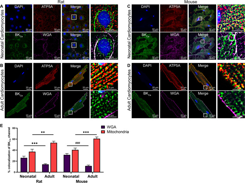
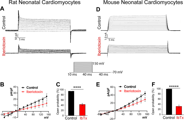
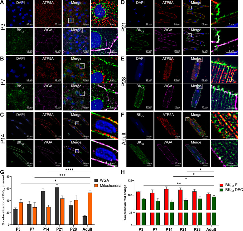
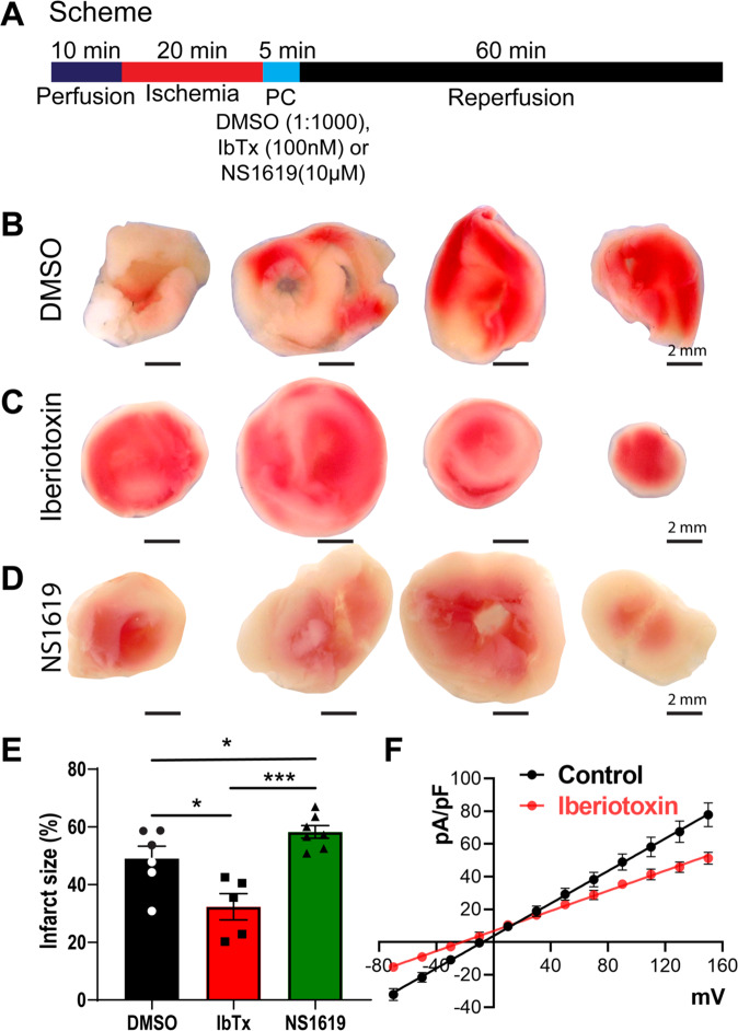
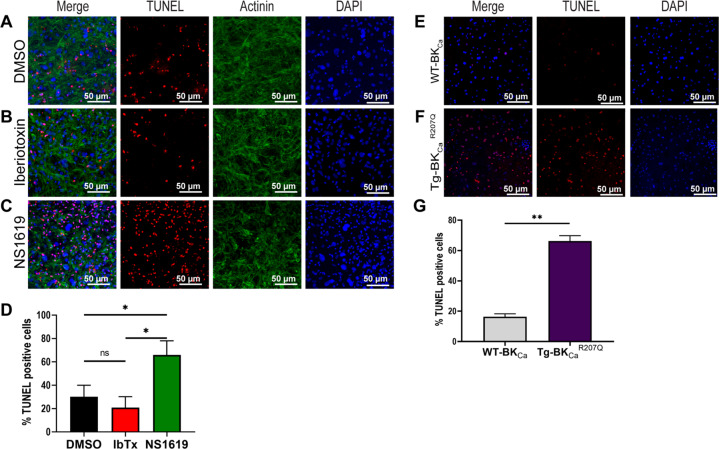
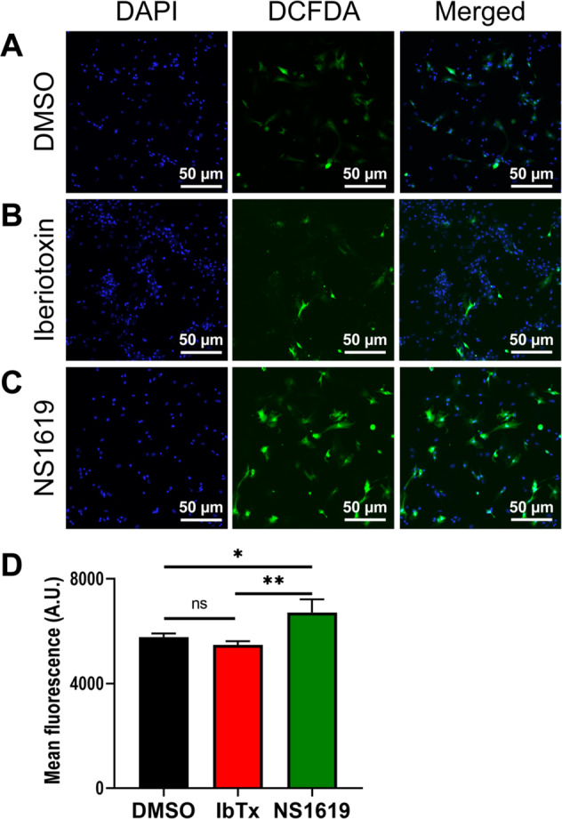
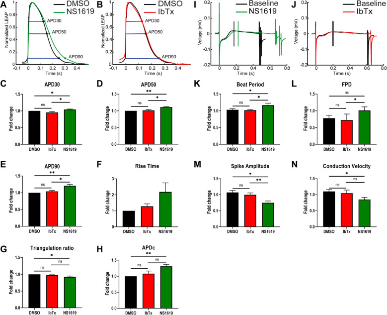
BKCa channels are large-conductance calcium and voltage-activated potassium channels that are heterogeneously expressed in a wide array of cells. Activation of BKCa channels present in mitochondria of adult ventricular cardiomyocytes is implicated in cardioprotection against ischemia-reperfusion (IR) injury. However, the BKCa channel's activity has never been detected in the plasma membrane of adult ventricular cardiomyocytes. In this study, we report the presence of the BKCa channel in the plasma membrane and mitochondria of neonatal murine and rodent cardiomyocytes, which protects the heart on inhibition but not activation. Furthermore, K+ currents measured in neonatal cardiomyocyte (NCM) was sensitive to iberiotoxin (IbTx), suggesting the presence of BKCa channels in the plasma membrane. Neonatal hearts subjected to IR when post-conditioned with NS1619 during reoxygenation increased the myocardial infarction whereas IbTx reduced the infarct size. In agreement, isolated NCM also presented increased apoptosis on treatment with NS1619 during hypoxia and reoxygenation, whereas IbTx reduced TUNEL-positive cells. In NCMs, activation of BKCa channels increased the intracellular reactive oxygen species post HR injury. Electrophysiological characterization of NCMs indicated that NS1619 increased the beat period, field, and action potential duration, and decreased the conduction velocity and spike amplitude. In contrast, IbTx had no impact on the electrophysiological properties of NCMs. Taken together, our data established that inhibition of plasma membrane BKCa channels in the NCM protects neonatal heart/cardiomyocytes from IR injury. Furthermore, the functional disparity observed towards the cardioprotective activity of BKCa channels in adults compared to neonatal heart could be attributed to their differential localization.


Conflict of interest statement

The authors declare no competing interests.

Figures

Fig. 1
Fig. 1. Localization of native BKCa channel in neonatal and cardiomyocytes isolated from the rodent heart.
A, B, C, and D Isolated cardiomyocytes loaded with WGA (magenta), fixed, permeabilized, and labeled with BKCa (green), and ATP5A (red), antibodies. Nuclei were stained with DAPI (blue). The right panels in A, B, C, and D show merged images at higher magnification. E The purple bar represents percentage colocalization between the BKCa channel and plasma membrane (WGA), which is significantly higher in neonatal cardiomyocytes compared to adult cardiomyocytes. The red bar represents percentage colocalization between the BKCa channel and mitochondria, which is lower in neonatal cardiomyocytes as compared to adult cardiomyocytes isolated from rodent hearts. All experiments were repeated independently at least four times and colocalization data are represented as mean ± SEM from 10 cells in each “n” number. P-values were determined by a one-tailed paired student’s t-test; **≤ 0.001, ***≤ 0.0001, ###≤ 1.0*10–8.
Fig. 2
Fig. 2. Rodent neonatal cardiomyocytes express functional BKCa channels.
Traces of whole-cell BKCa channel currents were recorded from A rat neonatal cardiomyocytes and D mouse neonatal cardiomyocytes with or without iberiotoxin (100 nM). Neonatal cardiomyocytes B rat and E mouse, membrane currents were activated by 40 ms voltage steps of 20 mV between −70 to +150 mV from resting −70 mV holding potential. The current versus voltage relationships are plotted with or without iberiotoxin. C and F Percentage block of BKCa current in the presence or absence of iberiotoxin. Cells were isolated from ten independent litters from mice and rats, and data are represented as mean ± SEM. P-values were determined by a one-tailed paired student’s t-test; ****≤ 0.00001, *****≤ 0.000001.
Fig. 3
Fig. 3. Mitochondrial targeting BKDEC splicing increases with age in neonatal cardiomyocytes.
AF Cardiomyocytes isolated from aging (P3, P7, P14, P21, P28, and adult) rats loaded with WGA (magenta), fixed, permeabilized, and labeled with BKCa (green) and ATP5A (red). The nucleus was stained with DAPI (blue). The right panels of AF show colocalization of merged images between BKCa to WGA and BKCa to ATP5A at higher magnification. G Protein proximity index analysis showing percentage colocalization of BKCa to WGA decreases, whereas BKCa to mitochondria increases in cardiomyocytes isolated from P3 to the adult stage. H Relative levels of BKCa FL and BKCa DEC mRNA expression in cardiomyocytes isolated from P3, P7, P14, P21, P28, and adult hearts were measured by qRT-PCR and were normalized to their relative GAPDH expression. Data represented as fold-change to P3 BKCa FL and P3 BKCa DEC; mean ± SEM; at least three independent experiments. P-values were determined by a one-tailed paired student’s t-test; *≤ 0.05, **≤ 0.001, ***≤ 0.0001, ****≤ 0.00001.
Fig. 4
Fig. 4. Inhibition of BKCa channel protects the neonatal heart from ischemia injury.
A Schematics of the ischemia-reperfusion protocol. Rat P6 pup hearts were subjected to 20 min ischemia, 5 min post-conditioned with DMSO, Iberiotoxin (100 nM), or NS1619 (10 μM) followed by 60 min of reperfusion. Hearts post-conditioned with C and E iberiotoxin exhibited significantly less infarction (white) as compared to the B and E DMSO control. In hearts post-conditioned with NS1619 displayed significantly higher infarction compared to DMSO control and IbTx treatment (E). The current versus voltage relationships are plotted with or without iberiotoxin (F). Data represented as mean ± SEM for percentage infarction from four independent experiments and electrophysiology data represented from four independent litters P6 rat pups. P-values were determined by a one-tailed paired student’s t-test; *≤ 0.05, ***≤ 0.0001.
Fig. 5
Fig. 5. Inhibition of BKCa channel protects the neonatal cardiomyocytes from apoptotic damage post hypoxia injury.
Isolated mouse neonatal cardiomyocytes were subjected to 6 h hypoxia at 1% O2, 5 min of post-conditioning with DMSO, Iberiotoxin (100 nM), or NS1619 (10 μM) followed by 12 h of reoxygenation at 21% O2. Cells were fixed and stained for TUNEL-positive nuclei (red), Actinin (green), and Nucleus (blue). A total of 100,000 cells were seeded per assay. B and D Cardiomyocytes post-conditioned with IbTx shows fewer TUNEL-positive nuclei compared to DMSO control (A and D). Cardiomyocytes post-conditioned with NS1619 exhibited a significantly higher number of TUNEL-positive nuclei compared to DMSO and IbTx (D). Cardiomyocytes isolated from E wild-type pups (WT-BKCa) or pups expressing F genetically activated BKCa (Tg-BKCaR207Q) were subjected to 6 h hypoxia at 1% O2 followed by 12 h of reoxygenation at 21% O2. Cells were fixed and stained for TUNEL-positive nuclei (red), and Nucleus (blue). Neonatal cardiomyocytes isolated from Tg-BKCaR207Q pups showed higher TUNEL-positive nuclei as compared to WT-BKCa (G). Data represented as percentage TUNEL-positive cells (D and G) mean ± SEM from eight out of ten independent experiments. P-values were determined by a one-tailed unpaired student’s t-test followed by a non-parametric Mann–Whitney test for comparison between the ranks; ns is not significant, *≤ 0.05, **≤ 0.001.
Fig. 6
Fig. 6. Inhibition of BKCa channel protects the neonatal cardiomyocytes from oxidative damage post hypoxia injury.
Isolated rat neonatal cardiomyocytes were subjected to 6 h hypoxia at 1% O2, 5 min of post-conditioning with DMSO, Iberiotoxin (100 nM), or NS1619 (10 μM) followed by 12 h of reoxygenation. Cells were stained with CM-H2DCFDA (green) for 45 min and NucBlue™ for nucleus (blue). A total of 100,000 cells were seeded per assay. B and D Cardiomyocytes post-conditioned with IbTx show less cellular ROS compared to DMSO control (A and D). C Cardiomyocytes post-conditioned with NS1619 exhibited significantly higher ROS generation as compared to IbTx and DMSO control and quantified (D). Data represented as mean fluorescence unit ± SEM from eight of ten independent experiments. P-values were determined by a one-tailed unpaired student’s t-test followed by a non-parametric Mann–Whitney test for comparison between the ranks; ns is not significant, *≤ 0.05, **≤ 0.001.
Fig. 7
Fig. 7. Functional analysis of activation and inhibition BKCa channel in p3 neonatal cardiomyocytes on multi-electrode array (MEA) systems.
Rat neonatal cardiomyocytes plated on classic MEA 24-well plates containing electrodes were dosed with DMSO, NS1619, or IbTX. Normalized LEAP waveforms for NS1619 (A; green trace) and IbTx (B; red trace) were overlaid with DMSO control (black trace). Bar plots represent C APD30, D APD50, E APD90, F rise time, G triangulation ratio, and H APDc for IbTX and NS1619 normalized to DMSO group and represented as fold-change mean ± SEM. Field potential duration waveform (I, J of neonatal cardiomyocytes post-treatment with NS1619 (I; green trace), and IbTx (J; red trace) overlaid with their respective pre-treatment baseline electrodes. Bar plots represent K beat period, L field potential duration, M spike amplitude, and N conduction velocity for DMSO, IbTx, and NS1619 were normalized to their respective pre-treatment electrodes and represented as fold-change mean ± SEM from three independent littles. P-values were determined by a one-tailed paired student’s t-test; ns is not significant, *≤ 0.05, **≤ 0.001.
Fig. 8
Fig. 8. Plasma membrane localization of BKCa channel in human infant hearts.
Human infant heart sections were fixed and labeled with A BKCa (green), B WGA (red), and C Nuclei (blue). The bottom panels A’–D’ are shown at higher magnification with colocalization of BKCa to WGA in D and D’.

Similar articles

Cited by

References

    1. Becker MN, Brenner R, Atkinson NS. Tissue-specific expression of a Drosophila calcium-activated potassium channel. J Neurosci: Off J Soc Neurosci. 1995;15:6250–9. doi: 10.1523/JNEUROSCI.15-09-06250.1995. - DOI - PMC - PubMed
    1. Gururaja Rao S, Bednarczyk P, Towheed A, Shah K, Karekar P, Ponnalagu D, et al. BKCa (Slo) channel regulates mitochondrial function and lifespan in drosophila melanogaster. Cells. 2019;8:945–64. - PMC - PubMed
    1. Butler A, Tsunoda S, McCobb DP, Wei A, Salkoff L. mSlo, a complex mouse gene encoding “maxi” calcium-activated potassium channels. Science. 1993;261:221–4. doi: 10.1126/science.7687074. - DOI - PubMed
    1. Bailey CS, Moldenhauer HJ, Park SM, Keros S, Meredith AL. KCNMA1-linked channelopathy. J Gen Physiol. 2019;151:1173–89. doi: 10.1085/jgp.201912457. - DOI - PMC - PubMed
    1. Li B, Jie W, Huang L, Wei P, Li S, Luo Z, et al. Nuclear BK channels regulate gene expression via the control of nuclear calcium signaling. Nat Neurosci. 2014;17:1055–63. doi: 10.1038/nn.3744. - DOI - PMC - PubMed