. 2022 Apr 7;12(1):5875.
doi: 10.1038/s41598-022-09589-9.

SH3-domain mutations selectively disrupt Csk homodimerization or PTPN22 binding

Affiliations

SH3-domain mutations selectively disrupt Csk homodimerization or PTPN22 binding

Ben F Brian 4th et al. Sci Rep. .

Abstract

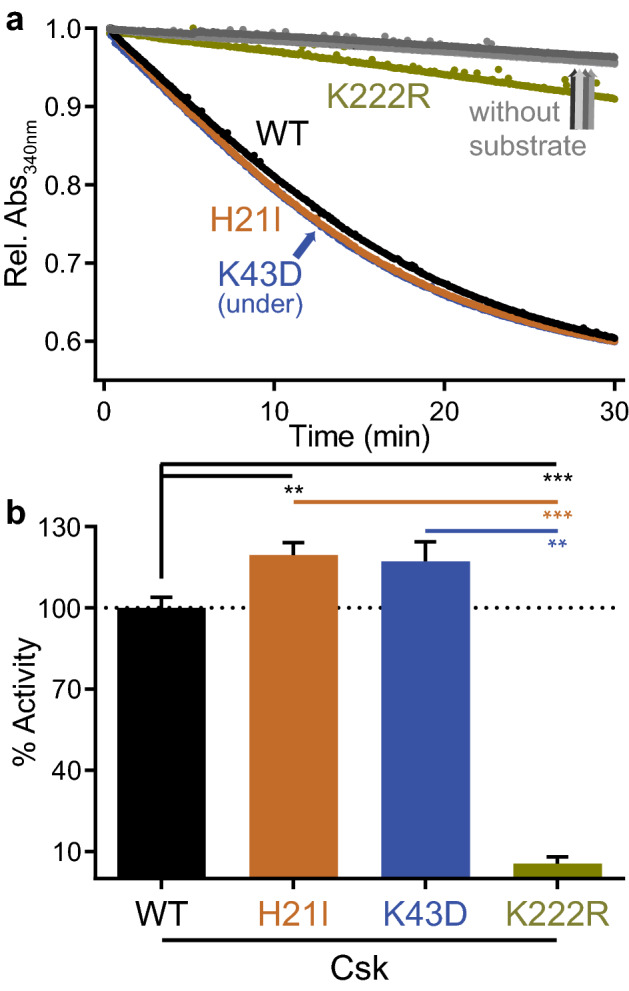
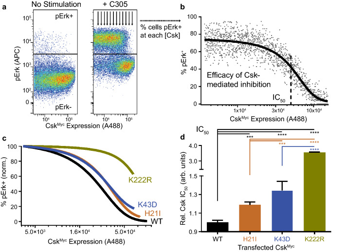
The kinase Csk is the primary negative regulator of the Src-family kinases (SFKs, e.g., Lck, Fyn, Lyn, Hck, Fgr, Blk, Yes), phosphorylating a tyrosine on the SFK C-terminal tail that mediates autoinhibition. Csk also binds phosphatases, including PTPN12 (PTP-PEST) and immune-cell PTPN22 (LYP/Pep), which dephosphorylate the SFK activation loop to promote autoinhibition. Csk-binding proteins (e.g., CBP/PAG1) oligomerize within membrane microdomains, and high local concentration promotes Csk function. Purified Csk homodimerizes in solution through an interface that overlaps the phosphatase binding footprint. Here we demonstrate that Csk can homodimerize in Jurkat T cells, in competition with PTPN22 binding. We designed SH3-domain mutations in Csk that selectively impair homodimerization (H21I) or PTPN22 binding (K43D) and verified their kinase activity in solution. Disruption of either interaction in cells, however, decreased the negative-regulatory function of Csk. Csk W47A, a substitution previously reported to block PTPN22 binding, had a secondary effect of impairing homodimerization. Csk H21I and K43D will be useful tools for dissecting the protein-specific drivers of autoimmunity mediated by the human polymorphism PTPN22 R620W, which impairs interaction with Csk and with the E3 ubiquitin ligase TRAF3. Future investigations of Csk homodimer activity and phosphatase interactions may reveal new facets of SFK regulation in hematopoietic and non-hematopoietic cells.


Conflict of interest statement

The authors declare no competing interests.

Figures

Figure 1
Figure 1
Csk self-associates when overexpressed in Jurkat cells. (a) Immunoblots of anti-Myc immunoprecipitates (IP) from transiently transfected Jurkat T cells. Transfection conditions include epitope-tagged Csk constructs (CskMyc, CskHA) and empty pEF6 vector (–) as indicated. Representative of at least three independent experiments. (b) Immunoblots showing transfected CskMyc and CskHA and endogenous Erk1/2 loading control in whole-cell lysate (w) and Myc-immunodepleted lysate (dep) from the immunoprecipitations above.
Figure 2
Figure 2
H21I substitution in the SH3 domain of Csk selectively disrupts self-association. (a) Position of the SH3 domain in full-length Csk (PDB ID: 1K9A). (b) Csk residue H21 in the SH3/SH3 homodimer interface (PDB ID 1CSK). The position of PTPN22 peptide binding to the SH3 domain was derived in PyMol using PDB IDs 1K9A 1CSK, 1JEG, 1QWE, and 2P6X. (c) Representative immunoblots of whole-cell or Myc-immunodepleted lysates from Jurkat cells transfected with CskHA and CskMyc constructs (both WT or both H21I). (d) Corresponding immunoblots of transfected Csk and endogenous PTPN22 in Myc immunoprecipitates. (e) Quantifications from immunoprecipitate blots, corrected for CskMyc pulldown in each sample, shown relative to WT. Error bars: standard error of the mean (SEM), n = 4 independent experiments. Significance: one-way ANOVA with Tukey’s multiple comparison test (Sig.ANOVA) ****p < 0.0001, ***p = 0.0001 or 0.0008.
Figure 3
Figure 3
Mutation of the homodimer interface decreases the apparent molecular weight of Csk in solution. (a) Colloidal blue staining (reducing SDS PAGE, inset) and migration through a Superdex 200 size-exclusion column of purified, recombinant Csk WT and H21I. (b) The apparent molecular weight of each Csk variant in solution calculated from column partitioning of proteins with known molecular weights.
Figure 4
Figure 4
Unlike W47A, K43D substitution in the SH3 domain of Csk selectively disrupts PTPN22 binding. (a) Csk residue K43D in the secondary binding interface between PTPN22 and the SH3 domain of Csk. The binding site for the proline (P) residues in the peptide PXXP motif, including Csk W47A, overlap with the dimer footprint; only one of the proline residues is engaged in this structure. (b) Representative immunoprecipitate blots from Jurkat cells transfected with CskHA and CskMyc constructs (both WT or both K43D). (c) Quantifications from immunoprecipitate blots, corrected for CskMyc pulldown in each sample, shown relative to WT. Error bars: SEM, n = 3. Sig.ANOVA *p = 0.0133 or 0.0451. (d) Representative immunoprecipitate blots from Jurkat cells transfected with CskHA and CskMyc constructs (both WT or both W47A). Boxed images cropped from non-adjacent lanes of the same blot with brightness/contrast corrections applied uniformly prior to cropping. (e) Quantifications from immunoprecipitate blots, corrected as above. Error bars: SEM, n = 4. Sig.ANOVA **p = 0.0010, *p = 0.0282.
Figure 5
Figure 5
H21I and K43D substitutions do not impair Csk kinase activity in solution. (a) Continuous spectrophotometric assay reporting ADP production during peptide phosphorylation. Reactions were initiated by adding purified Csk (WT, H21I, K43D, or K222R) to the reaction mix. Control reactions without substrate peptide are also shown. (b) Linear velocities of the initial kinase reaction relative to WT. Error bars: SEM, n = 4.
Figure 6
Figure 6
Loss of Csk dimerization may impair its function in T cells. (a) Jurkat T cells transiently transfected with CskMyc and rested (left) or TCR-stimulated for 2 min using C305 antibody (right) and stained for intracellular Myc and pErk; within the Myc+ population, the cutoff for pErk positivity is indicated (horizontal line). Arrows depict quantification of % pErk+ cells in bins of increasing CskMyc expression. (b) % Erk positivity vs. CskMyc expression. The apparent IC50 for Csk suppression (dotted line) was obtained by fitting to a sigmoidal dose–response curve. (c) Representative dose–response curves showing the relative efficacies of transfected CskMyc constructs (WT, H21I, K43D, or K222R) in suppressing TCR-induced Erk phosphorylation. (d) Relative IC50 values for Csk constructs Due to incomplete suppression of TCR signaling, K222R fits were constrained so the lower baseline and slope mirrored the average fit values from the other samples. Error bars: SEM, n = 3 (K222R), 6 (K43D), 21 (H21I), 27 (WT) independently transfected and stimulated samples. Mix-effects analysis was performed using GraphPad Prism software, with Tukey’s test for multiple comparisons ****p < 0.0001, ***p = 0.0001.

References

    1. Okada M. Regulation of the SRC family kinases by Csk. Int. J. Biol. Sci. 2012;8:1385–1397. doi: 10.7150/ijbs.5141. - DOI - PMC - PubMed
    1. Brown MT, Cooper JA. Regulation, substrates and functions of src. Biochim. Biophys. Acta. 1996;1287:121–149. - PubMed
    1. Chow LM, Fournel M, Davidson D, Veillette A. Negative regulation of T-cell receptor signalling by tyrosine protein kinase p50csk. Nature. 1993;365:156–160. doi: 10.1038/365156a0. - DOI - PubMed
    1. Davidson D, Bakinowski M, Thomas ML, Horejsi V, Veillette A. Phosphorylation-dependent regulation of T-cell activation by PAG/Cbp, a lipid raft-associated transmembrane adaptor. Mol. Cell Biol. 2003;23:2017–2028. doi: 10.1128/MCB.23.6.2017-2028.2003. - DOI - PMC - PubMed
    1. Kawabuchi M, et al. Transmembrane phosphoprotein Cbp regulates the activities of Src-family tyrosine kinases. Nature. 2000;404:999–1003. doi: 10.1038/35010121. - DOI - PubMed

MeSH terms

Substances