

Quantum dots based in-vitro co-culture cancer model for identification of rare cancer cell heterogeneity
- PMID: 35393460
- PMCID: PMC8991261
- DOI: 10.1038/s41598-022-09702-y
Quantum dots based in-vitro co-culture cancer model for identification of rare cancer cell heterogeneity
Abstract
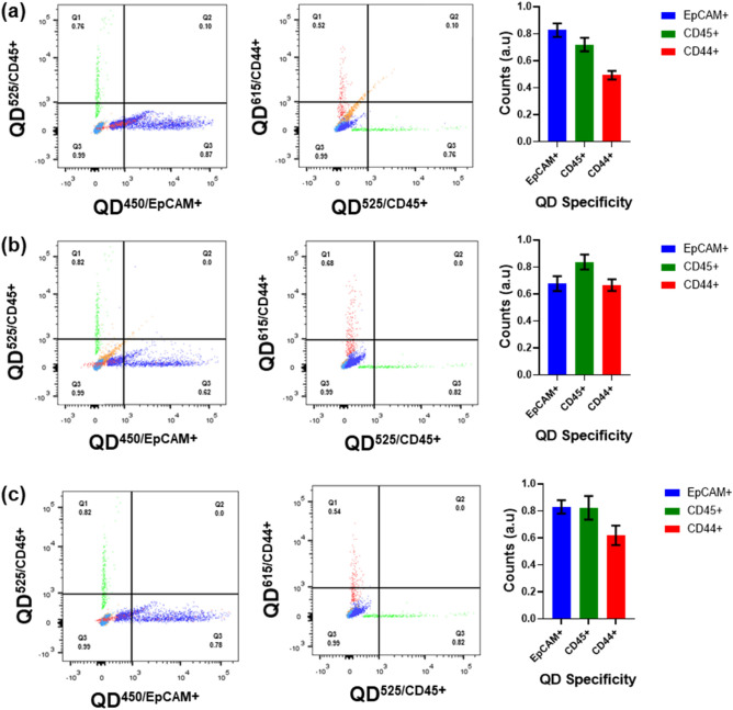
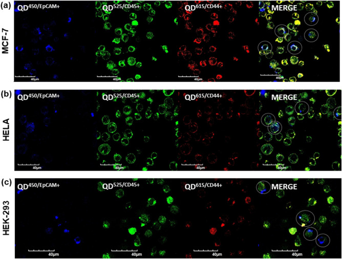
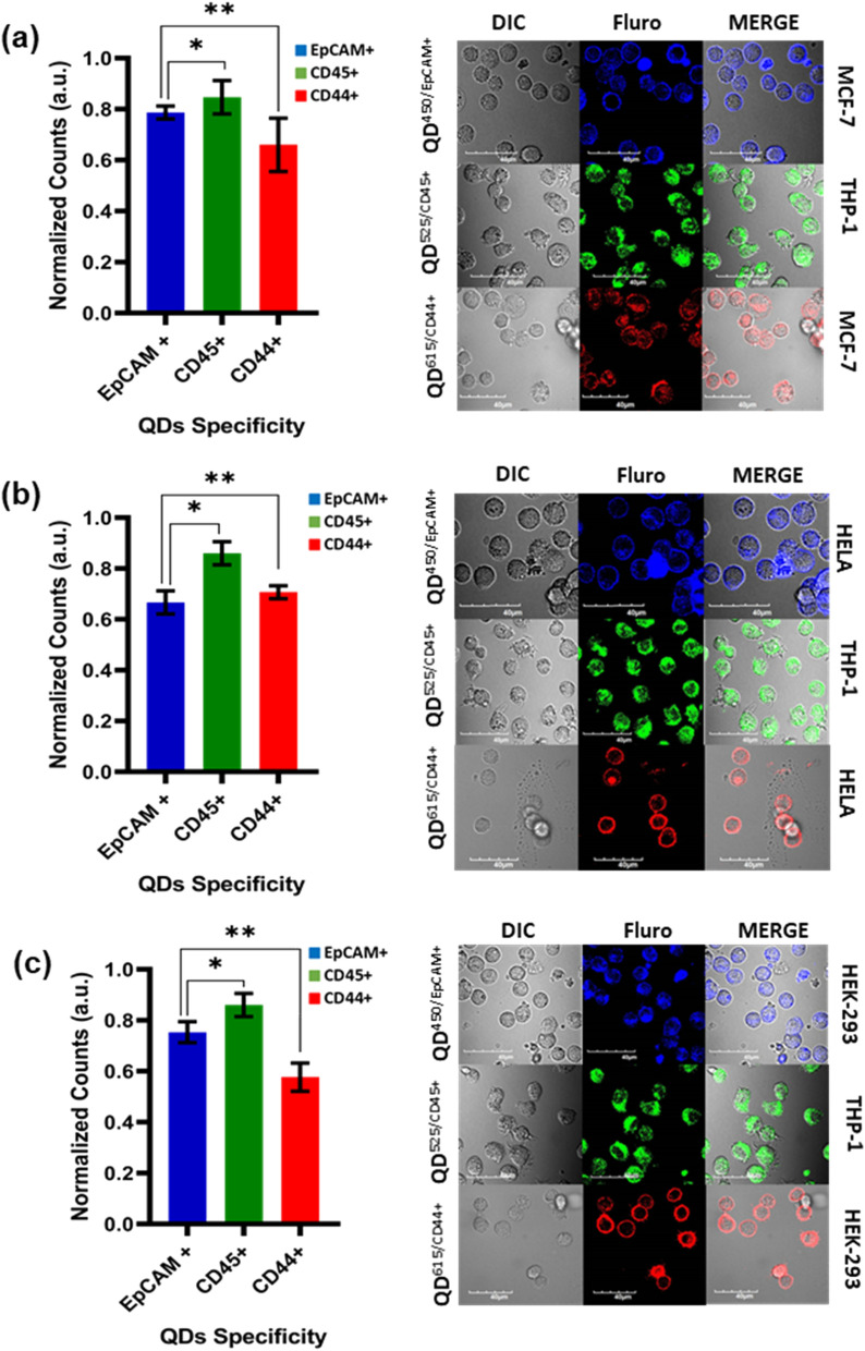
Cancer cell heterogeneity (CCH) is crucial in understanding cancer progression and metastasis. The CCH is one of the stumbling blocks in modern medicine's therapeutics and diagnostics . An in-vitro model of co-culture systems of MCF-7, HeLa, HEK-293, with THP-1 cells showed the occurrence of EpCAM positive (EpCAM+) and EpCAM negative (EpCAM-) heterogenetic cancer cell types labeled with the Quantum Dot antibody conjugates (QDAb). This in-vitro model study could provide insights into the role of rare cancer cells manifestation and their heterogeneity in metastatic progression and risk for severe infections in these patients. We successfully report the presence of CCH based on the fluorescence ratios of the co-cultured cancer cells when treated with the QDAb. These short-term mimic co-cultures give a compelling and quite associated model for assessing early treatment responses in various cancers.
© 2022. The Author(s).
Conflict of interest statement
The authors declare no competing interests.
Figures
Similar articles
-
Hybrid silica-coated Gd-Zn-Cu-In-S/ZnS bimodal quantum dots as an epithelial cell adhesion molecule targeted drug delivery and imaging system.Int J Pharm. 2019 Oct 30;570:118645. doi: 10.1016/j.ijpharm.2019.118645. Epub 2019 Aug 26. Int J Pharm. 2019. PMID: 31465835
-
Optical Aptamer Probes of Fluorescent Imaging to Rapid Monitoring of Circulating Tumor Cell.Sensors (Basel). 2016 Nov 23;16(11):1909. doi: 10.3390/s16111909. Sensors (Basel). 2016. PMID: 27886058 Free PMC article.
-
A fluorescence turn-on biosensor based on graphene quantum dots (GQDs) and molybdenum disulfide (MoS2) nanosheets for epithelial cell adhesion molecule (EpCAM) detection.Biosens Bioelectron. 2017 Jul 15;93:182-188. doi: 10.1016/j.bios.2016.09.012. Epub 2016 Sep 4. Biosens Bioelectron. 2017. PMID: 27614683
-
Revisiting the Roles of Pro-Metastatic EpCAM in Cancer.Biomolecules. 2020 Feb 7;10(2):255. doi: 10.3390/biom10020255. Biomolecules. 2020. PMID: 32046162 Free PMC article. Review.
-
Shedding light on the EpCAM: An overview.J Cell Physiol. 2019 Aug;234(8):12569-12580. doi: 10.1002/jcp.28132. Epub 2019 Jan 9. J Cell Physiol. 2019. PMID: 30628064 Review.
Cited by
-
Synergistic anti-diabetic effect of phloroglucinol and total procyanidin dimer isolated from Vitisvinifera methanolic seed extract potentiates via suppressing oxidative stress: in-vitro evaluation studies.3 Biotech. 2024 Mar;14(3):76. doi: 10.1007/s13205-024-03929-4. Epub 2024 Feb 14. 3 Biotech. 2024. PMID: 38371900 Free PMC article.
-
Applications of Quantum Dots in Preventive Oncology.Asian Pac J Cancer Prev. 2024 Mar 1;25(3):747-756. doi: 10.31557/APJCP.2024.25.3.747. Asian Pac J Cancer Prev. 2024. PMID: 38546057 Free PMC article. Review.
-
Isolation and anticancer activity of quercetin from Acalypha indica L. against breast cancer cell lines MCF-7 and MDA-MB-231.3 Biotech. 2023 Aug;13(8):289. doi: 10.1007/s13205-023-03705-w. Epub 2023 Aug 2. 3 Biotech. 2023. PMID: 37547624 Free PMC article.
References
Publication types
MeSH terms
Substances
LinkOut - more resources
Full Text Sources
Medical
Miscellaneous
