Review
. 2022 Jul;22(7):414-430.
doi: 10.1038/s41568-022-00466-1. Epub 2022 Apr 7.

Tumour-infiltrating B cells: immunological mechanisms, clinical impact and therapeutic opportunities

Affiliations
Review

Tumour-infiltrating B cells: immunological mechanisms, clinical impact and therapeutic opportunities

Céline M Laumont et al. Nat Rev Cancer. 2022 Jul.

Abstract

Although immunotherapy research to date has focused largely on T cells, there is mounting evidence that tumour-infiltrating B cells and plasma cells (collectively referred to as tumour-infiltrating B lymphocytes (TIL-Bs)) have a crucial, synergistic role in tumour control. In many cancers, TIL-Bs have demonstrated strong predictive and prognostic significance in the context of both standard treatments and immune checkpoint blockade, offering the prospect of new therapeutic opportunities that leverage their unique immunological properties. Drawing insights from autoimmunity, we review the molecular phenotypes, architectural contexts, antigen specificities, effector mechanisms and regulatory pathways relevant to TIL-Bs in human cancer. Although the field is young, the emerging picture is that TIL-Bs promote antitumour immunity through their unique mode of antigen presentation to T cells; their role in assembling and perpetuating immunologically 'hot' tumour microenvironments involving T cells, myeloid cells and natural killer cells; and their potential to combat immune editing and tumour heterogeneity through the easing of self-tolerance mechanisms. We end by discussing the most promising approaches to enhance TIL-B responses in concert with other immune cell subsets to extend the reach, potency and durability of cancer immunotherapy.


Conflict of interest statement

Competing interests

The authors declare no competing interests.

Figures

Fig. 1 |
Fig. 1 |. B cells in health and malignancy.
a | Heat maps comparing abundance of B cells and plasma cells (PCs) in normal and tumour tissues. Data from Genotype-Tissue Expression consortium (GTEx v. 8, n = 16,704 samples) and The Cancer Genome Atlas (TCGA) consortium (harmonized dataset, n = 9,922 primary solid tumours) were analysed using xCell to generate cell type enrichment scores for naive B cells (B naive), memory B cells (B memory) and PCs. Scores averaged across samples of the same origin, scaled across columns and ranked by decreasing order. Asterisk indicates an identical normal tissue comparator was not available, so a closely related tissue was used instead. Log-fold change for B cell (average of naive B cells and memory B cells) and PC scores between tumours and their normal counterparts are reported (right). Red, tumour tissue > normal tissue; no colour, tumour tissue ~ normal tissue; blue, tumour tissue < normal tissue. b | Heat map summarizing prognostic associations for intratumoural B cells, PCs, IgG and IgA based on an update of previous literature searches,. Tumour sites are deemed positive, neutral, or negative based on the majority of cohorts. Supplementary Tables 1 and 2 provide supporting data and references. ACC, adrenocortical carcinoma; BLCA, bladder urothelial carcinoma; BRCA, breast invasive carcinoma; CESC, cervical squamous cell carcinoma and endocervical adenocarcinoma; CHOL, cholangiocarcinoma; COAD, colon adenocarcinoma; ESCA, oesophageal carcinoma; GBM, glioblastoma multiforme; HNSC, head and neck squamous cell carcinoma; KICH, kidney chromophobe; KIRC, kidney renal clear cell carcinoma; KIRP, kidney renal papillary cell carcinoma; LGG, brain lower grade glioma; LIHC, liver hepatocellular carcinoma; LUAD, lung adenocarcinoma; LUSC, lung squamous cell carcinoma; MESO, mesothelioma; OV, ovarian serous cystadeno-carcinoma; PAAD, pancreatic adenocarcinoma; PCPG, pheochromocytoma and paraganglioma; PRAD, prostate adenocarcinoma; READ, rectum adenocarcinoma; SARC, sarcoma; SKCM, skin cutaneous melanoma and squamous cell carcinoma; STAD, stomach adenocarcinoma; TGCT, testicular germ cell tumours; THCA, thyroid carcinoma; THYM, thymoma; UCEC, uterine corpus endometrial carcinoma; UCS, uterine carcinosarcoma; UVM, uveal melanoma.
Fig. 2 |
Fig. 2 |. B cell differentiation and tolerance.
a | Stages of B cell activation, clonal expansion, somatic hypermutation, affinity selection, class switch recombination and differentiation to plasma cells (PCs). These events can involve germinal centre reactions or extrafollicular responses, with the former generally being the better source of long-lived PCs. All of these B cell subsets are found in human tumours, with the most commonly reported subsets being activated B cells, memory B cells and PCs. b | Central tolerance takes place in bone marrow and eliminates immature self-reactive B cells via clonal deletion; receptor editing of immunoglobulin light chains (L chains) offers one chance at rescue. Peripheral tolerance maintains weakly and moderately self-reactive B cells in a hypo-responsive or inactive state through several mechanisms. B cell responses to foreign antigens (or potentially neoantigens in cancer) can occur through conventional activation or clonal redemption. Disruption of peripheral tolerance during inflammation, autoimmunity or cancer can license self-reactive B cells and PCs to participate in immune responses. With the exception of human papillomavirus (HPV) antigens and tumour protein p53 (p53) neoantigens (although most p53-specific autoantibodies appear to recognize non-mutated epitopes), studies to date indicate that TIL-Bs generally recognize self-antigens with varying degrees of tumour-specific expression. BCR, B cell receptor; H chain, heavy chain; NA, not applicable; TIL-B, tumour-infiltrating B lymphocyte; Treg cell, regulatory T cell.
Fig. 3 |
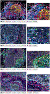
Fig. 3 |. TIL-B neighbourhoods.
ad | Multiplex immunofluorescence staining of untreated human high-grade serous ovarian cancer (parts ac) and medullary breast cancer tissues (part d) showing examples of a secondary follicular tertiary lymphoid structure (part a), astromal lympho-myeloid aggregate (LMA) with infiltration of a neighbouring epithelial region by CD4 and CD8 T cells and B cells (part b), a dense infiltrate of B cells restricted to the stromal compartment despite infiltration of adjacent epithelium by T cells and macrophages (part c) and a dense stromal infiltrate of plasma cells (PCs) accompanied by intra-epithelial B cells (part d). Left column: low-magnification views with relevant structural zones labelled. Right column: high-magnification views of boxed regions from left column showing examples of CD8 T cell (CD8+CD3+), presumptive CD4 T cell (CD8CD3+), dendritic cell (CD201+), follicular dendritic cell (FDC; CD21+), high endothelial venule (HEV; PNAd+), B cell (CD20+), PC (CD20CD79A+), macrophage (CD68+) and regulatory T cells (Treg cells; CD3+FOXP3+). Markers and associated colours specified below each image. Epithelium, tumour epithelium; FOXP3, forkhead box protein P3; PanCK, pan cytokeratin; PNAd, peripheral node addressin; stroma, tumour stroma; TIL-B, tumour-infiltrating B lymphocyte. Image courtesy of Katy Milne, Tashi Rastogi and Karanvir Singh, BC Cancer.
Fig. 4 |
Fig. 4 |. Model for intramolecular epitope spreading from neo-epitopes to self-epitopes in cancer.
Top panel: ah | Antigen released by tumour cells (part a) is taken up by professional antigen presenting cells (APCs) (part b) which then prime neo-epitope-specific CD4 T follicular helper (TFH) cells and/or T peripheral helper (TPH) cells as well as CD8 T cells. TFH cells and/or TPH cells prime a B cell response against a self-epitope on the same antigen (part c), triggering clonal expansion and plasma cell (PC) differentiation (part d). PC-derived autoantibodies form immune complexes with antigen, which facilitates antigen uptake and presentation by APCs (part e). Additional self-reactive CD4 and CD8 T cells undergo priming, expansion and differentiation (part f). TFH cells and/or TPH cells prime and amplify self-reactive B cells (part g), leading to further epitope spreading (part h). Bottom panel: neo-epitope-specific CD8 T cell (cell 1) eliminates neoantigen-expressing tumour cells. Without epitope spreading, this can result in outgrowth of variant tumour cells that no longer express the neoantigen. With epitope spreading, CD4 and CD8 T cells (cell 2, 3 and 4) can additionally target self-epitopes which may drive responses less susceptible to immune editing. Moreover, orthogonal antibody-mediated effector mechanisms can be engaged, including complement-dependent cytotoxicity (CDC), antibody-dependent cellular cytotoxicity (ADCC) and antibody-dependent cellular phagocytosis (ADCP), resulting in better tumour control. BCR, B cell receptor; MHC, major histocompatibility complex; TCR, T cell receptor.
Fig. 5 |
Fig. 5 |. TIL-B effector mechanisms.
a–e | Five major categories of effector mechanism. ADCC, antibody-dependent cellular cytotoxicity; ADCP, antibody-dependent cellular phagocytosis; ADO, adenosine; 4–1BBL, 4–1BB ligand; CCL, C–C motif chemokine ligand; CDC, complement-dependent cytotoxicity; FASLG, FAS ligand; GABA, γ-aminobutyric acid; GZMB, granzyme B; ICD, immunogenic cell death; ICOS, inducible T cell co-stimulatory; ICOSL, ICOS ligand; IFNγ, interferon-γ; LMA, lympho-myeloid aggregate; LTαβ, lymphotoxin-αβ; mAb, monoclonal antibody; MAC, membrane attack complex; MHC, major histocompatibility complex; NK cell, natural killer cell; OX40L, OX40 ligand; PD-L1, programmed cell death 1 ligand 1; pIgR, polymeric immunoglobulin receptor; TCR, T cell receptor; TFH cell, T follicular helper cell; TGFβ, tumour growth factor-β; TH1 cell, T helper 1 cell; TIL-B, tumour-infiltrating B lymphocyte; TLS, tertiary lymphoid structure; TNF, tumour necrosis factor; TPH cell, T peripheral helper cell; TRAIL (or TNFSF10), TNF superfamily member 10; Treg cell, regulatory T cell; TRIM21, tripartite motif containing 21.

References

    1. Thommen DS et al. A transcriptionally and functionally distinct PD-1+CD8+ T cell pool with predictive potential in non-small-cell lung cancer treated with PD-1 blockade. Nat. Med 24, 994–1004 (2018). - PMC - PubMed
    1. Workel HH et al. A transcriptionally distinct CXCL13+CD103+CD8+ T-cell population is associated with B-cell recruitment and neoantigen load in human cancer. Cancer Immunol. Res 7, 784–796 (2019). - PubMed
    1. Li H et al. Dysfunctional CD8 T cells form a proliferative, dynamically regulated compartment within human melanoma. Cell 176, 775–789 (2019). - PMC - PubMed
    1. Sautès-Fridman C, Petitprez F, Calderaro J & Fridman WH Tertiary lymphoid structures in the era of cancer immunotherapy. Nat. Rev. Cancer 19, 307–325 (2019). - PubMed
    1. Jhunjhunwala S, Hammer C & Delamarre L Antigen presentation in cancer: insights into tumour immunogenicity and immune evasion. Nat. Rev. Cancer 21, 298–312 (2021). - PubMed

Publication types