Review
. 2022 Jun;27(6):2674-2688.
doi: 10.1038/s41380-022-01531-9. Epub 2022 Apr 7.

Dissecting the clinical heterogeneity of early-onset Alzheimer's disease

Affiliations
Review

Dissecting the clinical heterogeneity of early-onset Alzheimer's disease

Daniel W Sirkis et al. Mol Psychiatry. 2022 Jun.

Abstract

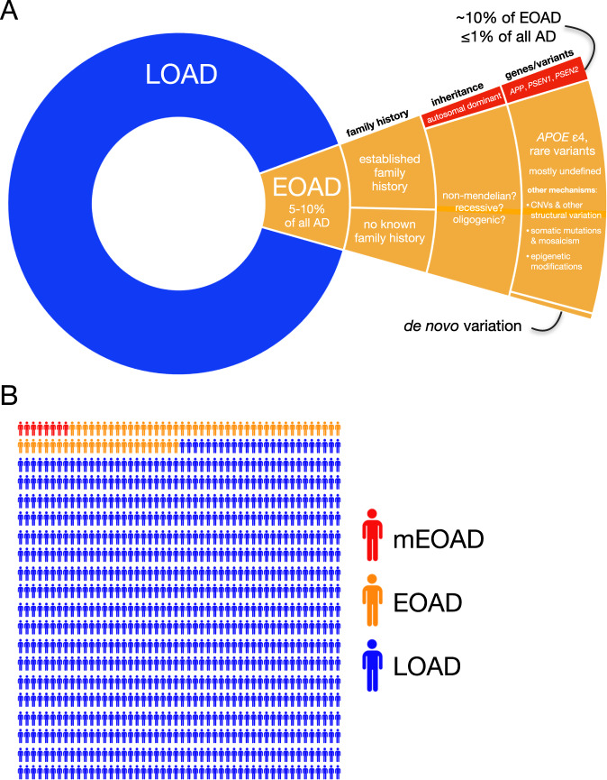
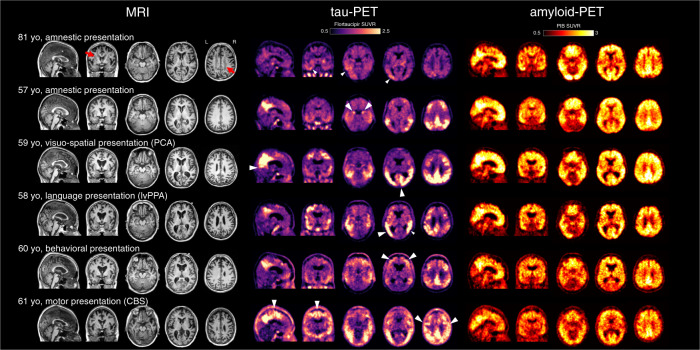
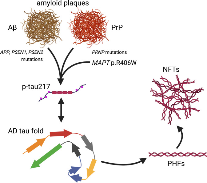
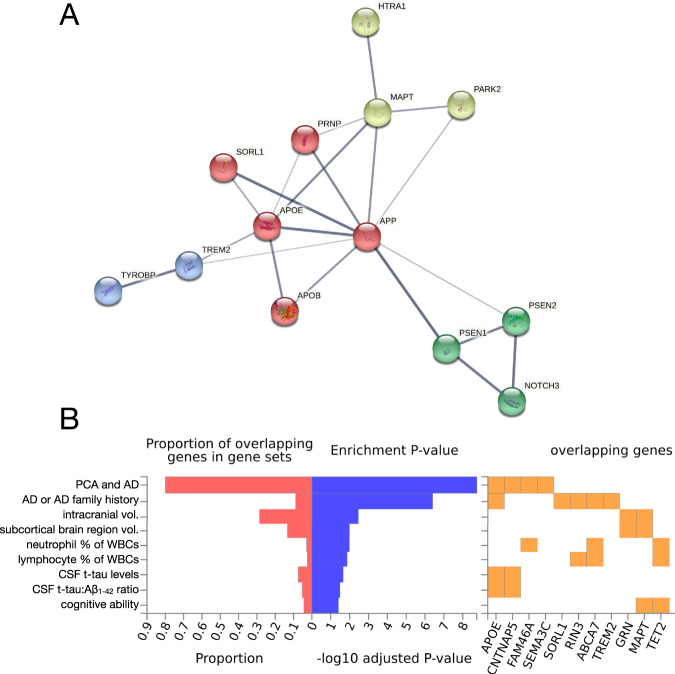
Early-onset Alzheimer's disease (EOAD) is a rare but particularly devastating form of AD. Though notable for its high degree of clinical heterogeneity, EOAD is defined by the same neuropathological hallmarks underlying the more common, late-onset form of AD. In this review, we describe the various clinical syndromes associated with EOAD, including the typical amnestic phenotype as well as atypical variants affecting visuospatial, language, executive, behavioral, and motor functions. We go on to highlight advances in fluid biomarker research and describe how molecular, structural, and functional neuroimaging can be used not only to improve EOAD diagnostic acumen but also enhance our understanding of fundamental pathobiological changes occurring years (and even decades) before the onset of symptoms. In addition, we discuss genetic variation underlying EOAD, including pathogenic variants responsible for the well-known mendelian forms of EOAD as well as variants that may increase risk for the much more common forms of EOAD that are either considered to be sporadic or lack a clear autosomal-dominant inheritance pattern. Intriguingly, specific pathogenic variants in PRNP and MAPT-genes which are more commonly associated with other neurodegenerative diseases-may provide unexpectedly important insights into the formation of AD tau pathology. Genetic analysis of the atypical clinical syndromes associated with EOAD will continue to be challenging given their rarity, but integration of fluid biomarker data, multimodal imaging, and various 'omics techniques and their application to the study of large, multicenter cohorts will enable future discoveries of fundamental mechanisms underlying the development of EOAD and its varied clinical presentations.


Conflict of interest statement

The authors declare no competing interests.

Figures

Fig. 1
Fig. 1. The prevalence of early-onset Alzheimer’s disease.
A A hierarchical pie chart illustrates the prevalence of EOAD in relation to LOAD. EOAD is thought to represent ~5–10% of all AD [1]. While only ~10% of EOAD cases are thought to be due to autosomal-dominant inheritance [3, 4, 11], a substantial proportion (perhaps a majority) of EOAD cases have a positive family history [19]. The major genes implicated in mEOAD are APP, PSEN1, and PSEN2, while the primary risk factor for non-mendelian EOAD is the APOE ε4 allele [4]. Rare variants in ~20 additional genes have been implicated in risk for EOAD and its clinical variants. In addition, a small number of apparently sporadic EOAD cases with an unusually young age of onset have been shown to harbor de novo PSEN1 variants [186]. Given the estimated ~90–100% heritability for EOAD [11], additional variants mediating EOAD risk are likely to be discovered. Additional genetic mechanisms likely to be studied with increasing intensity in the coming years include copy number variants (CNVs) and other structural variation, somatic variation and mosaicism, and epigenetic modifications. B In a random sample of 1000 individuals with AD, we might expect ~50–100 people to have EOAD; fewer than 10 would be expected to have mEOAD.
Fig. 2
Fig. 2. Structural MRI, tau-PET, and amyloid-PET in representative cases with LOAD and EOAD subtypes.
Patients with typical amnestic LOAD (top row) show brain atrophy with white matter lesions (red arrows), mild to moderate tau-PET signal predominantly in the temporal and parietal lobes (white arrowheads), and global cortical amyloid-PET signal. Additional clinical subtypes of EOAD are illustrated (rows 2–6). Tau-PET signal is greater in EOAD compared to LOAD and the regional distribution mirrors the clinical syndromes; white arrowheads indicate phenotype-specific features. For example, medial temporal binding is observed in amnestic variant, parieto-occipital binding in visuospatial variant (PCA), a left-predominant pattern in language variant (lvPPA), higher frontal binding in behavioral presentations, and high perirolandic binding in motor presentations (CBS). Atrophy can be observed on the MRI in regions with high tau-PET signal. Amyloid-PET does not show robust association with age of onset or clinical features. Images courtesy of Gil Rabinovici, UCSF Memory and Aging Center.
Fig. 3
Fig. 3. Alzheimer’s disease tau pathology can be induced by distinct amyloids and specific MAPT variants.
The AD tau fold is normally thought to occur downstream of Aβ fibril or plaque formation. However, neuropathological analyses of rare cases with pathogenic PRNP variants and prion disease suggest that amyloids composed of prion protein (rather than Aβ) may also be sufficient to induce the tau fold characteristic of AD [199], phosphorylation of tau at threonine 217 (p-tau217; [199]), and the formation of PHFs and NFTs [197, 199]. While extracellular amyloid is generally assumed to be required for the initiation of this pathogenic cascade, rare individuals harboring the MAPT p.R406W variant and presenting with clinical AD suggest that this variant may circumvent the requirement for upstream amyloid [192, 193] and therefore may represent an alternative starting point for the production of p-tau217 [193] and generation of PHFs [188, 192] and NFTs [192] characteristic of AD. It is currently unclear precisely how p-tau217 is related to the formation of PHFs and NFTs. The AD tau fold drawn here is represented as in Hallinan et al., 2021.
Fig. 4
Fig. 4. Interaction network and GWAS enrichment analyses of early-onset Alzheimer’s disease-associated genes.
A A list of 26 EOAD- and PCA-associated genes1 was submitted for analysis via the STRING database (v 11.5; [238]) to visualize potential physical and functional interactions between the encoded proteins. The analysis recapitulated well-known interactions while also revealing an interaction between HTRA1 and MAPT, reflecting HTRA1’s ability to degrade aggregated and fibrillar tau [239, 240]. Further review of the literature reveals that HTRA1 is capable of degrading APP and APOE in addition to tau [241, 242]. This suggests that rare, deleterious variation within HTRA1 might increase EOAD risk (as suggested in [225]) not only via mechanisms related to CARASIL/CADASIL (i.e., in a manner analogous to NOTCH3 pathogenic variants), but also potentially via effects on tau, APP, or APOE metabolism. Querying the full STRING network with this gene set and limiting active interaction sources to experiments and databases, we obtained a protein–protein interaction enrichment p value of 1.1 × 10−16. Network edge thickness indicates the strength of the supporting data; nodes are colored according to cluster identity resulting from MCL clustering. B The same gene set was submitted to the FUMA GWAS platform (GENE2FUNC function; [243]) to determine whether EOAD- and PCA-associated genes were enriched in sets of significantly associated genes for a large number of GWAS phenotypes. The analysis revealed significant (pFDR < 0.05) enrichment for expected phenotypes (e.g., AD and family history of AD, PCA, CSF t-tau levels), but also revealed significant enrichments for phenotypes like intracranial volume, subcortical brain region volumes, relative neutrophil and lymphocyte abundance, and cognitive ability. A subset of the significant FUMA GWAS results were selected for display. 1Gene list: APP, PSEN1, PSEN2, APOE, APOB, SEMA3C, CNTNAP5, FAM46A, CCL11, MAPT, PRNP, GRN, C9orf72, SORL1, ABCA7, TREM2, TYROBP, PSD2, TCIRG1, RIN3, RUFY1, NOTCH3, HTRA1, CHCHD10, PARK2, and TET2 (all references provided within the main text).

References

    1. Alzheimer’s Association. 2021 Alzheimer’s disease facts and figures. Alzheimers Dement J Alzheimers Assoc. 2021;17:327–406. doi: 10.1002/alz.12328. - DOI - PubMed
    1. Zhu X-C, Tan L, Wang H-F, Jiang T, Cao L, Wang C, et al. Rate of early onset Alzheimer’s disease: a systematic review and meta-analysis. Ann Transl Med. 2015;3:38. - PMC - PubMed
    1. Kunkle BW, Vardarajan BN, Naj AC, Whitehead PL, Rolati S, Slifer S, et al. Early-Onset Alzheimer Disease and Candidate Risk Genes Involved in Endolysosomal Transport. JAMA Neurol. 2017;74:1113–22. doi: 10.1001/jamaneurol.2017.1518. - DOI - PMC - PubMed
    1. Reitz C, Rogaeva E, Beecham GW. Late-onset vs nonmendelian early-onset Alzheimer disease: a distinction without a difference? Neurol Genet. 2020;6:e512. doi: 10.1212/NXG.0000000000000512. - DOI - PMC - PubMed
    1. Mendez MF. Early-Onset Alzheimer Disease. Neurol Clin. 2017;35:263–81. doi: 10.1016/j.ncl.2017.01.005. - DOI - PMC - PubMed

Publication types