. 2022 Apr 7;5(1):333.
doi: 10.1038/s42003-022-03290-3.

RhopH2 and RhopH3 export enables assembly of the RhopH complex on P. falciparum-infected erythrocyte membranes

Affiliations

RhopH2 and RhopH3 export enables assembly of the RhopH complex on P. falciparum-infected erythrocyte membranes

Michał Pasternak et al. Commun Biol. .

Abstract

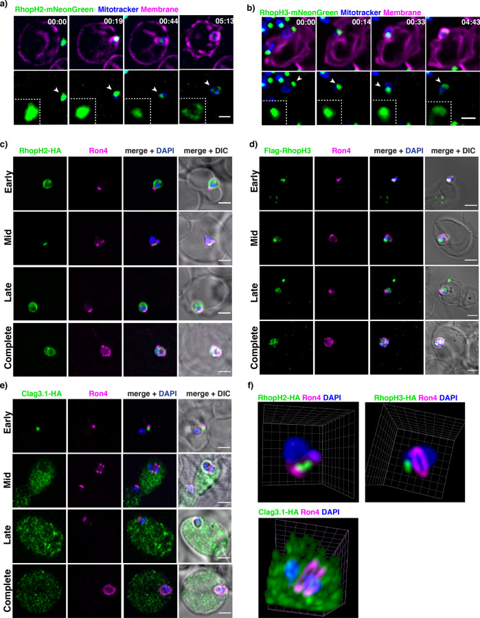
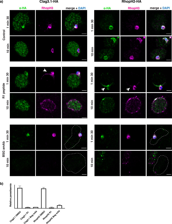
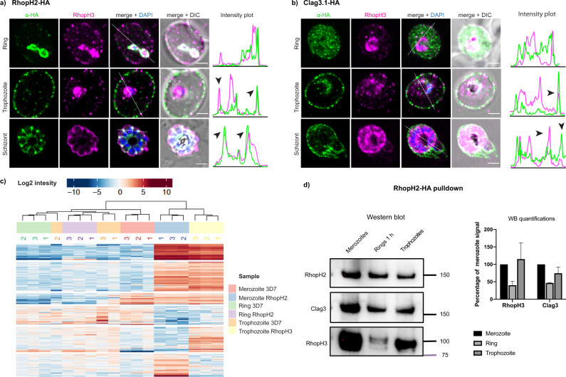
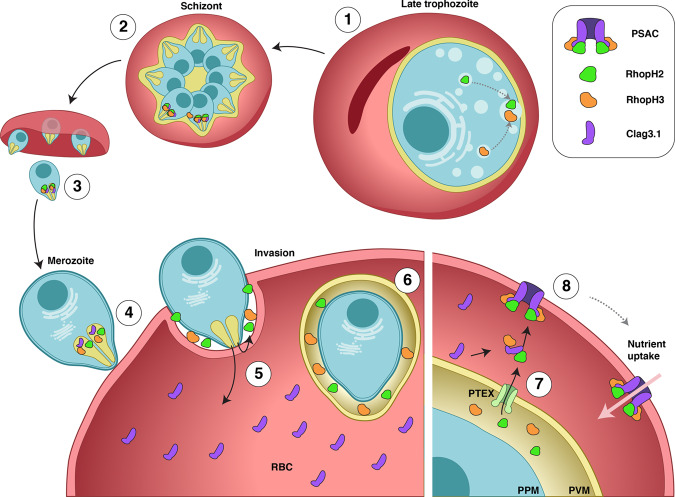
RhopH complexes consists of Clag3, RhopH2 and RhopH3 and are essential for growth of Plasmodium falciparum inside infected erythrocytes. Proteins are released from rhoptry organelles during merozoite invasion and trafficked to the surface of infected erythrocytes and enable uptake of nutrients. RhopH3, unlike other RhopH proteins, is required for parasite invasion, suggesting some cellular processes RhopH proteins function as single players rather than a complex. We show the RhopH complex has not formed during merozoite invasion. Clag3 is directly released into the host cell cytoplasm, whilst RhopH2 and RhopH3 are released into the nascent parasitophorous vacuole. Export of RhopH2 and RhopH3 from the parasitophorous vacuole into the infected erythrocyte cytoplasm enables assembly of Clag3/RhopH2/RhopH3 complexes and incorporation into the host cell membrane concomitant with activation of nutrient uptake. This suggests compartmentalisation prevents premature channel assembly before intact complex is assembled at the host cell membrane.


Conflict of interest statement

The authors declare no competing interests.

Figures

Fig. 1
Fig. 1. Super-resolution live imaging of RhopH2 and RhopH3.
a Live RhopH2-mNeonGreen expressing trophozoites and schizont parasites with SiR-DNA-stained nuclei (blue). mNeonGreen signal accumulation in forming rhoptries - white arrows. b Live RhopH3-mNeonGreen expressing trophozoites and schizont parasites with SiR-DNA-stained nuclei (blue). mNeonGreen signal accumulation in forming rhoptries (white arrows) and membrane association (yellow arrows). c Live RhopH2-mNeonGreen expressing trophozoites and schizont parasites with membrane dye (purple). d Live RhopH3-mNeonGreen expressing trophozoites and schizont parasites with membrane dye (purple). Each panel is a single z section; MIP – maximum intensity projections with a scale bar 2 µm. Time stamps in the upper-right corner represent time points of the overnight time-lapse experiment in the hours: minutes format.
Fig. 2
Fig. 2. Subcellular localisation of RhopH proteins during parasite invasion.
a Live RhopH2-mNeonGreen merozoites invading human erythrocytes imaged using lattice-light sheet microscopy. mNeonGreen signal (white arrows) visible as a bright spot on the merozoite apical end corresponding to RhopH2. The signal became diffused upon successful invasion as the parasitophorous vacuole formed. b Live RhopH3-mNeonGreen merozoites invading human erythrocytes imaged using lattice-light sheet microscopy. mNeonGreen signal (white arrows) visible as a bright spot on the merozoite apical end corresponding to RhopH3. The signal became diffused upon successful invasion as the parasitophorous vacuole formed. c Localisation of RhopH2-HA and RON4 in fixed merozoites at early, mid, late and complete stages of erythrocyte invasion. RON4 was a marker for the tight junction to assess the stage of invasion. Scale bar 2 µm. d Flag-RhopH3 and RON4 in fixed merozoites at early, mid, late and complete stages of erythrocyte invasion. RON4 was a marker for the tight junction to assess the stage of invasion. Scale bar 2 µm. e Clag3.1-HA and RON4 in fixed merozoites at early, mid, late and complete stages of erythrocyte invasion. RON4 was a marker for the tight junction to assess the stage of invasion. Scale bar 2 µm. f Super-resolution 3D reconstructions of invading merozoites. RON4 labelled in magenta marks the tight junction. HA-tagged Clag3.1, RhopH2, or RhopH3 are labelled in green. Clag3.1 signal appears in the host cytoplasm while RhopH2 and RhopH3 remain concentrated at the apical end of the invading merozoite.
Fig. 3
Fig. 3. RhopH proteins have different cellular localizations during invasion.
a Super-resolution imaging of Clag3.1-HA, RhopH2-HA and RhopH3 (DMSO control). RhopH proteins in the presence of invasion inhibitors with DMSO control (top two panels), R1 peptide (middle two panels) and anti-BSG (bottom two panels). Arrows point to membrane localization of RhopH2 and RhopH3. A dashed line outlines the erythrocyte. Scale bar 2 µm. b Invasion efficiency in > 2000 cells per group from two independent experiments for each of the conditions. Error bars show SD.
Fig. 4
Fig. 4. RhopH complex reassociates following RhopH2 and RhopH3 export in trophozoite stage.
a Super-resolution images showing colocalization of RhopH2-HA and RhopH3 during parasite development. Intensity plots along the white broken line show lack of colocalization at the ring stage, black arrows point at the points of colocalization on the erythrocyte surface and in the rhoptry. Scale bar 2 µm. b Super-resolution images showing colocalization of Clag3.1-HA with RhopH3 during parasite development. Intensity plots are shown along the white broken line that shows lack of colocalization at the ring stage, black arrows point at the points of colocalization on the erythrocyte surface and in the rhoptry. Scale bar 2 µm. c Heat map of the mass-spectrometry analysis from RhopH2-HA immunoprecipitation from developmental stages. d Immuno-blot (left panel) and quantification (right panel) of RhopH3 and Clag3 from RhopH2-HA immunoprecipitation of developmental stages. Error bars show SD from three biological replicates. Full blots shown (Supplementary Fig. 8).
Fig. 5
Fig. 5. Purified RhopH complex spontaneously associates with RBC membranes.
a RhopH complex purified from trophozoites expressing Clag3.1-Flag using an anti-Flag resin. Eluates were further purified by size-exclusion chromatography and analysed by negative-stain electron microscopy and mass spectrometry (left panel). b RhopH complex added to human erythrocytes resulted in lysis and haemoglobin release. This lysis activity was abolished if the protein was denatured for 5 min at 95 °C. Quantifications from three independent replicates. c Purified RhopH complex can associate with human erythrocytes and ghost membranes. Full blots shown (Supplementary Fig. 8).
Fig. 6
Fig. 6. Model for RhopH proteins trafficking onto the RBC surface.
a RhopH2 and RhopH3 colocalize in late schizonts (1 and 2). Upon egress (3) free merozoites attach to new red blood cells (4). During invasion, rhoptry content is discharged and RhopH2 and RhopH3 are released into the nascent parasitophorous vacuole while Clag3 is predominantly injected into the host cell cytoplasm (5). Upon successful invasion, RhopH2 and RhopH3 are confined within the parasitophorous vacuole but show very little interaction (6). In trophozoites, RhopH2 and RhopH3 are exported via the PTEX translocon (7). In the host cell cytoplasm, both RhopH2 and RhopH3 associate with Clag3 and the RhopH complex consisting of RhopH2, RhopH3 and Clag3 can spontaneously insert into the erythrocyte membrane, where it forms the nutrient channel (8).

References

    1. Cowman A, Crabb B. Invasion of red blood cells by malaria parasites. Cell. 2006;124:755–766. - PubMed
    1. Marti M, Spielmann T. Protein export in malaria parasites: many membranes to cross. Curr. Opin. Microbiol. 2013;16:445–451. - PMC - PubMed
    1. Parsons M, Myler PJ. Illuminating parasite protein production by ribosome profiling. Trends Parasitol. 2016;32:446–457. - PMC - PubMed
    1. Cowman AF, Healer J, Marapana D, Marsh K. Malaria: Biology and disease. Cell. 2016;167:610–624. - PubMed
    1. Desai SA. Why do malaria parasites increase host erythrocyte permeability? Trends Parasitol. 2014;30:151–159. - PMC - PubMed

Publication types

MeSH terms

Substances