. 2022 Apr 8;7(7):e152271.
doi: 10.1172/jci.insight.152271.

Gene therapy of Csf2ra deficiency in mouse fetal monocyte precursors restores alveolar macrophage development and function

Affiliations

Gene therapy of Csf2ra deficiency in mouse fetal monocyte precursors restores alveolar macrophage development and function

Fengqi Li et al. JCI Insight. .

Abstract

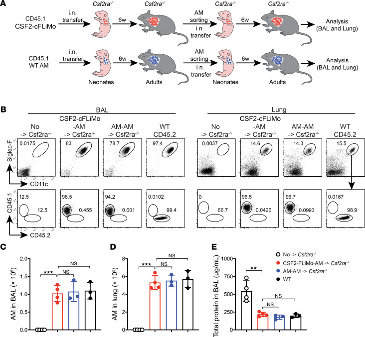
Tissue-resident macrophage-based immune therapies have been proposed for various diseases. However, generation of sufficient numbers that possess tissue-specific functions remains a major handicap. Here, we showed that fetal liver monocytes cultured with GM-CSF (CSF2-cFLiMo) rapidly differentiated into a long-lived, homogeneous alveolar macrophage-like population in vitro. CSF2-cFLiMo retained the capacity to develop into bona fide alveolar macrophages upon transfer into Csf2ra-/- neonates and prevented development of alveolar proteinosis and accumulation of apoptotic cells for at least 1 year in vivo. CSF2-cFLiMo more efficiently engrafted empty alveolar macrophage niches in the lung and protected mice from severe pathology induced by respiratory viral infection compared with transplantation of macrophages derived from BM cells cultured with M-CSF (CSF1-cBMM) in the presence or absence of GM-CSF. Harnessing the potential of this approach for gene therapy, we restored a disrupted Csf2ra gene in fetal liver monocytes and demonstrated their capacity to develop into alveolar macrophages in vivo. Altogether, we provide a platform for generation of immature alveolar macrophage-like precursors amenable for genetic manipulation, which will be useful to dissect alveolar macrophage development and function and for pulmonary transplantation therapy.

Keywords: Gene therapy; Immunology; Influenza; Macrophages.


Figures

Figure 1
Figure 1. Fetal liver monocytes can proliferate in vitro with GM-CSF and further develop into mature functional alveolar macrophages in vivo.
(A) Illustration of experimental scheme. (B) F4/80 and Ly6C expression on fetal liver monocytes cultured in vitro with GM-CSF (CSF2-cFLiMo) at the indicated time points. Shown are representative dot plots pre-gated on viable CD45+ single cells (left panel) and a column graph with percentages of F4/80+Ly6C and F4/80Ly6C+ cells (right panel). (CF) FLiMo were isolated from E14.5 embryos (CD45.1) and grown for 2 weeks with GM-SCF prior to i.n. transfer of 5 × 10⁴ cells to neonatal Csf2ra–/– (CD45.2) mice within the first 3 days after birth. (C) Shown are representative dot plots pre-gated on viable CD45.1 donor-derived cells from the bronchoalveolar lavage (BAL) of recipients (n = 3) at indicated time points after transfer. Untreated (UT) Csf2ra–/– recipient mice (6 weeks old) are included as a control (n = 3). (D) Total number of donor-derived alveolar macrophages (AMs) in the lung determined in recipients at the indicated time points (n = 3/time point). (E) Cell surface expression of AM markers on CSF2-cFLiMo prior to transfer and CSF2-cFLiMo–derived AMs 6 weeks after transfer to Csf2ra–/– neonates as well as endogenous AMs from age-matched control mice. Shown are representative histograms with the black line indicating specific staining and gray areas depicting fluorescence minus one controls (n = 3/group). (F) Total protein in the BAL of indicated groups of mice. The data are representative of 3 independent experiments. Data are presented as mean ± SD. ANOVA (1-way) was used in F. NS, not significant; *P < 0.05, **P < 0.01, ***P < 0.001, ****P < 0.0001.
Figure 2
Figure 2. CSF2-cFLiMo–derived alveolar macrophages have the ability to self-renew in vivo.
(A) Illustration of experimental regimen. CSF2-cFLiMo (CD45.1) or mature alveolar macrophages (AMs) isolated from adult mice were transferred i.n. to neonatal CD45.2 Csf2ra–/– mice. After 6 weeks, donor-derived AMs were sorted and 5 × 10⁴ of cells were transferred i.n. again to neonatal Csf2ra–/– mice. BAL and lung were analyzed 6 weeks after second-round transfer in BE. (B) Representative dot plots showing the phenotype of CD45.1 donor-derived AMs in the BAL and lung, pre-gated as viable CD45+ single cells (n = 3 to 4/group). (C and D) Numbers of donor-derived AMs and WT AMs in the BAL (C) and lung (D) (n = 3 to 4/group). (E) Total protein in the BAL (n = 3 to 4/group). Age-matched Csf2ra–/– (n = 4) and CD45.2 WT (n = 3) mice were included as negative and positive controls, respectively. The data are representative of 3 experiments. Data are presented as mean ± SD. ANOVA (1-way) was used in CE. NS, not significant; **P < 0.01, ***P < 0.001.
Figure 3
Figure 3. Gene expression profiles of transferred CSF2-cFLiMo in Csf2ra–/– mice.
(A) Illustration of experimental regimen. Primary fetal liver monocytes (FLiMo) from E14.5 embryos, CSF2-cFLiMo cultured 2 weeks prior to transfer, ex vivo CSF2-cFLiMo–derived mature alveolar macrophages (AMs) from Csf2ra–/– recipients 6 weeks after transfer, and AMs from adult naive mice were sorted using flow cytometry. RNA-Seq was performed (2 biological replicates per group). (B) Principal component analysis (PCA) and (C) matrix clustering of the transcriptomes of all samples are shown. (D) Heatmaps showing expression of monocyte and AM markers. (E) The numbers of upregulated and downregulated genes by CSF2 or niche. (F) Heatmap showing the top 100 differentially expressed genes and representative genes of CSF2 and niche regulated. (G) Venn diagram of differentially expressed genes. Intersections of CSF2-upregulated or CSF2-downregulated versus niche-upregulated or niche-downregulated genes. The absolute gene numbers and percentages in the intersections are shown.
Figure 4
Figure 4. CSF2-cFLiMo–derived alveolar macrophages are functional in phagocytosis and efferocytosis.
(A) Illustration of experimental regimen. Different numbers of CSF2-cFLiMo (CD45.1) after 2 weeks (2w) or 4 months (4m) of culturing were transferred i.n. to neonatal CD45.2 Csf2ra–/– mice and analyzed 6 weeks later in B and C. (B) Total numbers of donor-derived alveolar macrophages (AMs) in the lung of recipient Csf2ra–/– mice or WT mice (n = 3/group). (C) Total protein levels in the BAL (n = 3/ group). (D) Efferocytosis of i.t. instilled apoptotic thymocytes by AMs at the indicated time points. Values shown depict percentages of efferocytotic AMs (n = 2 to 3/group). (E) Illustration of experimental regimen. CD45.1 CSF2-cFLiMo were generated from E14.5 embryos and cultured 2 weeks in vitro. Different numbers of CSF2-cFLiMo were transferred i.t. to 10-week-old adult Csf2ra–/– mice and analyzed 10 weeks later in F and G. (F) Total numbers of donor-derived AMs in the BAL and lung of recipient Csf2ra–/– mice or AMs in the BAL and lung of WT mice (n = 3/group). (G) Total protein levels in the BAL. Age-matched Csf2ra–/– (n = 3) and WT (n = 3) mice were included as negative and positive controls, respectively. Data are presented as mean ± SD and the results are representative of 3 experiments. Student’s t test (unpaired) was used in D and ANOVA (1-way) was used in B, C, F, and G. NS, not significant; *P < 0.05, **P < 0.01, ***P < 0.001.
Figure 5
Figure 5. Influenza virus–induced disease severity is increased in Csf2ra–/– mice containing CSF1-cBMM–derived alveolar macrophages compared with Csf2ra–/– mice containing CSF2-cFLiMo.
(A) Illustration of experimental regimen. Fetal liver monocytes were isolated from E14.5 embryos (CD45.1) and cultured 2 weeks in vitro to generate CSF2-cFLiMo. BM was isolated from adult mice (CD45.1CD45.2) and cultured 7 days with M-CSF in vitro to generate CSF1-cBMM. CSF2-cFLiMo and CSF1-cBMM were pooled in 1:1 ratio and transferred i.n. to neonatal Csf2ra–/– mice (CD45.2). (BE) Recipients were analyzed 10 weeks later. (B) Representative dot plots and (C) percentages of donor-derived alveolar macrophages (AMs) in BAL and lung of the recipients (n = 3/group). (D and E) Shown are representative histograms (D) and MFI (E) of AM markers on CSF2-cFLiMo– and CSF1-cBMM–derived AMs in BAL of recipients, as well as AMs from BAL of untreated WT mice (n = 3/group). (F) Illustration of experimental scheme. Csf2ra–/– neonates (CD45.2) were transferred with CD45.1 CSF2-cFLiMo or CSF1-cBMM and analyzed in GM. (GI) The phenotype of donor-derived AMs in the BAL (pre-gated as viable CD45.1+ singlets) (G), the number of donor-derived AMs (H), and proteinosis in recipients (I) was determined 10 weeks after transfer prior infection (n = 3 to 4/group). (JM) Mice were infected with 10 PFU influenza virus (PR8) and morbidity was analyzed at indicated days (n = 9–10/group); p.i., post-infection. Shown are percentage body weight relative to the day of infection (J), body temperature (K), survival curve (L), and O2 saturation at 7 days after infection (M). Age-matched Csf2ra–/– and WT mice were included as negative and positive controls (n = 8/group). Data are presented as mean ± SD and the results are representative of 3 experiments. Student’s t test (unpaired) was used in C and ANOVA (1-way) was used in E, H, I, and M. NS, not significant; **P < 0.01, ***P < 0.001, ****P < 0.0001.
Figure 6
Figure 6. Rejection of allogenic CSF2-cFLiMo–derived alveolar macrophages.
(A) Illustration of experimental regimen. CD45.2 BALB/c and CD45.1 B6 CSF2-cFLiMo were generated from E14.5 embryos and cultured 2 weeks in vitro. BALB/c CSF2-cFLiMo were transferred to neonatal CD45.2 Csf2ra–/– mice (B6 background) either separately or in a 1:1 ratio with B6 CSF2-cFLiMo and analyzed 10 weeks later in B and C. (B) Representative dot plots showing the phenotype of donor-derived alveolar macrophages (AMs) in the BAL, pre-gated on viable CD45+ single cells (n = 3/group). (C) Percentage of donor-derived AMs in BAL of cotransferred recipients (n = 3). (D) Illustration of experimental regimen. GM-CSF cultured neonatal liver monocytes (CSF2-cNLiMo) generated from CD45.1 WT male neonates after 2-week culture were i.n. transferred to neonatal CD45.2 Csf2ra–/– mice and analyzed after 10 weeks in E and F. Mice were grouped according to sex (n = 6 to 7/group). (E and F) Numbers of donor-derived and WT AMs in the BAL (E) and lung (F) are shown. Age-matched Csf2ra–/– and CD45.2 WT mice were included as negative and positive controls (n = 3/group). Data are presented as mean ± SD and the results are representative of 3 experiments. Student’s t test (unpaired) was used in C and ANOVA (1-way) was used in E and F. *P < 0.05, **P < 0.01, ***P < 0.001, ****P < 0.0001.
Figure 7
Figure 7. Gene therapy of Csf2ra deficiency by gene transfer to CSF2-cFLiMo.
(A) Illustration of experimental regimen. Fetal liver monocytes (FLiMo) were purified from E14.5 CD45.2 Csf2ra–/– or CD45.1 WT embryos and spin-infected with a retrovirus encoding for Csf2ra and GFP (RVCsf2ra-gfp) or GFP alone (control RVgfp). Cells were cultured with CSF2 for 7 days. RVCsf2ra-gfp-transduced Csf2ra–/– CSF2-cFLiMo (RVCsf2ra-gfp-FLiMo) or identically treated CD45.1 WT CSF2-cFLiMo were transferred i.n. to neonatal CD45.2+ Csf2ra–/– mice and evaluated after 8 weeks. (B) Efficiency of spin infection (GFP+) and survival of cultured cells at days 3 and 7 after infection. (C) GM-CSF receptor alpha protein expression levels on the cell surface of Csf2ra–/– and WT FLiMo 3 and 7 days after transduction with RVCsf2ra-gfp or RVgfp, respectively. (D) Representative dot plots showing the phenotype of donor-derived cells in the BAL and lung, pre-gated on viable CD45+ single cells. (E and F) Numbers of donor-derived alveolar macrophages (AMs) and WT AMs in the BAL (E) and lung (F). (G) Total protein levels in the BAL. (DG) Age-matched Csf2ra–/– and CD45.2 WT mice were included as negative and positive controls. (H) Representative histograms showing cell surface expression of characteristic proteins on AMs harvested from GM-CSFRa and GFP on RVCsf2ra-gfp-FLiMo–derived AMs and WT AMs showing fluorescence minus one control (gray) and specific antibodies against indicated markers (black line). (BH) n = 3–4/group. Data are presented as mean ± SD and the results are representative of 3 experiments. ANOVA (1-way) was used in EG. NS, not significant; *P < 0.05, **P < 0.01, ***P < 0.001, ****P < 0.0001.

Similar articles

Cited by

References

    1. Davies LC, et al. Tissue-resident macrophages. Nat Immunol. 2013;14(10):986–995. doi: 10.1038/ni.2705. - DOI - PMC - PubMed
    1. Hoeffel G, Ginhoux F. Ontogeny of tissue-resident macrophages. Front Immunol. 2015;6:486. - PMC - PubMed
    1. Hussell T, Bell TJ. Alveolar macrophages: plasticity in a tissue-specific context. Nat Rev Immunol. 2014;14(2):81–93. doi: 10.1038/nri3600. - DOI - PubMed
    1. Kopf M, et al. The development and function of lung-resident macrophages and dendritic cells. Nat Immunol. 2015;16(1):36–44. doi: 10.1038/ni.3052. - DOI - PubMed
    1. Guilliams M, et al. Alveolar macrophages develop from fetal monocytes that differentiate into long-lived cells in the first week of life via GM-CSF. J Exp Med. 2013;210(10):1977–1992. doi: 10.1084/jem.20131199. - DOI - PMC - PubMed

Publication types

Substances

Supplementary concepts