. 2022 Apr 15;11(4):bio059146.
doi: 10.1242/bio.059146. Epub 2022 Apr 8.

NANOS3 suppresses premature spermatogonial differentiation to expand progenitors and fine-tunes spermatogenesis in mice

Affiliations

NANOS3 suppresses premature spermatogonial differentiation to expand progenitors and fine-tunes spermatogenesis in mice

Hiroki Inoue et al. Biol Open. .

Abstract

In the mouse testis, sperm originate from spermatogonial stem cells (SSCs). SSCs give rise to spermatogonial progenitors, which expand their population until entering the differentiation process that is precisely regulated by a fixed time-scaled program called the seminiferous cycle. Although this expansion process of progenitors is highly important, its regulatory mechanisms remain unclear. NANOS3 is an RNA-binding protein expressed in the progenitor population. We demonstrated that the conditional deletion of Nanos3 at a later embryonic stage results in the reduction of spermatogonial progenitors in the postnatal testis. This reduction was associated with the premature differentiation of progenitors. Furthermore, this premature differentiation caused seminiferous stage disagreement between adjacent spermatogenic cells, which influenced spermatogenic epithelial cycles, leading to disruption of the later differentiation pathway. Our study suggests that NANOS3 plays an important role in timing progenitor expansion to adjust to the proper differentiation timing by blocking the retinoic acid (RA) signaling pathway.

Keywords: Mouse; Nanos3; Retinoic acid; Spermatogenesis; Testis.


Conflict of interest statement

Competing interests The authors declare no competing or financial interests.

Figures
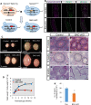
Fig. 1.
Fig. 1.
Testicular abnormalities observed in BAC-cKO mice. (A) Experimental scheme to obtain BAC-cKO males. Nanos3−/−; BAC-Tg females were crossed with Nanos3cre/+ males. The Nanos3-Rfp sequence is removed by Nanos3-Cre during germ cell development from E14.5. A Nanos3−/−; BAC-Tg male was used as the control. (B) Wholemount immunostaining of seminiferous tubules in 8-week-old testes. The signals of anti-RFP and anti-CDH1 are shown in magenta and green, respectively. The white dotted lines represent the outline of seminiferous tubules. Scale bars: 100 μm. (C) Testes from 1, 2, 4 or 8-week-old control and BAC- cKO mice. Scale bars: 1 mm. (D) Body and testis weights were measured in control and cKO mice at 1, 2, 4, 8 and 12 weeks of age. The testis weight was normalized by body weight. Values represent the mean±s.e.m. *P<0.05, **P<0.01. (E–J) Hematoxylin and Eosin (H&E)-stained cross-sections of control and mutant testes from 8- or 12-week-old mice (E–H) and epididymides from 12-week-old mice (I,J). Scale bars: 50 μm (E–H), 100 μm (I,J). (K) Counts of spermatozoa in a 12-week-old cauda epididymis (control: n=6, mutant: n=3). Values represent the mean±s.e.m. *P<0.05.
Fig. 2.
Fig. 2.
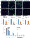
Quantification of undifferentiated and differentiating spermatogonia in BAC-cKO mice. (A–H) Immunostaining of 8-week-old testes with germ cell markers to distinguish developmental states; anti-GFRA1 (stem-state cells including population among undifferentiated spermatogonia, A,A′), anti-PLZF (undifferentiated spermatogonia, C,C′), anti-KIT (differentiated spermatogonia, E,E′) or anti-SYCP3 (spermatocytes, G,G′), and Sertoli cell markers, anti-GATA4 or anti-SOX9. The signals of germ cell markers are shown in green and signals for Sertoli cells are shown in magenta. Nuclei were counterstained with DAPI (blue). Scale bars: 100 μm. The number of germ cells was normalized by the number of Sertoli cells (B,D,F,H). Germ cell marker-positive cells are indicated by arrowheads. Values represent the mean±s.e.m. **P<0.001. (control: n=3, mutant: n=3). (I) Quantification of the clusters detected by wholemount immunofluorescence for CDH1. The horizontal axis represents the number of cells in a cluster. Cluster counts were normalized by the As spermatogonia number (n=3). Values represent the mean±s.e.m. *P<0.05, **P<0.01.
Fig. 3.
Fig. 3.
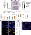
Quantitative analyses of undifferentiated spermatogonia in endo-cKO mice. (A) The number of PLZF-positive undifferentiated spermatogonia was normalized by the number of Sertoli cells (GATA4-positive cells). Values represent the mean±s.e.m. (B,C) Histological comparison between BAC-cKO and endo-cKO adult testes. H&E-stained cross-sections are shown (B). Scale bar: 100 μm. (C) The tubules were quantified based on the most differentiated spermatogenic cells (elongated, round-spermatids, spermatocytes or spermatogonia) contained in each tubule (BAC-cKO: n=2, endo-cKO: n=3). (D) The number of GFRA1-positive stem cells was normalized by the number of SOX9-positive Sertoli cells. Each cell type was counted by immunostaining sections of 4-, 8- and 12-week-old testes (n=3). Values represent the mean±s.e.m. (E) The ratio of pH3-positive proliferating spermatogonia to CDH1-positive undifferentiated spermatogonia in control (n=3) and endo-cKO (n=3). Values represent the mean±s.e.m. (F) The percentage of tubules that had c-PARP positive cells among counted tubules in control (n=3) and endo-cKO (n=3). Values represent the mean±s.e.m. (G) Immunostaining of 4-week-old testes with anti-STRA8 (red) and anti-CDH1 (green) antibodies. Nuclei were counterstained with DAPI (blue). Yellow arrows indicate STRA8-positive undifferentiating spermatogonia. White arrows indicate STRA8-negative undifferentiated spermatogonia. (H) The percentage of STRA8-positive cells among CDH1-positive undifferentiating spermatogonia. STRA8-positive cells significantly increased in endo-cKO testes. Values represent the mean±s.e.m. (I) Immunostaining of 4-week-old testes of endo-cKO mice with anti-GFRA1 (green), STRA8 (magenta) and PLZF (gray) antibodies. Nuclei were counterstained with DAPI (blue). GFRA1 and PLZF-double-positive stem cells indicated by arrows did not have STRA8 signals. Scale bar: 100 μm. (J) The percentage of STRA8-positive cells among the RARγ-positive spermatogonial progenitor population. STRA8-positive cells significantly increased in endo-cKO testes. Values represent the mean±s.e.m.
Fig. 4.
Fig. 4.
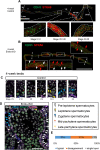
Asynchronous STRA8 upregulation during spermatogenesis in the endo-cKO. (A,B) The spliced images of multiple wholemount immunostaining images of testicular tubules from 4-week-old control (A) and endo-cKO mice (B). STRA8 and CDH1 signals are shown in red and green, respectively. The boundary of different STRA8 expression patterns is indicated by dashed lines. Bottom panels show the standard expression pattern of each domain. In the endo-cKO, there were intermittent STRA8-positive regions. STRA8-positive (indicated by red arrows) and -negative (indicated by white arrows) Aal clusters were intermingled. (C,D) Immunostaining of 4-week-old testes with anti-CDH1, anti-STRA8 and anti-SYCP1, which is a marker of zygotene to mid-pachytene spermatocytes. Nuclei were counterstained with DAPI (gray). Scale bar: 20 μm (C) or 50 μm (D). (E) Seminiferous stage distribution in endo-cKO mice. Seminiferous tubules were classified into three types: the tubules exhibiting typical cell associations (typical), those containing disagreeing cells (disagreement) and those having only a single layer (single layer).
Fig. 5.
Fig. 5.
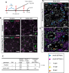
The progenitor number was partially recovered by RA signal inhibition. (A) Scheme of the experiment. RA was injected 8.6 days before 12 weeks of age, followed by daily RAi injection for 8 days. One of two testes was collected on the day of 12 weeks of age and then RA was injected again. At 1, 5 or 10 days or 5 days after the second RA injection, the other testis was collected. (B) Immunostaining of the RA-Rai-treated and untreated testes of control and endo-cKO. GFRA1, PLZF and STRA8 are shown in white, green and magenta, respectively. Scale bar: 100 μm. (C) The actual number and rate of increase of progenitors (GFRA1, PLZF+) per stem cell (GFRA1+, PLZF+) are shown. (D,E) Immunostaining of testes at day 0 or 10. CDH1, SYCP1 and STRA8 are shown in blue, green and magenta, respectively. Nuclei were counterstained with DAPI (gray). Seminiferous stages are indicated in Roman numerals. Tubules containing atypical combinations of spermatogenic cells are labeled as ‘Dis’ (Disagreement). Scale bar: 50 μm.
Fig. 6.
Fig. 6.
Stage disagreement occurred even in the synchronized testicular tubules. (A) Scheme of the experiment. RAi was daily injected for 7 days from P2 to P8, and then RA was injected. The testes were collected just before RA injection (P9), 1, 5, 11 and 21 days after RA (P10, P14, P20 and P30). (B,C) Immunostaining of testes at P9 to P30. CDH1 and STRA8 are shown in green and magenta, respectively. Nuclei were counterstained with DAPI (gray). (B) White and magenta arrowheads indicate STRA8-negative and -positive intermediate spermatogonia, respectively. (C) White arrows indicate undifferentiated spermatogonia. Spermatogenic stages are indicated by Int (intermediate spermatogonia), L (leptotene spermatocytes), Z (zygotene spermatocytes), eP (early pachytene spermatocytes), mP (mid or late spermatocytes) and Spt (round spermatids). Seminiferous stages are indicated in Roman numerals. Tubules containing atypical combinations of spermatogenic cells are labeled as ‘Dis’ (Disagreement). Scale bars: 100 μm in B and C, or 20 μm in magnified images in C. Asterisks indicate non-specific interstitial somatic signal.
Fig. 7.
Fig. 7.
NANOS3 target mRNAs differ from those of NANOS2. (A–D) Immunostaining of 4-week-old testes with anti-FLAG-M2 (green) and anti-KIT (magenta) antibodies. Nuclei were counterstained with DAPI (blue). Magnified images of epididymides are shown in A′ and C′. Scale bars: 50 μm. White, green and yellow arrowheads indicate type A spermatogonia, type B spermatogonia and pre-leptotene spermatocytes, respectively. Asterisks indicate FLAG-positive spermatogonia. (E) RT-qPCR analyses of mRNAs co-precipitated with anti-FLAG antibody from testis extracts of 4-week-old Flag-tagged Nanos3-OE mice. The fold enrichment of each mRNA in IP of anti-FLAG compared with IP of IgG was calculated (ratio of each mRNA level in FLAG IP to IgG IP). Actinβ was used to normalize the mRNA enrichment level. Values represent the mean±s.d. **P<0.01. ***P<0.001.

Similar articles

Cited by

References

    1. Ashley, T., Gaeth, A. P., Creemers, L. B., Hack, A. M. and de Rooij, D. G. (2004). Correlation of meiotic events in testis sections and microspreads of mouse spermatocytes relative to the mid-pachytene checkpoint. Chromosoma 113, 126-136. 10.1007/s00412-004-0293-5 - DOI - PubMed
    1. Beedle, M. T., Stevison, F., Zhong, G., Topping, T., Hogarth, C., Isoherranen, N. and Griswold, M. D. (2019). Sources of all-trans retinal oxidation independent of the aldehyde dehydrogenase 1A isozymes exist in the postnatal testis. Biol. Reprod 100, 547-560. 10.1093/biolre/ioy200 - DOI - PMC - PubMed
    1. Chakraborty, P., Buaas, F. W., Sharma, M., Snyder, E., de Rooij, D. G. and Braun, R. E. (2014). LIN28A Marks the spermatogonial progenitor population and regulates its cyclic expansion. Stem Cells 32, 860-873. 10.1002/stem.1584 - DOI - PMC - PubMed
    1. Chen, Y., Zheng, Y., Gao, Y., Lin, Z., Yang, S., Wang, T., Wang, Q., Xie, N., Hua, R., Liu, M.et al. (2018). Single-cell RNA-seq uncovers dynamic processes and critical regulators in mouse spermatogenesis. Cell Res. 28, 879-896. 10.1038/s41422-018-0074-y - DOI - PMC - PubMed
    1. Endo, T., Romer, K. A., Anderson, E. L., Baltus, A. E., De Rooij, D. G. and Page, D. C. (2015). Periodic retinoic acid-STRA8 signaling intersects with periodic germ-cell competencies to regulate spermatogenesis. Proc. Natl. Acad. Sci. U. S. A 112, E2347-E2356. 10.1073/pnas.1505683112 - DOI - PMC - PubMed

Publication types