Review
. 2022 Jun;100(6):817-828.
doi: 10.1007/s00109-022-02195-2. Epub 2022 Apr 8.

Neurovascular abnormalities in retinopathy of prematurity and emerging therapies

Affiliations
Review

Neurovascular abnormalities in retinopathy of prematurity and emerging therapies

Chang Dai et al. J Mol Med (Berl). 2022 Jun.

Abstract

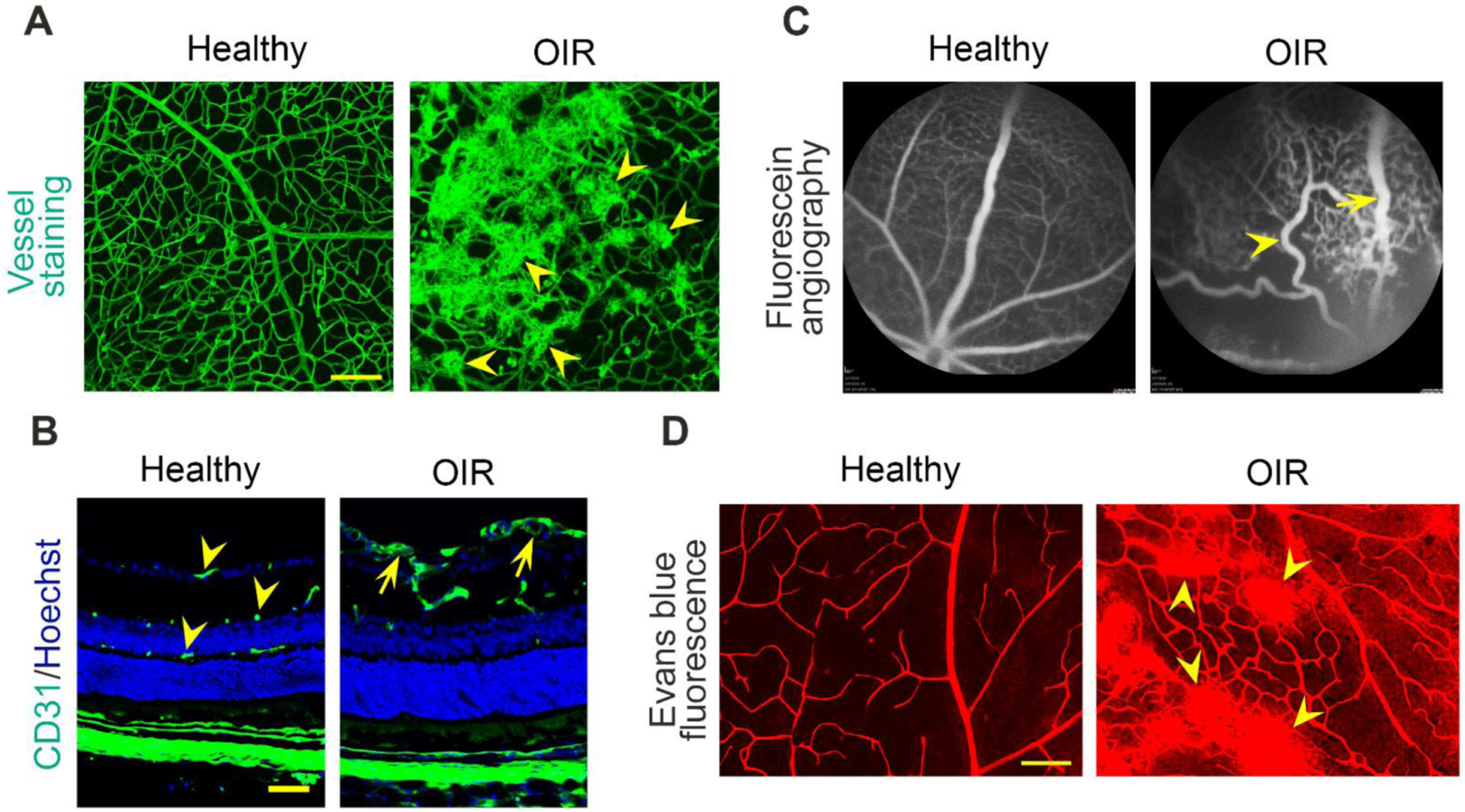
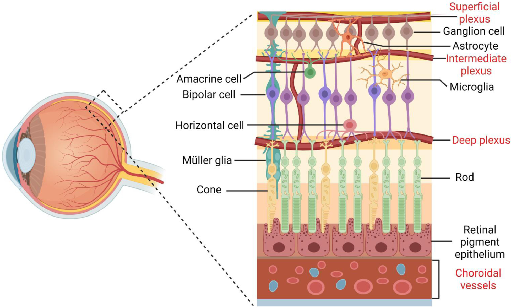
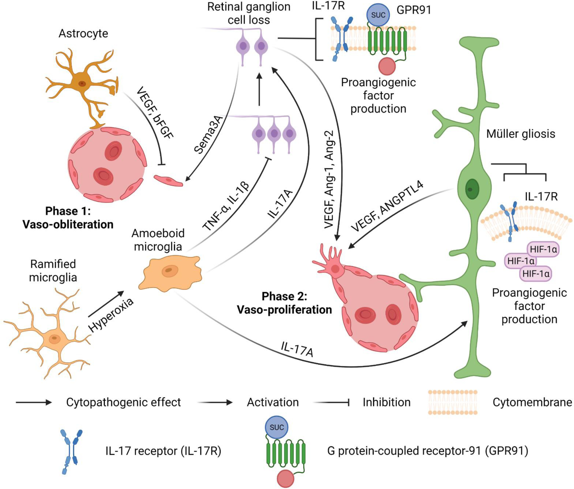
Blood vessels in the developing retina are formed in concert with neural growth, resulting in functional neurovascular network. Disruption of the neurovascular coordination contributes to the pathogenesis of retinopathy of prematurity (ROP), a potentially blinding retinal neovascular disease in preterm infants that currently lacks an approved drug therapy in the USA. Despite vasculopathy as predominant clinical manifestations, an increasing number of studies revealed complex neurovascular interplays among neurons, glial cells and blood vessels during ROP. Coordinated expression of glia-derived vascular endothelial growth factor (VEGF) in spatio-temporal gradients is pivotal to the formation of well-organized vascular plexuses in the healthy retina, whereas uncoordinated VEGF expression triggers pathological angiogenesis with disorganized vascular tufts in ROP. In contrast with VEGF driving both pathological and physiological angiogenesis, neuron-derived angiogenic factor secretogranin III (Scg3) stringently regulates ROP but not healthy retinal vessels in animal models. Anti-VEGF and anti-Scg3 therapies confer similar high efficacies to alleviate ROP in preclinical studies but are distinct in their disease selectivity and safety. This review discusses neurovascular communication among retinal blood vessels, neurons and glial cells during retinal development and ROP pathogenesis and summarizes the current and emerging therapies to address unmet clinical needs for the disease.

Keywords: Anti-angiogenic therapy; Neurovascular interaction; Oxygen-induced retinopathy; Retinopathy of prematurity; Secretogranin III; Vascular endothelial growth factor.


Conflict of interest statement

Conflict of interest. WL is the shareholder of Everglades Biopharma, LLC and LigandomicsRx, LLC. WL is an inventor of issued and pending patents. The remaining authors declare no conflict of interest.

Figures

Fig. 1
Fig. 1
Vascular characteristics of ROP. A Flat-mount retinas of healthy and OIR mice stained with Alexa Flour 488-conjugated isolectin B4. Scale bar= 100 μm. Arrowheads indicate pathological retinal neovascularization. B Retinal sections of healthy vs. OIR mice stained with anti-CD31 monoclonal antibody and Hoechst. Scale bar= 50 μm. Arrowheads indicate normal superficial, intermediate and deep retinal plexuses. Arrows indicate pathological neovascularization in the superficial layer. C Fluorescein angiography images of healthy vs. OIR mice. The arrowhead indicates a tortuous artery. The arrow indicates a dilated vein. D Images showing the leakage of fluorescent Evans blue dye from retinal vessels in healthy and OIR mice. Arrowheads indicate the leaking sites in the OIR retina. Scale bar= 100 μm.
Fig. 2
Fig. 2
Cellular organization of the neuronal and vascular retina. There are six major types of neurons in the retina: rod and cone photoreceptors, bipolar cells, retinal ganglion cells, horizontal cells and amacrine cells. These neurons form complex neural circuities to detect light, generate electric impulse and transmit visual signals. Three basic types of glial cells are present in the human retina: Müller cells, astrocytes and microglia. The processes of astrocytes and microglia contribute to the blood-retina barrier together with pericytes and endothelial cells of the superficial, intermediate and deep vascular plexuses. Retinal pigmental epithelial cells are at the outer most layer of the retina, beneath which are choroidal vessels.
Fig. 3
Fig. 3
Neurovascular interactions in ROP. Phase 1: Vaso-obliteration. Activated microglia transform from ramified to amoeboid shapes. The tumour necrosis factor-α (TNF-α) and interleukin-1β (IL-1β) secreted from activated amoeboid microglia induce pronounced retinal ganglion cell (RGC) loss and further stimulate the secretion of semaphorin 3A (Sema3A) from adjacent neurons to mediate microvascular injury. Vascular endothelial growth factor (VEGF) and basic fibroblast growth factor (bFGF) from astrocytes minimize vascular damage. Phase 2: Vaso-proliferation. Accumulated succinate stimulates G protein-coupled receptor-91 (GPR91) on RGCs to facilitate the production of proangiogenic factors, such as VEGF, angiopoietin-1 (Ang-1) and angiopoietin-2 (Ang-2). Stabilized HIF-1α in hypoxic Müller cells promotes the expression of VEGF and angiopoietin-like 4 (ANGPTL4) to induce neovascularization and vascular leakage. Furthermore, interleukin-17A (IL-17A) from amoeboid microglial cells targets interleukin-17 receptors (IL-17Rs) in Müller cells and RGCs to enhance the production of VEGF.

References

    1. Hartnett ME (2015) Pathophysiology and Mechanisms of Severe Retinopathy of Prematurity. Ophthalmology 122:200–210. 10.1016/j.ophtha.2014.07.050 - DOI - PMC - PubMed
    1. Blencowe H, Lawn JE, Vazquez T, et al. (2013) Preterm-associated visual impairment and estimates of retinopathy of prematurity at regional and global levels for 2010. Pediatr Res 74 (Suppl 1):35–49. 10.1038/pr.2013.205 - DOI - PMC - PubMed
    1. Lepore D, Quinn GE, Molle F, et al. (2014) Intravitreal Bevacizumab versus Laser Treatment in Type 1 Retinopathy of Prematurity. Ophthalmology 121:2212–2219. 10.1016/j.ophtha.2014.05.015 - DOI - PubMed
    1. Vogel RN, Strampe M, Fagbemi OE, et al. (2018) Foveal Development in Infants Treated with Bevacizumab or Laser Photocoagulation for Retinopathy of Prematurity. Ophthalmology 125:444–452. 10.1016/j.ophtha.2017.09.020 - DOI - PubMed
    1. Clark A, Wright T, Isaac M, et al. (2017) Macular morphology following unilateral bevacizumab injection for retinopathy of prematurity: an OCT study. J AAPOS 21:499–501.e1. 10.1016/j.jaapos.2017.06.024 - DOI - PubMed

Publication types

MeSH terms

Substances

LinkOut - more resources