

Short-term changes in Bruch's membrane opening-based morphometrics during the first week after trabeculectomy
- PMID: 35394209
- PMCID: PMC8990274
- DOI: 10.1007/s00417-022-05644-3
Short-term changes in Bruch's membrane opening-based morphometrics during the first week after trabeculectomy
Abstract
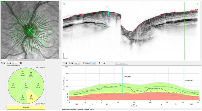
Purpose: To evaluate the dynamics of Bruch's membrane opening-based morphometrics of the optic nerve head (ONH) using spectral-domain optical coherence tomography (SD-OCT) during the first week after glaucoma surgery by trabeculectomy with mitomycin C.
Methods: Prospective, longitudinal analysis of 25 eyes of 25 patients treated by trabeculectomy. Twenty-four eyes had evaluable postoperative SD-OCT examinations. Bruch's membrane opening minimum rim width (BMO-MRW) and peripapillary retinal nerve fiber layer (RNFL) thickness were analyzed at baseline before surgery, 1 day, 2 to 3 days, and 1 week after surgery. Changes compared to baseline were correlated to intraocular pressure (IOP).
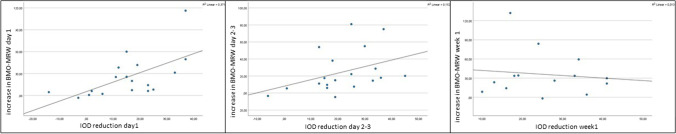
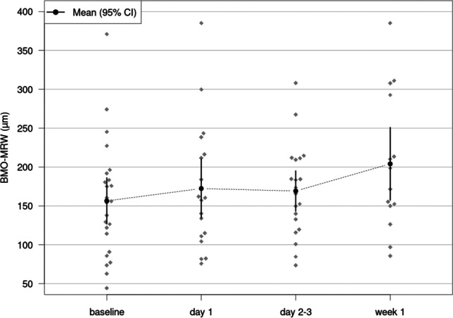
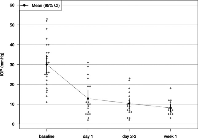
Results: One day after surgery, the mean BMO-MRW changed by + 26.17 µm, p = 0.001 (mean IOP reduction by 17.01 mmHg). This increase persisted on day 2-3 with a mean increase of BMO-MRW of + 25.33 µm, p = 0.001 (mean IOP reduction by 20.46 mmHg) and by week 1 with a mean BMO-MRW increase of + 33.17 µm, p < 0.001 (mean IOP reduction by 22.55 mmHg). The increase in BMO-MRW correlated significantly with the reduction of IOP on day 1 (Spearman's rho ρ = 0.656, p = 0.003) and d2-3 (Spearman's rho ρ = 0.479, p = 0.038). There was no statistically significant correlation found between the IOP and the increase in BMO-MRW in week 1. RNFL thickness showed no significant changes at day 1 as well as days 2-3 (p ≥ 0.078, respectively). It showed a small but significant increase in week 1 by 3.94 µm, p = 0.015.
Conclusions: Structural reversal of disc cupping in BMO-MRW occurs as early as 1 day after trabeculectomy and correlates to the extent of the IOP reduction. During the whole first week after surgery, a strong increase in BMO-MRW can be noted. The changes in BMO-based parameters need to be considered when evaluating patients' longitudinal follow-up.
Keywords: Glaucoma; Optic coherence tomography; Optic nerve head; Trabeculectomy.
© 2022. The Author(s).
Conflict of interest statement
The authors declare no competing interests.
Figures
References
-
- Chauhan BC, O’Leary N, Almobarak FA, Reis AS, Yang H, Sharpe GP, Hutchison DM, Nicolela MT, Burgoyne CF. Enhanced detection of open-angle glaucoma with an anatomically accurate optical coherence tomography-derived neuroretinal rim parameter. Ophthalmology. 2013;120(3):535–543. doi: 10.1016/j.ophtha.2012.09.055. - DOI - PMC - PubMed
-
- Chauhan BC, Danthurebandara VM, Sharpe GP, Demirel S, Girkin CA, Mardin CY, Scheuerle AF, Burgoyne CF. Bruch’s membrane opening minimum rim width and retinal nerve fiber layer thickness in a normal white population: a multicenter study. Ophthalmology. 2015;122(9):1786–1794. doi: 10.1016/j.ophtha.2015.06.001. - DOI - PMC - PubMed
-
- Danthurebandara VM, Sharpe GP, Hutchison DM, Denniss J, Nicolela MT, McKendrick AM, Turpin A, Chauhan BC. Enhanced structure-function relationship in glaucoma with an anatomically and geometrically accurate neuroretinal rim measurement. Invest Ophthalmol Vis Sci. 2015;56(1):98–105. doi: 10.1167/iovs.14-15375. - DOI - PubMed
MeSH terms
Substances
Grants and funding
LinkOut - more resources
Full Text Sources
