. 2022 May;36(5):e22310.
doi: 10.1096/fj.202101803R.

Controlled dual release of dihydrotestosterone and flutamide from polycaprolactone electrospun scaffolds accelerate burn wound healing

Affiliations

Controlled dual release of dihydrotestosterone and flutamide from polycaprolactone electrospun scaffolds accelerate burn wound healing

Huaikai Shi et al. FASEB J. 2022 May.

Abstract

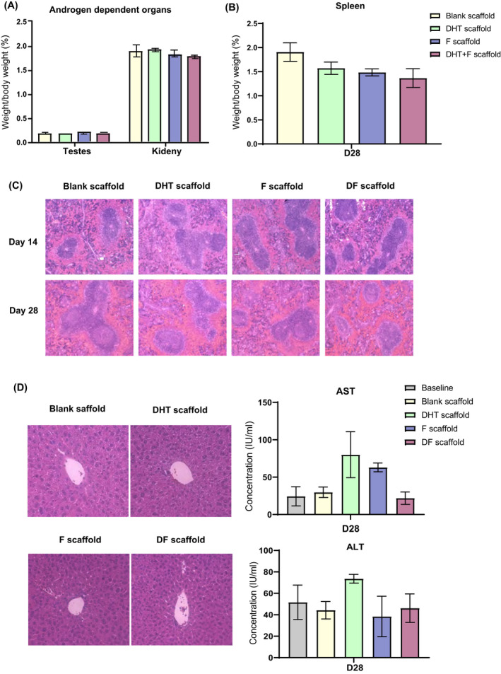
Wound healing is a complex process involving multiple independent and overlapping sequential physiological mechanisms. In addition to cutaneous injury, a severe burn stimulates physiological derangements that induce a systemic hypermetabolic response resulting in impaired wound healing. Topical application of the anti-androgen drug, flutamide accelerates cutaneous wound healing, whereas paradoxically systemic dihydrotestosterone (DHT) improves burn wound healing. We developed and characterized a PCL scaffold that is capable of controlled release of androgen (DHT) and anti-androgen (F) individually or together. This study aims to investigate whether local modification of androgen actions has an impact on burn injury wound healing. In a full-thickness burn wound healing, mouse model, DHT/F-scaffold showed a significantly faster wound healing compared with F-scaffold or DHT-scaffold. Histology analysis confirmed that DHT/F-scaffold exhibited higher re-epithelization, cell proliferation, angiogenesis, and collagen deposition. Dual release of DHT and F from PCL scaffolds promoted cell proliferation of human keratinocytes and alters the keratinocyte cell cycle. Lastly, no adverse effects on androgen-dependent organs, spleen and liver were observed. In conclusion, we demonstrated DHT plus F load PCL scaffolds accelerated burn wound healing when loading alone did not. These findings point to a complex role of androgens in burn wound healing and open novel therapeutic avenues for treating severe burn patients.

Keywords: PCL scaffold; androgens; burn injury; controlled drug delivery; flutamine; wound healing.


Figures

FIGURE 1
FIGURE 1
(A) SEM analysis of drug loaded electrospun PCL scaffolds before and after drug delivery. (B) both DHT and F were released constantly into PBS at 37°C from the PCL scaffolds without initial burst release over 31 days. Data are shown as mean ± SEM; N = 3. (C) Burst release of DHT into the circulation was detected on day 7 by LC‐MS when PCL scaffolds were applied on day 0 post‐burn injury. (D) Local DHT delivery was achieved with PCL scaffolds applied on day 7 post‐burn injury. N = 6 per group per time point, *p < .05
FIGURE 2
FIGURE 2
(A) Scaffold treatments were applied to the burn wound and assessments were performed according to the wound healing process. (B) DF scaffold displayed fastest wound recovery after scaffold treatments. (C) F and DF mixed scaffold treatments promote burn injury wound healing with the highest percentage of wound healed found in DF mixed scaffold at days 14 and 21. *DF versus F, p < .05, **F versus DHT, p < .01, ##DF versus DHT, p < .001, N = 6 per group per time point, error bar = SEM. (D) DF scaffold‐treated wound displayed smallest remaining wound area (black arrow) by H&E at day 14. (E) DF and F scaffold‐treated wound demonstrated significant higher length of re‐epithelization compared with DHT scaffold. *p < .05, N = 6 per group per time point, error bar = SEM. (F) and (G) DF scaffold‐treated wound demonstrated significant higher number of PCNA+ epithelial cells compared with F scaffold and blank scaffold‐treated wound. Black stars indicate the PCNA+ epithelial cells. *p < .05, N = 6 per group per time point, error bar = SEM
FIGURE 3
FIGURE 3
(A) IHC staining images of CD146, F4/80, PCNA at day 14 and PSR/PSR polarization light microscopy at day 28 (B) DF scaffold‐treated wound demonstrated significant higher number of CD146+ blood vessels compared with F scaffold, indicating enhanced angiogenesis at day 14. N = 6 per group, *p > .05, error bar = SEM (C) no significant difference was found for F4/80+ macrophage at wound site in all scaffold‐treated animals. N = 6 per group, error bar = SEM (D) DF scaffold‐treated mice demonstrated a significantly higher number of PCNA+ fibroblast at day 14, N = 6 per group, *p < .05, **p < .01, ***p < .001, error bar = SEM (E) collagen deposition also enhanced in DF scaffold‐treated wound at day 28. *p < .05, **p < .01, ***p < .001, N = 6 per group per time point
FIGURE 4
FIGURE 4
No significant change in the weight (w/w) of (A) androgen‐dependent organ testes and kidney and (B) spleen was observed at the endpoint of scaffold treatments (day 28) compared with the blank control. N = 6 per group, error bar = SEM (C). H&E staining demonstrated normal spleen histology with distinct white pulp and red pulp post scaffold treatments at day 14 and 28. (D) No histological changes were observed in liver samples after scaffold treatments. Furthermore, no differences in serum AST or ALT concentrations were observed compared with untreated controls. N = 6 per group, error bar = SEM
FIGURE 5
FIGURE 5
(A) Proliferation of HaCaT under scaffold treatments, DF scaffold treatment significantly enhanced HaCaT proliferation compared with that of DHT and F treatments. N = 3 per group, *p < .05, **p < .01, ***p < .001, ****p < .0001 (B) PCL scaffold treatments showed similar HaCaT migration distance post 24 and 48 h. N = 3 per group per time points (C) and (D) HaCaT cell cycle analysis revealed that a synergistic positive effect of F (reduce Sub‐G1) and DHT (extent G2‐M), correlated to the significant enhanced HaCaT proliferation observed from DF scaffold treatment. N = 3 per group, *p < .05, **p < .01, ***p < .001. (E) Flow cytometry analysis of HaCaT cell cycle
FIGURE 6
FIGURE 6
Illustration of dual‐drug scaffold accelerate burn injury wound healing via enhanced keratinocytes proliferation and migration with altered keratinocyte cell cycle

Similar articles

Cited by

References

    1. Jeschke MG. Post‐burn hypermetabolism: past, present and future. J Burn Care Res. 2016;37:86‐96. - PMC - PubMed
    1. Jeschke MG, Chinkes DL, Finnerty CC, et al. Pathophysiologic response to severe burn injury. Ann Surg. 2008;248:387‐401. - PMC - PubMed
    1. Ahn CS, Maitz PK. The true cost of burn. Burns. 2012;38:967‐974. - PubMed
    1. Ashcroft GS, Mills SJ. Androgen receptor‐mediated inhibition of cutaneous wound healing. J Clin Invest. 2002;110:615‐624. - PMC - PubMed
    1. Lai JJ, Lai KP, Chuang KH, et al. Monocyte/macrophage androgen receptor suppresses cutaneous wound healing in mice by enhancing local TNF‐alpha expression. J Clin Invest. 2009;119:3739‐3751. - PMC - PubMed

Publication types