. 2022 Apr 8;7(70):eabn6660.
doi: 10.1126/sciimmunol.abn6660. Epub 2022 Apr 8.

Human enteric viruses autonomously shape inflammatory bowel disease phenotype through divergent innate immunomodulation

Affiliations

Human enteric viruses autonomously shape inflammatory bowel disease phenotype through divergent innate immunomodulation

Fatemeh Adiliaghdam et al. Sci Immunol. .

Abstract

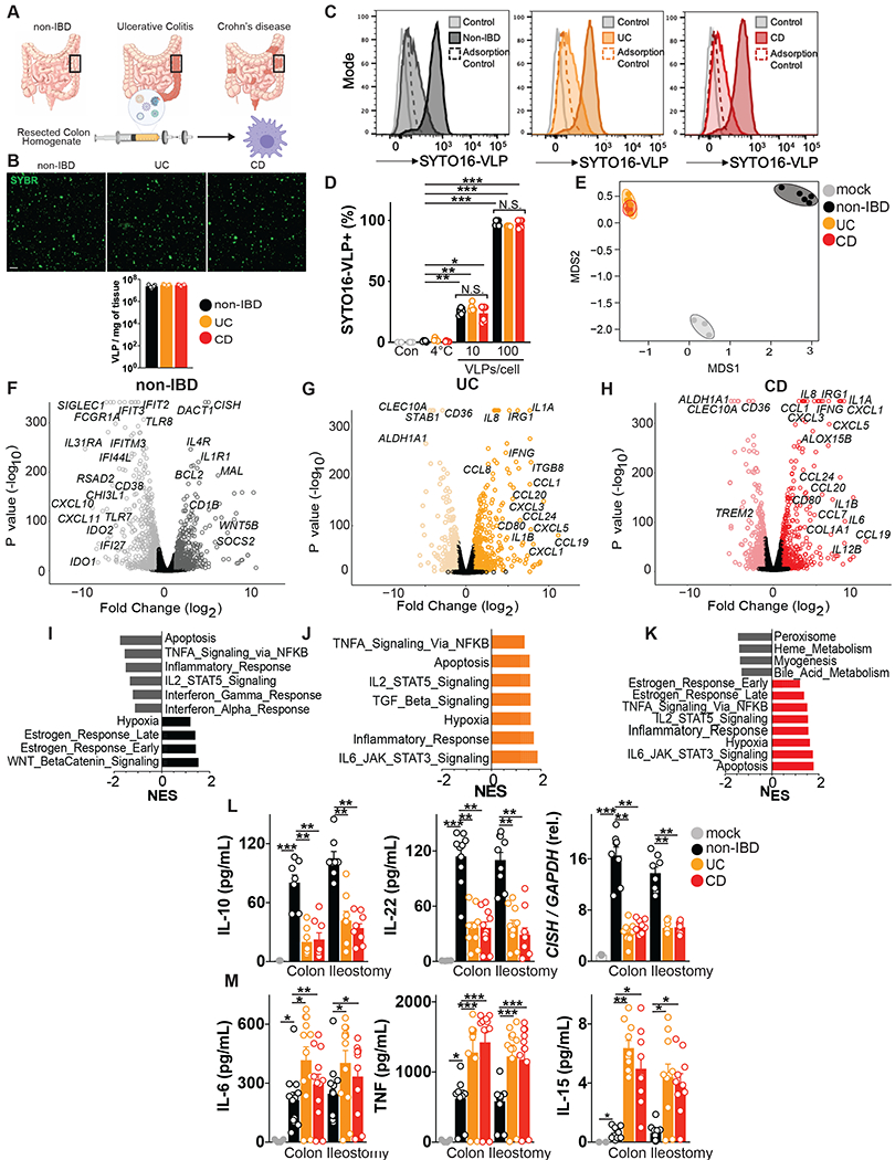
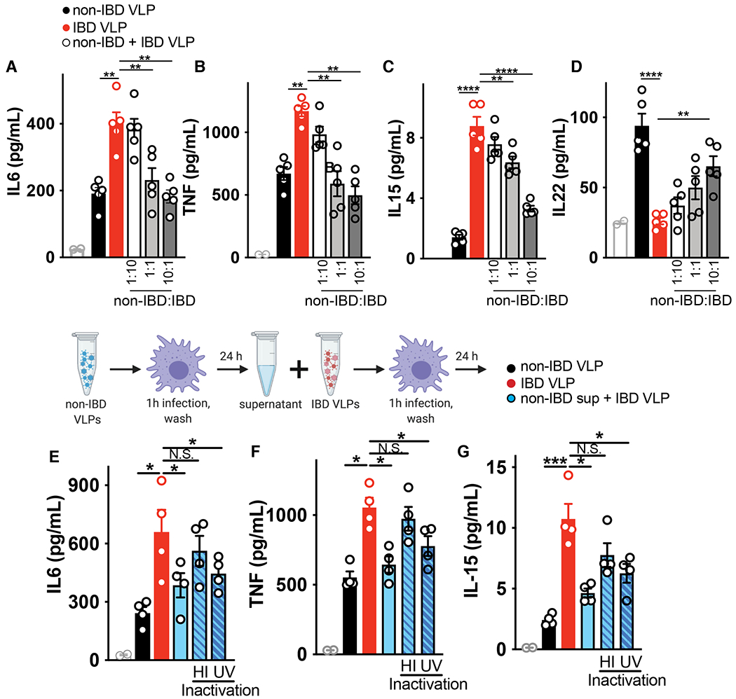
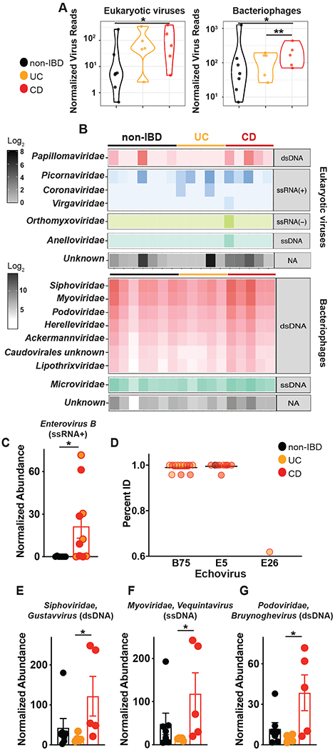
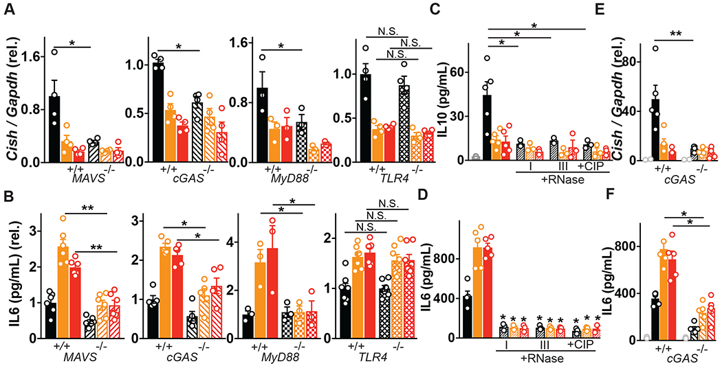
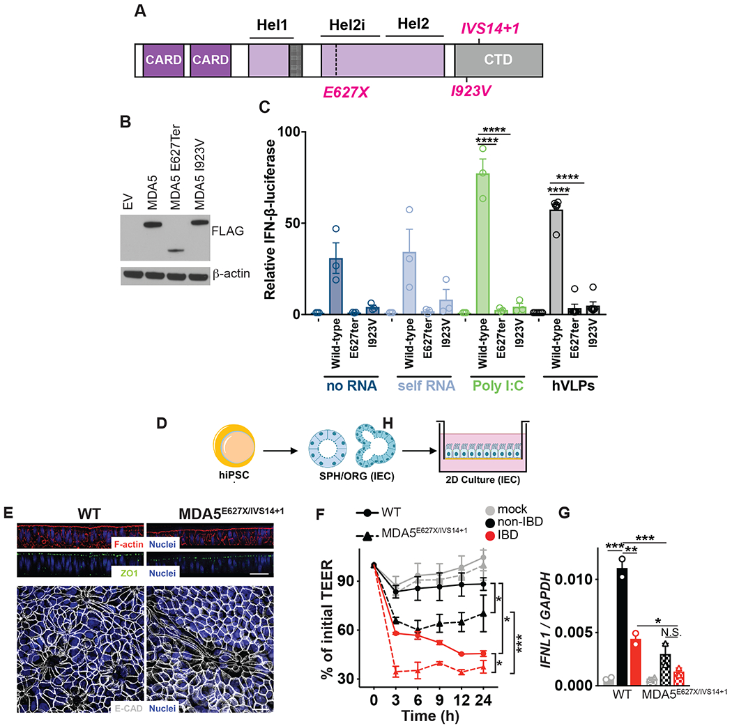
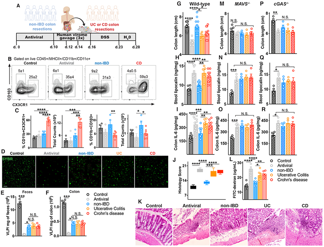
Altered enteric microorganisms in concert with host genetics shape inflammatory bowel disease (IBD) phenotypes. However, insight is limited to bacteria and fungi. We found that eukaryotic viruses and bacteriophages (collectively, the virome), enriched from non-IBD, noninflamed human colon resections, actively elicited atypical anti-inflammatory innate immune programs. Conversely, ulcerative colitis or Crohn's disease colon resection viromes provoked inflammation, which was successfully dampened by non-IBD viromes. The IBD colon tissue virome was perturbed, including an increase in the enterovirus B species of eukaryotic picornaviruses, not previously detected in fecal virome studies. Mice humanized with non-IBD colon tissue viromes were protected from intestinal inflammation, whereas IBD virome mice exhibited exacerbated inflammation in a nucleic acid sensing-dependent fashion. Furthermore, there were detrimental consequences for IBD patient-derived intestinal epithelial cells bearing loss-of-function mutations within virus sensor MDA5 when exposed to viromes. Our results demonstrate that innate recognition of IBD or non-IBD human viromes autonomously influences intestinal homeostasis and disease phenotypes. Thus, perturbations in the intestinal virome, or an altered ability to sense the virome due to genetic variation, contribute to the induction of IBD. Harnessing the virome may offer therapeutic and biomarker potential.


Conflict of interest statement

Competing interests: A provisional patent related to this work has been filed with F.A and K.L.J as inventors. K.L.J. is an employee of Moderna Inc., 200 Technology Square, Cambridge MA 02138, since November 2021. K.L.J and S.H. are members of the scientific advisory board for Ancilia Biosciences. None of these relationships influenced the work performed in this study. The other authors declare no competing interests.

Figures

Fig 1.
Fig 1.. Human colon tissue resident viruses trigger anti-inflammatory while IBD viromes divergently provoke pro-inflammatory macrophage responses.
(A) Virus-like particles (VLPs) isolated from colon resections post-surgery from non-IBD (n=5), ulcerative colitis (UC, n=5) or Crohn’s disease (CD, n=5) patients. (B) Visualization and quantification of VLP isolates per mg of colon tissue using confocal microscopy for RNA/DNA using SYBR Gold. Scale bar is 1 μm. (C, D) Flow cytometric quantification of uptake of SYTO-16 labeled human colon-resection derived VLPs by human peripheral blood-derived macrophages (1 or 10 VLPs per cell). Adsorption control is fluorescently labeled VLPs delivered to human macrophages at 4°C. (E) Multidimensional scaling (MDS) plot of human macrophage transcriptional profiles induced by non-IBD or UC or CD derived VLPs (10 VLPs/cell, 24h). (F-H) Volcano plots showing differentially expressed genes comparing F) non-IBD delivery to mock adsorption, (G) UC delivery to mock adsorption or (H) CD delivery to mock adsorption. (I-K) Hallmark gene sets significantly (P value< 0.05, FDR <0.20) represented in (I) non-IBD, (J) UC or, (K) CD VLP induced transcriptional programs as assessed by Gene Set Enrichment Analysis (GSEA). NES is normalized enrichment score. (L) Production of anti-inflammatory mediators interleukin (IL)-10, IL-22 or CISH or (M) pro-inflammatory mediators TNF, IL-6, IL-15 as measured by ELISA or qPCR following delivery of VLPs (10 VLPs/cell, 24h) extracted from non-IBD, ulcerative colitis (UC) or Crohn’s disease (CD) colon resections (n=8-12 per group) or ileostomy content (n=8 per group) to primary human peripheral blood-derived macrophages. Data are mean ± s.e.m. of biological replicates. *P<0.05, **P<0.01, ***P<0.001, ****P<0.0001 as determined by one-way ANOVA with Tukey’s multiple comparison test.
Fig 2.
Fig 2.. Suppression of IBD virome-induced inflammation by healthy human enteric viromes.
Primary human peripheral blood-derived macrophages were delivered colon resection-derived non-IBD VLPs (10 VLPs/cell), IBD VLPs (10 VLPs/cell) or indicated ratios of non IBD to IBD VLPs (total of 10 VLPs/cell) and (A) IL-6, (B) TNF, (C) IL-15 and (D) IL-22 levels were measured at 24h by ELISA. Schematic for incubation of human primary macrophages with IBD VLPs and 24h supernatant from non-IBD VLP-exposed macrophages. (E) IL-6, (F) TNF and (G) IL-15 levels were measured by ELISA following IBD VLP delivery to macrophages with the addition of non-IBD macrophage supernatant that was heat inactivated (95°C for 10 min) or UV crosslinked (three times 200 mJ/cm2 for 15 minutes) as controls. Data are mean ± s.e.m. of 5 biological replicates. *P<0.05, **P<0.01, ***P<0.001, ****P<0.0001 as determined by one-way ANOVA with Tukey’s multiple comparison test.
Fig 3.
Fig 3.. Detection and characterization of VLPs in colon surgical resections from non-IBD, Ulcerative Colitis and Crohn’s disease patients.
VLPs were assessed using VLP DNA and RNA metagenomic sequence data. Sequences were assigned a eukaryotic viral or phage taxonomic lineage using primary searches against virus sequence databases and subsequently confirmed in secondary searches using reference databases with additional non-viral taxonomic lineages. Specific eukaryotic viral families were selected for subsequent analysis by gating on the alignment length and percent identity for each family that separated the high-quality alignments from potentially spurious alignments (Table S3). (A) Violin plots represent the distribution of individual datasets for normalized reads aligning to eukaryotic viruses or bacteriophages from non-IBD, UC or CD cohorts; samples were compared using two-sided Wilcoxon rank sum tests, *P<0.05. (B) Taxonomic assignments of VLP sequences in non-IBD, UC or CD colon tissue. (C) Significantly elevated abundance of eukaryotic virus, Picornaviridae>Enterovirus B in UC and CD colon resections. (D) Identification of Echovirus B75, Echovirus E5 and Echovirus E26 Enterovirus B serotypes in UC or CD colon tissue by bbmap. All potential Picornavirales reads were re-mapped one at a time to six Enterovirus B genomes (KF874627.1, NC_001472.1, AF083069.1, AY302539.1, D00627.1, NC_038307.1), using the bbmap tool, mapPacBio.sh with the parameters recommended for remote homologies (vslow k=8 maxindel=200 minratio=0.1). The subset of these reads that mapped to Enterovirus B, along with the previously identified Enterovirus B reads, were plotted, split into Serotypes on the x-axis and percent identity on the y-axis. Each read found is plotted and grouped and colored by the type of sample in which it was found (Non-IBD, UC or CD). Non-IBD versus UC and non-IBD versus CD were significantly different (p-adj = 0.029, 0.001). (E) Top significantly increased genera of Siphoviridae, (F) Myoviridae or (G) Podoviridae bacteriophages in CD colon resections. Data are the mean of 5-7 biological replicates. *P<0.05, Kruskal–Wallis test with a Dunn’s post-hoc test.
Fig 4.
Fig 4.. Host viral receptor requirement for sensing human enteric viruses.
(A) Cish or (B) IL-6 levels following exposure of primary bone marrow derived macrophages (BMDMs) from wild-type, MAVS−/−, cGAS−/−, MyD88−/− or TLR4-deficient mice to non-IBD, Ulcerative Colitis (UC) or Crohn’s disease (CD) colon resection-derived VLPs as measured by qPCR and ELISA. (C) IL-10 or (D) IL-6 production 24h following transfection of THP1 cells with 2.5 μg of VLP RNA or VLP RNA pre-treated with RNase I (5 μg/mL, 1h), RNase III (5 μg/mL, 1h) or Calf Intestinal Phosphatase (CIP, 250U, 1h). (E) Cish levels or (F) IL-6 production 24h following transfection of WT or cGAS−/− BMDMs with 2.5 μg of VLP DNA as measured by qPCR or ELISA. Data are mean ± s.e.m. of 2-5 biological replicates repeated twice. *P<0.05, **P<0.01, ***P<0.001, ****P<0.0001 as determined by one-way ANOVA with Tukey’s multiple comparison test.
Fig. 5.
Fig. 5.. Detrimental consequences for IBD patient intestinal epithelial cells bearing MDA5 loss-of-function variants in the context of the virome.
(A) Schematic of human MDA5 and mapped IBD-associated MDA5 variants rs35744605/E627X, rs35667974/I923V and rs35732034/IVS14+1. (B) Western Blot of ectopically expressed, FLAG-tagged MDA5 in HEK293T cells. (C) Relative activation of IFNβ luciferase reporter in HEK293T cells transfected with Empty vector (EV), FLAG-tagged WT MDA5, MDA5E627Ter or MDA5I923V and transfected with 1μg/mL RNA isolated from the cytoplasm of HEK293T cells (self RNA), poly I:C or RNA isolated from human colon resection virus like particles (VLPs) for 24 hours. (D) Schematic of patient lymphoblastoid cell line (LBL) derived human induced pluripotent stem cells (hiPSCs) transdifferentiated into intestinal epithelial cell monolayers. (E) Differentiated mature monolayers evaluated for structural integrity markers. Upper panel, orthogonal view of F-actin (phalloidin, red), tight junction protein ZO-1 (green) and nuclei (DAPI, blue) by confocal microscopy. Scale bar represents 50μm. Lower panel, maximal intensity projection images of epithelial adherens junction marker E-cadherin (gray) and nuclei (DAPI, blue). Scale bar is 20 μm. (F) Transepithelial electrical resistance (TEER) over indicated time or (G) IFNL1 levels at 24h as measured by qPCR of WT or MDA5 E627Ter/IVS14+1 patient hiPSC-derived intestinal epithelial cell monolayers and exposed to VLPs (10 VLPs/cell) extracted from non-IBD or IBD colon resections. Data are mean ± s.e.m. of 2-5 biological replicates repeated twice. *P<0.05, **P<0.01, ***P<0.001, ****P<0.0001 as determined by one-way ANOVA with Tukey’s multiple comparison test.
Fig 6.
Fig 6.. Mice with a “humanized” non-IBD virome had attenuated intestinal inflammation while those with a “humanized” IBD-derived virome exhibited intestinal inflammation.
(A) Schematic of mouse gut virome depletion using an antiviral cocktail (acyclovir 20 mg/kg, lamivudine 10 mg/kg, ribavirin 30 mg/kg, oseltamivir 10 mg/kg, gray, daily gavage for 10 days), and reconstitution with 200 μL of human non-IBD or IBD VLPs pool (4×10⁸ VLPs per mouse) by gavage on day 10, 12 and 14. 2.5% DSS colitis model was commenced on day 16. (B) Flow cytometry plots of CD45+MHCII+CD11c+CD11b+ cells from the colonic lamina propria, and percentages and counts of (C) CX3CR1+ or CD103+ mononuclear phagocytes in control, antiviral depleted (AV), non-IBD or IBD humanized virome mice on day 17. (D-F) Confocal images and quantification of viral-like particles (VLPs) using SYBR Gold in feces or colon tissue of humanized virome mice. (G) Colon length, (H) stool lipocalin, (I) IL-6 levels in colonic explant supernatants cultured for 24 hours following 12 days of induced DSS colitis measured by ELISA. (J,K) Representative hematoxylin and eosin–stained sections and blinded histologic scores in mouse colon tissue (scale bar, 50 μm). (L) FITC-dextran level in serum 4 hours after oral gavage following 7 days of DSS. (M-O) Colon length, stool lipocalin or colonic explant IL-6 levels following induction of DSS colitis in antiviral-treated MAVS−/− mice or (P-R) cGAS−/− mice administered human non-IBD, UC or CD colon-derived viromes. Data are mean ± s.e.m. of 4-8 animals. Representative of 2 independent experiments. *P<0.05, **P<0.01, ***P<0.001, ****P<0.0001, two-tailed unpaired t-test or one-way ANOVA with Tukey’s multiple comparison test.

References

    1. Norman JM et al. Disease-specific alterations in the enteric virome in inflammatory bowel disease. Cell 160, 447–460 (2015). - PMC - PubMed
    1. Liang G et al. The stepwise assembly of the neonatal virome is modulated by breastfeeding. Nature 581, 470–474 (2020). - PMC - PubMed
    1. Edwards RA et al. Global phylogeography and ancient evolution of the widespread human gut virus crAssphage. Nat Microbiol 4, 1727–1736 (2019). - PMC - PubMed
    1. Camarillo-Guerrero LF, Almeida A, Rangel-Pineros G, Finn RD, Lawley TD, Massive expansion of human gut bacteriophage diversity. Cell 184, 1098–1109 e1099 (2021). - PMC - PubMed
    1. Barr JJ et al. Bacteriophage adhering to mucus provide a non-host-derived immunity. Proc Natl Acad Sci U S A 110, 10771–10776 (2013). - PMC - PubMed

Publication types