. 2022 Apr 8;8(14):eabm8501.
doi: 10.1126/sciadv.abm8501. Epub 2022 Apr 8.

Translational recoding by chemical modification of non-AUG start codon ribonucleotide bases

Affiliations

Translational recoding by chemical modification of non-AUG start codon ribonucleotide bases

Yoshihiko Fujita et al. Sci Adv. .

Abstract

In contrast to prokaryotes wherein GUG and UUG are permissive start codons, initiation frequencies from non-AUG codons are generally low in eukaryotes, with CUG being considered as strongest. Here, we report that combined 5-cytosine methylation (5mC) and pseudouridylation (Ψ) of near-cognate non-AUG start codons convert GUG and UUG initiation strongly favored over CUG initiation in eukaryotic translation under a certain context. This prokaryotic-like preference is attributed to enhanced NUG initiation by Ψ in the second base and reduced CUG initiation by 5mC in the first base. Molecular dynamics simulation analysis of tRNAiMet anticodon base pairing to the modified codons demonstrates that Ψ universally raises the affinity of codon:anticodon pairing within the ribosomal preinitiation complex through partially mitigating discrimination against non-AUG codons imposed by eukaryotic initiation factor 1. We propose that translational control by chemical modifications of start codon bases can offer a new layer of proteome diversity regulation and therapeutic mRNA technology.


Figures
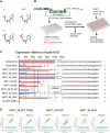
Fig. 1.
Fig. 1.. Double mRNA chemical modification alters start codon accuracy.
(A) Atomic structure of chemically modified nucleotides (5mC and Ψ) is shown along with natural nucleotides C and U. Cyan, carbon; blue, nitrogen; red, oxygen; yellow, phosphorus. (B) Experimental scheme. Oligos containing wild-type or mutant versions of 5′UTR of NAT1/eIF4G2 or control synthetic GFP mRNA were synthesized to generate capped poly(A) GFP mRNA. The mRNA is cotransfected with iRFP670 mRNA as internal standard. GFP/iRFP(670) expression ratio was quantified by flow cytometry. (C) Translation of natural or 5mC:Ψ-modified GFP mRNA with different non-AUG start codons was quantified in 293FT, relative to the value from GFP mRNA bearing the AUG start codon under a typical Kozak context (columns 1). 5′UTR nucleotide sequences of the constructs used in this figure are shown besides the graph. A (black) of the Kozak AUG codon or G (blue) of the first position of NAT1 start codon was altered to G (red) or A, C, and U (red), respectively. Mutated residues in other constructs were also labeled red. Bars indicate SD (n = 3 except Kozak GUG, n = 2). *P < 0.003 and **P = 0.06 (n = 2). (D) The plot of GFP versus iRFP expression in 10,000 cells cotransfected with indicated GFP mRNAs and Kozak AUG iRFP670 mRNA. For each mRNA shown, left shows the plot using natural mRNA (in orange), while the right, using double-modified mRNA (in red). For each panel, the plot using control Kozak AUG mRNA is shown in green.
Fig. 2.
Fig. 2.. Effect of 5mC, 5hmC, Ψ, and 1mΨ on start codon specificity during translation initiation.
(A) GFP mRNA derivatives with indicated 5′UTR were synthesized in the presence of 5mCTP (blue bars), 5hmCTP (cyan bars), ΨTP (green bars), 1mΨTP (purple bars), both 5mCTP and ΨTP (red bars), and unmodified nucleotides (orange bars) and subjected for expression assay. Median GFP/iRFP expression ratio was normalized to the value from Kozak AUG GFP mRNA with the same modification. Bars indicate SD (n = 3, except n = 2 for 1mΨ). *P < 0.01, **P ≤ 0.05, and ***P = 0.06. (B) The plot of GFP versus iRFP expression is shown for indicated mRNA species as in Fig. 1D; natural RNA in orange, specific modification in red or blue, and control mRNA in green.
Fig. 3.
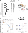
Fig. 3.. Determination of codon:anticodon affinity by the ABF method.
(A) Estimated binding free energy. ΔGbinding score (see the schematics in fig. S4B) of all codons are shown. The scores were obtained from P(d1, d2, d3) averaged over five simulation trials for each model. Data for AUG, GUG, and CUG are taken from (29). Table to the right lists ΔG, and the energetic penalty compared with AUG. (B) Base pairing penalty relative to the free energy obtained for AUG in kJ/mol (ΔΔG=ΔGbindingΔGbindingAUG) was plotted against initiation frequencies from indicated codons relative to one from AUG in Homo sapiens HEK293–derived cells, which was determined here using NAT1_24 mRNA derivatives (Fig. 1C) (orange circles) or previously using firefly luciferase reporters initiated by equivalent codons (blue circles) (13). (C) Average structure of the bound state (d1~,d2~,d3~)=(4.5,4.5,4.5) of eukaryotic P-site bearing AUG start codon (stick models in orange). Locations of anticodon (black) and eIF1 β-hairpin loop (stick model with atomic colorcode as in Fig. 1A except R33 and K37 in red) are highlighted. (D) Inter–base pair pitch calculated from the average structure of the bound state (d1~,d2~,d3~)=(4.5,4.5,4.5).
Fig. 4.
Fig. 4.. Schematics of the base pair binding dynamics.
The base pairing dynamics are described for AUG (A), CΨG (B), or GΨG (C) start codons. Conformational changes inferred from the free energy landscape shown in the graphs in panel 1 (also see fig. S5) are summarized in panel 2. The transition path Rn (• is AUG, CΨG, or GΨG) is shown by black arrows. Dotted line, base-pairing distance; intermediate distance, close but not base pairing; large distance, weak or no interaction.
Fig. 5.
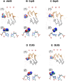
Fig. 5.. Average structures of start codon paired with anticodon in the P-site predicted by ABF MD simulation.
Panels 1 and 2, averaged structures corresponding to (d1~,d2~,d3~)=(4.5,4.5,4.5) computed for AUG (A), CΨG (B), GΨG (C), CUG (D), and GUG (E) are presented. In panel 1, nucleotides of the codon (orange) and anticodon (gray) are drawn by thick lines. Thin red and blue lines are parts of eIF1 and eIF1A, respectively, with eIF1-N34 highlighted in a spherical model (with the same color code as Fig. 1A). In panel 2, the pair of bases at the first position is shown by stick models with the atomic color code as in Fig. 1A, along with the spherical model of eIF1-N34. In (A), (D), and (E), averaged structures corresponding to (d1~,d2~,d3~)=(4.5,4.5,4.5) are presented for AUG, CUG, and GUG, based on previously reported simulation study (29).
Fig. 6.
Fig. 6.. The mCUG pairing dynamics.
(A) Base pair binding dynamics for mCUG. Panel 1, free energy landscape. Panel 2, deduced pairing pathway. (B) Average structure of the bound states of mCUG (orange) pairing to anticodon bases (gray) with eIF1 highlighted in panel 1 as in Fig. 5 (panel 1). Panel 2, the base pair at the first position.
Fig. 7.
Fig. 7.. The possible role of eIF1-N34 in discrimination of the first codon base by size.
Codon-specific effect of eIF1 mutations on initiation frequencies in yeast is presented in a graph. Fold increase compared with the average expression level relative to AUG in WT (see fig. S7) is presented for indicated eIF1 mutants. Yeast strains used are KAY1057 (SUI1 LEU2 ura3) and its isogenic derivatives, H4563 (sui1-K60E), H4564 (sui1-L96P), H4944 (sui1-N34A), and H4945 (sui1-N34E) (19, 31). Bars indicate SEM. P values are shown for significant differences obtained with indicated pairs (experiments with eIF1-K60E, n = 8 for AUU and CUG and n = 6 for GUG and ACG; eIF1-L96P, n = 8 for each start codon; eIF1-N34E, n = 10 for CUG and GUG and n = 8 for AUU and ACG; eIF1-N34A, n = 14 for each start codon).

References

    1. Hinnebusch A. G., Ivanov I. P., Sonenberg N., Translational control by 5′-untranslated regions of eukaryotic mRNAs. Science 352, 1413–1416 (2016). - PMC - PubMed
    1. A. G. Hinnebusch, T. E. Dever, K. Asano, in Translational Control in Biology and Medicine, M. B. Mathews, N. Sonenberg, J. W. B. Hershey, Eds. (Cold Spring Harbor Lab Press, Cold Spring Harbor, NY, 2007), pp. 225–268.
    1. Gilbert W. V., Bell T. A., Schaening C., Messenger RNA modifications: Form, distribution and function. Science 352, 1408–1412 (2016). - PMC - PubMed
    1. Asano K., Why is start codon selection so precise in eukaryotes? Translation 2, e28387 (2014). - PMC - PubMed
    1. Zang L., Lui K. O., von Gise A., Ma Q., Ebina W., Ptaszek L. M., Später D., Xu H., Tabebordbar M., Gorbatov R., Sena B., Nahrendorf M., Briscoe D. M., Li R. A., Wagers A. J., Rossi D. J., Pu W. T., Chien K. R., Modified mRNA directs the fate of heart progenitor cells and induces vascular regeneration after myocardial infarction. Nat. Biotechnol. 31, 898–907 (2013). - PMC - PubMed