. 2022 Apr 8;8(14):eabh2445.
doi: 10.1126/sciadv.abh2445. Epub 2022 Apr 8.

The microdissected gene expression landscape of nasopharyngeal cancer reveals vulnerabilities in FGF and noncanonical NF-κB signaling

Affiliations

The microdissected gene expression landscape of nasopharyngeal cancer reveals vulnerabilities in FGF and noncanonical NF-κB signaling

Joshua K Tay et al. Sci Adv. .

Abstract

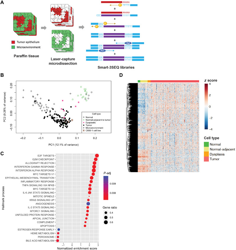
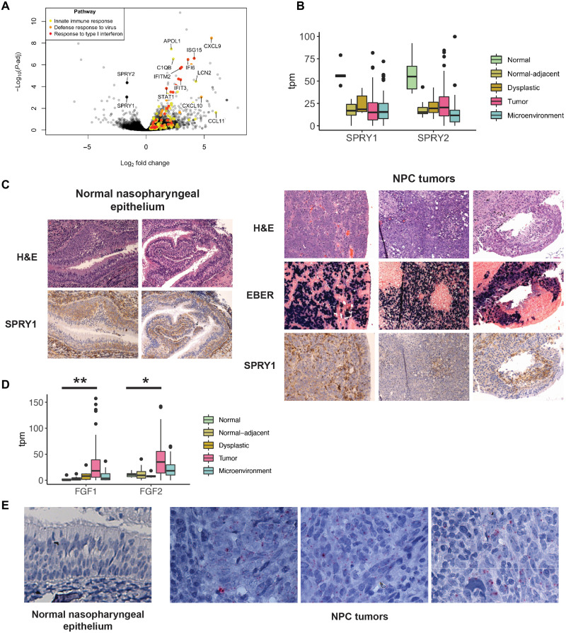
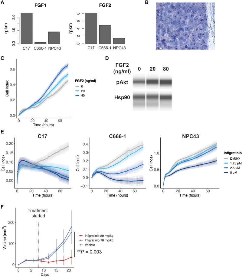
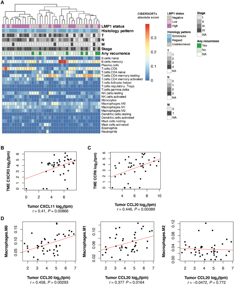
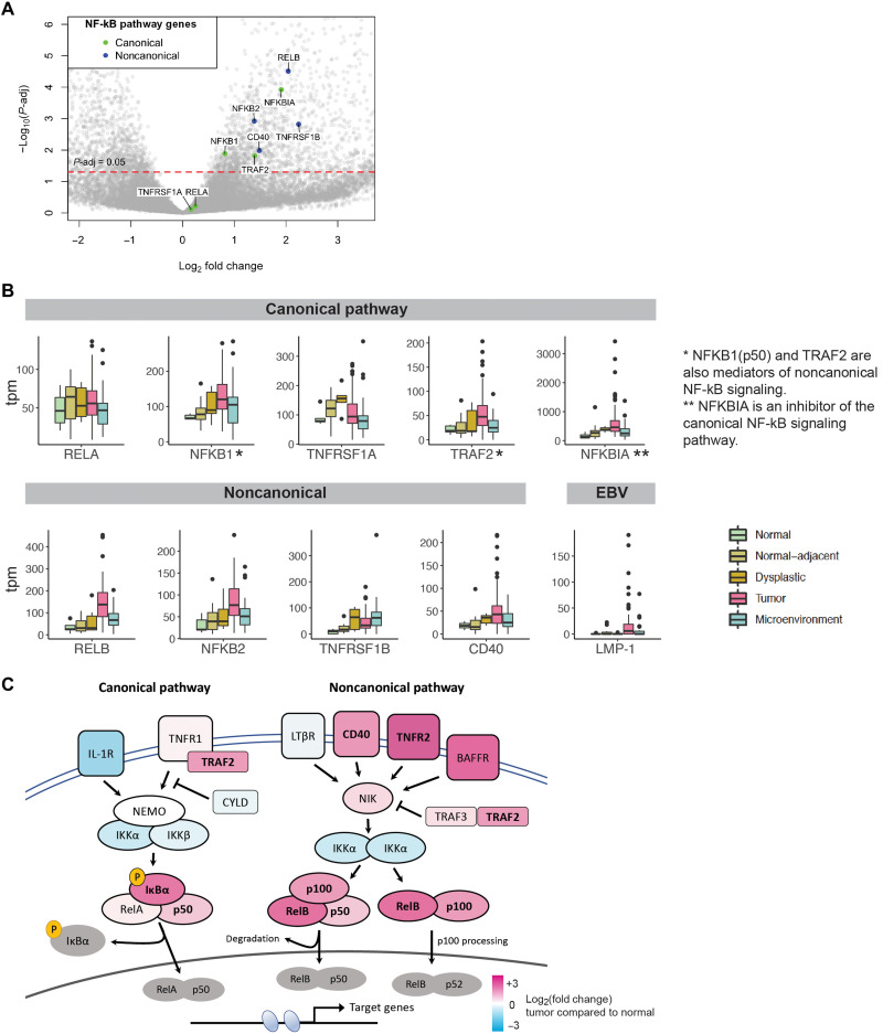
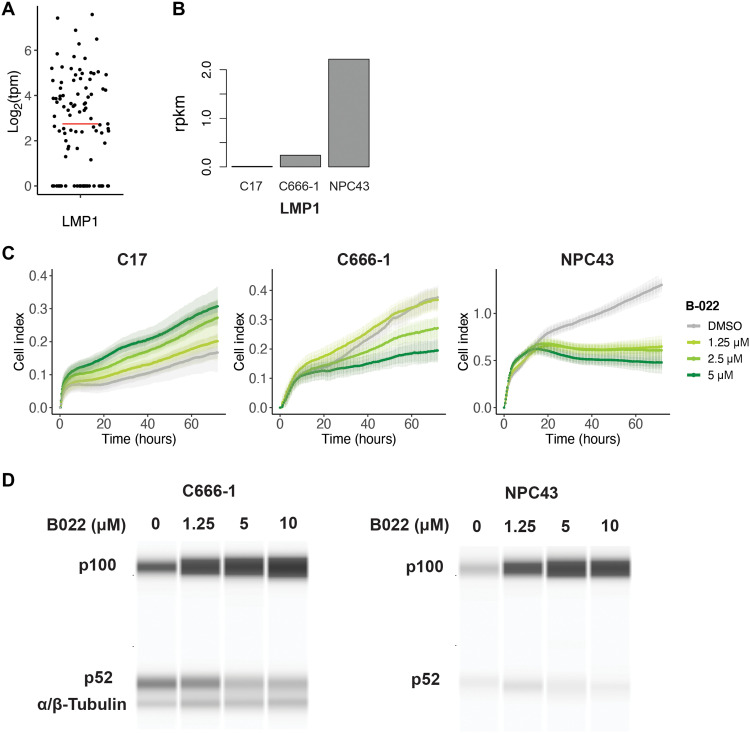
Nasopharyngeal cancer (NPC) is an Epstein-Barr virus (EBV)-positive epithelial malignancy with an extensive inflammatory infiltrate. Traditional RNA-sequencing techniques uncovered only microenvironment signatures, while the gene expression of the tumor epithelial compartment has remained a mystery. Here, we use Smart-3SEQ to prepare transcriptome-wide gene expression profiles from microdissected NPC tumors, dysplasia, and normal controls. We describe changes in biological pathways across the normal to tumor spectrum and show that fibroblast growth factor (FGF) ligands are overexpressed in NPC tumors, while negative regulators of FGF signaling, including SPRY1, SPRY2, and LGALS3, are down-regulated early in carcinogenesis. Within the NF-κB signaling pathway, the critical noncanonical transcription factors, RELB and NFKB2, are enriched in the majority of NPC tumors. We confirm the responsiveness of EBV-positive NPC cell lines to targeted inhibition of these pathways, reflecting the heterogeneity in NPC patient tumors. Our data comprehensively describe the gene expression landscape of NPC and unravel the mysteries of receptor tyrosine kinase and NF-κB pathways in NPC.


Figures

Fig. 1.
Fig. 1.. Hallmark biological processes are significantly altered in NPC.
(A) LCM of tumor epithelial cells and the microenvironment separately, followed by gene expression libraries prepared by Smart-3SEQ. (B) Principal components analysis of human nasopharyngeal tissue gene expression from tumor and normal biopsies (n = 175 libraries). (C) Significantly altered Hallmark biological processes comparing tumor epithelial cells (n = 54) and normal nasopharyngeal epithelium (n = 5). (D) Heatmap of 2570 genes from Hallmark biological processes found to be significantly altered (P-adj < 0.05) across the normal tumor spectrum (n = 120 libraries).
Fig. 2.
Fig. 2.. FGF signaling is enriched in NPC.
(A) Volcano plot of early gene expression changes in NPC showing differentially expressed genes between normal-adjacent epithelium (n = 14 tumors) and normal nasopharyngeal epithelium samples (n = 5 controls). (B) Gene expression of SPRY1 and SPRY2 across the normal tumor spectrum (n = 171 libraries). (C) Immunohistochemistry of SPRY1 in normal nasopharyngeal epithelium and NPC tumor samples. (D) Expression of FGF1 and FGF2 across the normal tumor spectrum (n = 171 libraries) **P-adj = 0.00326; *P-adj = 0.0158. (E) FGF2 RNA ISH in normal nasopharyngeal epithelium and NPC tumor specimens.
Fig. 3.
Fig. 3.. In vitro and in vivo inhibition of FGF signaling in NPC.
(A) Gene expression of FGF1 and FGF2 in EBV-positive NPC cell lines (n = 1 per cell line). (B) RNA ISH of FGF2 in the C666-1 NPC cell line grown in organoid. (C) Proliferation of the C666-1 cell line in media supplemented with FGF2. (D) Western blot of AKT expression in C666-1 cells after treatment with FGF2. (E) In vitro proliferation of EBV-positive NPC cell lines when treated with infigratinib, an FGFR inhibitor, as measured on the xCELLigence real-time cell analyzer. Error bars represent SD. (F) Growth of C666-1 xenografts in NSG mice when treated with infigratinib.
Fig. 4.
Fig. 4.. Tumor microenvironment relationships in NPC.
(A) Heatmap of CIBERSORTx deconvoluted immune cell fractions in the microenvironment (n = 40 paired tumor epithelial and microenvironment libraries). (B) Correlation analysis of tumor CXCL11 expression and its receptor CXCR3 in the microenvironment (n = 41 paired libraries). (C) Correlation analysis of tumor CCL20 expression and its receptor CCR6 in the microenvironment (n = 41 paired libraries). (D) Correlation analysis of tumor epithelial CCL20 and CIBERSORTx deconvoluted M0, M1, and M2 macrophage fractions in the microenvironment (n = 41 paired libraries).
Fig. 5.
Fig. 5.. Key pathway members of noncanonical NF-κB signaling are up-regulated in NPC.
(A) Volcano plot of differentially expressed genes comparing NPC tumors (n = 54 tumors) and normal nasopharyngeal epithelium (n = 5 controls), with key members of the NF-κB pathway highlighted. (B) Gene expression of key members of canonical and noncanonical NF-κB signaling (n = 171 libraries). (C) Heatmap based on fold change expression of key mediators of the NF-κB signaling pathway in NPC tumors compared to normal nasopharyngeal epithelium, with significant differentially expressed genes highlighted in bold.
Fig. 6.
Fig. 6.. LMP1-expressing tumors are sensitive to inhibition of noncanonical NF-κB signaling.
(A) Distribution of LMP1 gene expression in NPC tumors (n = 99 libraries, median log2tpm = 2.51, represented by red bar). (B) LMP1 gene expression in EBV-positive NPC cell lines (n = 1 per cell line). (C) In vitro proliferation of EBV-positive NPC cell lines when treated with B022, an NIK inhibitor targeting the noncanonical NF-κB pathway. Error bars represent SD. (D) Western blot of the noncanonical p100/p52 subunits in the C666-1 and NPC43 cell lines when treated with increasing concentrations of B022.

References

    1. Chang E. T., Adami H. O., The enigmatic epidemiology of nasopharyngeal carcinoma. Cancer Epidemiol. Biomarkers Prev. 15, 1765–1777 (2006). - PubMed
    1. Yu M. C., Yuan J. M., Epidemiology of nasopharyngeal carcinoma. Semin. Cancer Biol. 12, 421–429 (2002). - PubMed
    1. Lanier A., Bender T., Talbot M., Wilmeth S., Tschopp C., Henle W., Henle G., Ritter D., Terasaki P., Nasopharyngeal carcinoma in Alaskan Eskimos Indians, and Aleuts: A review of cases and study of Epstein-Barr virus, HLA, and environmental risk factors. Cancer 46, 2100–2106 (1980). - PubMed
    1. Chien Y. C., Chen J. Y., Liu M. Y., Yang H. I., Hsu M. M., Chen C. J., Yang C. S., Serologic markers of Epstein-Barr virus infection and nasopharyngeal carcinoma in Taiwanese men. N. Engl. J. Med. 345, 1877–1882 (2001). - PubMed
    1. Tay J. K., Siow C. H., Goh H. L., Lim C. M., Hsu P. P., Chan S. H., A comparison of EBV serology and serum cell-free DNA as screening tools for nasopharyngeal cancer: Results of the Singapore NPC screening cohort. Int. J. Cancer 146, 2923–2931 (2019). - PubMed

MeSH terms