. 2022 Apr 12;119(15):e2200068119.
doi: 10.1073/pnas.2200068119. Epub 2022 Apr 8.

Aster proteins mediate carotenoid transport in mammalian cells

Affiliations

Aster proteins mediate carotenoid transport in mammalian cells

Sepalika Bandara et al. Proc Natl Acad Sci U S A. .

Abstract

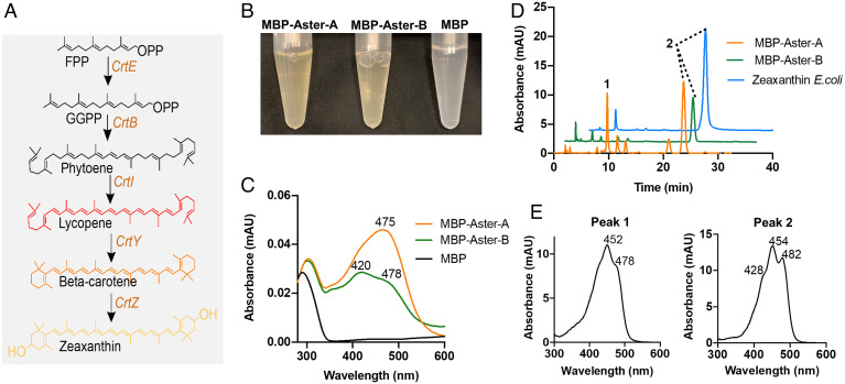
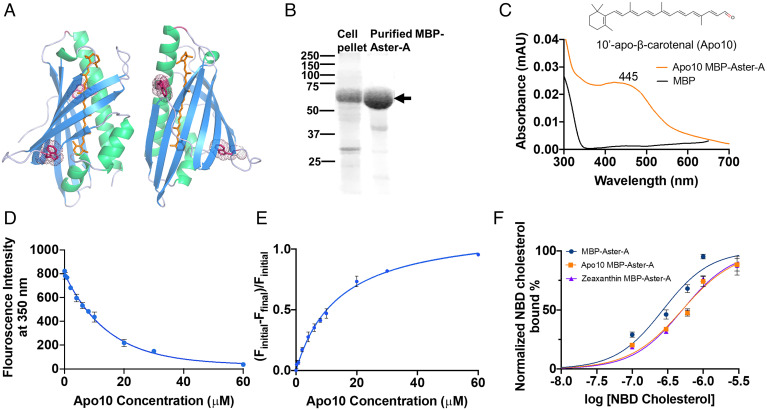
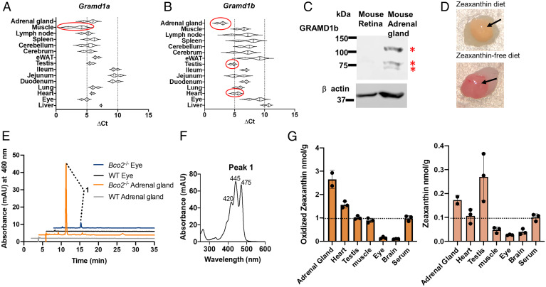
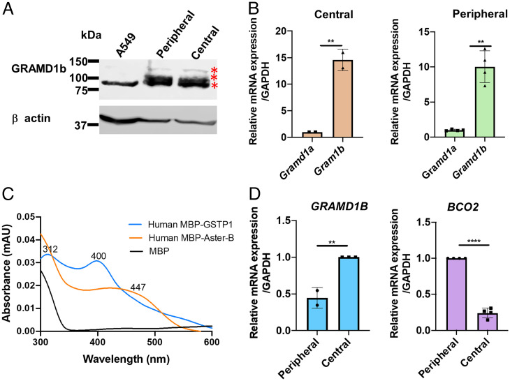
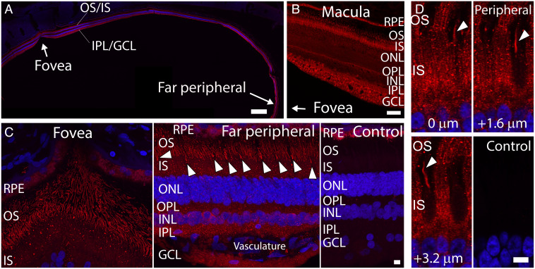
Some mammalian tissues uniquely concentrate carotenoids, but the underlying biochemical mechanism for this accumulation has not been fully elucidated. For instance, the central retina of the primate eyes displays high levels of the carotenoids, lutein, and zeaxanthin, whereas the pigments are largely absent in rodent retinas. We previously identified the scavenger receptor class B type 1 and the enzyme β-carotene-oxygenase-2 (BCO2) as key components that determine carotenoid concentration in tissues. We now provide evidence that Aster (GRAM-domain-containing) proteins, recently recognized for their role in nonvesicular cholesterol transport, engage in carotenoid metabolism. Our analyses revealed that the StART-like lipid binding domain of Aster proteins can accommodate the bulky pigments and bind them with high affinity. We further showed that carotenoids and cholesterol compete for the same binding site. We established a bacterial test system to demonstrate that the StART-like domains of mouse and human Aster proteins can extract carotenoids from biological membranes. Mice deficient for the carotenoid catabolizing enzyme BCO2 concentrated carotenoids in Aster-B protein-expressing tissues such as the adrenal glands. Remarkably, Aster-B was expressed in the human but not in the mouse retina. Within the retina, Aster-B and BCO2 showed opposite expression patterns in central versus peripheral parts. Together, our study unravels the biochemical basis for intracellular carotenoid transport and implicates Aster-B in the pathway for macula pigment concentration in the human retina.

Keywords: BCO2; GRAMD1; carotenoids; cholesterol; retina.


Conflict of interest statement

The authors declare no competing interest.

Figures

Fig. 1.
Fig. 1.
A bacterial test system for carotenoid binding proteins. (A) Bacterial zeaxanthin biosynthesis pathway. (B) Color comparison of the eluates of purified recombinant MBP-Aster-A, MBP-Aster-B, and MBP. (C) UV/Visible absorption spectra of recombinant murine MBP-Aster-A (orange trace), MBP-Aster-B (green trace), and MBP (black trace) (D) HPLC traces at 460 nm of organic extracts from the zeaxanthin biosynthesizing E.coli cells (blue trace) and purified MBP-Aster-A (orange trace) and MBP-Aster-B (green trace). (E) UV/Vis absorption spectra for the indicated peak 1 (β-cryptoxanthin) and peak 2 (zeaxanthin) in the chromatograms. The absorption maximum of each peak is indicated in the panels.
Fig. 2.
Fig. 2.
Recombinant MBP-Aster-A binds carotenoids and sterols. (A) Two-side view (in 180° angle) of the structure of the StART-domain of murine Aster A (PDB ID- 6GQF) with a bound carotenoid (orange) modeled into the binding cavity. Tryptophan residues in close vicinity to binding cavity are shown as dark-pink color sticks. (B) Sodium dodecyl sulfate polyacrylamide gel electrophoresis (SDS-PAGE) of the protein purification of recombinant MBP-Aster-A. Cell pellet (insoluble) fraction and affinity purified MBP-Aster-A were resolved by SDS-PAGE. The black arrow indicates the size of the MBP-Aster-A protein (67 kDa). (C) UV/Vis absorption spectrum of the β-apo-10′-carotenal (Apo10) bound to recombinant MBP-Aster-A and MBP. (D) Tryptophan fluorescence quenching of MBP-Aster-A in the presence of increasing concentrations of Apo10. (E) Bound over free fraction of binding assays with MBP-Aster-A and Apo10. (F) Competition assays with MBP-Aster-A with NBD cholesterol and carotenoids. MBP-Aster-A was incubated in the presence of increasing amounts of NBD cholesterol (black curve); the purified Apo10-MBP-Aster-A caroteneoprotein complex was incubated in the presence of increasing amounts of NBD cholesterol (orange curve). The purified zeaxanthin-MBP-Aster A caroteneoprotein complex was incubated in the presence of increasing amounts of NBD cholesterol (purple curve).
Fig. 3.
Fig. 3.
Carotenoids accumulate in Gramd1-expressing tissues of Bco2−/− mice. (A and B) qRT-PCR analysis of mRNA expression of (A) Gramd1a gene (encoding Aster A) and (B) Gramd1b (Aster-B) in tissues of wild-type mice. The ΔCt values are normalized to the β-actin housekeeping gene (lower ΔCt values indicate higher mRNA expression levels). The tissues with the highest mRNA expression are highlighted by red circles. (C) Western blot with protein extracts (20 μg per lane) of mouse retina and mouse adrenal gland with anti-GRAMD1b antibody. β-actin was used as the loading control. Three different isoforms (red asterisks) were detected in the adrenal gland. (D) Comparison of the color difference between freshly dissected adrenal glands collected from Bco2−/− mice fed with the zeaxanthin diet (upper photograph) and with zeaxanthin-free diet (lower photograph). The glandular parts are indicated with an arrow. (E) HPLC traces at 460 nm of lipid extracts of adrenal glands and eyes of wild-type (WT) and Bco2−/− mice subjected to feeding with zeaxanthin-rich diet. (F) UV/Vis spectra of the eluted oxidized zeaxanthin (peak 1). (G) The contents of oxidized zeaxanthin (Left) and zeaxanthin (Right) of different tissues of Bco2−/− mice (n = 3) fed with zeaxanthin. The dashed lines indicate the serum concentration of the carotenoids.
Fig. 4.
Fig. 4.
Opposite expression patterns of GRAMD1B and BCO2 in the human retina. (A) Western blot for GRAMD1B with protein extracts of A549 cells (positive control), and human peripheral and central retina. 20 μg of protein was separated in each lane. β-actin was used as loading control. (B) Relative mRNA expression of GRAMD1A and GRAMD1B in central and peripheral parts of human retinas (n = 3 individual donor retinas). (C) UV-Vis absorption spectrum of purified recombinant purified human MBP-ASTER-B and MBP-GSTP1 as well as MBP expressed in zeaxanthin synthesizing E. coli cells. (D) Relative mRNA expression of GRAMD1B and BCO2 in peripheral and central parts of the human retinas (n = 3 individual donor retinas). In (B) and (D), GAPDH was used to normalize the expression.
Fig. 5.
Fig. 5.
Immunofluorescence localization of GRAMD1B (Aster B) in the human retina. (A) Low magnification image of human retina labeled with anti-GRAMD1B antibody (red). Foveal and far peripheral regions are indicated by arrows. (B) High magnification image of macula region labeled with anti-GRAMD1B antibody (red). The direction of the foveal region is indicated by an arrow. (C) High magnification images of the foveal region (Left) and far peripheral retina (Middle). Cones in the far peripheral region are indicated by arrowheads. As control, IgG from unimmunized rabbits was applied to human retina (Right). Significant immunofluorescence signals were not detected throughout the retina. RPE demonstrated autofluorescence. (D) In the outer segment layer of the peripheral retina, anti-GRAMD1B antibody labeled rod and cone (arrowhead) outer segments (Top Row and Bottom Left). Images at three different z-heights (relative positions of 0, 1.6, and 3.2 µm) are shown. Control rabbit IgG does not demonstrate significant signal within the photoreceptor inner and outer segments (Bottom Right). In (A), (C), and (D), nuclei were labeled with hoechst33342 dye (blue). Scale bars are 600 µm for (A), 60 µm for (B), and 6 µm for (C and D). Abbreviations: RPE, retinal pigment epithelium; OS, photoreceptor outer segments; IS, photoreceptor inner segments; ONL, outer nuclear layer; OPL, outer plexiform layer, INL, inner nuclear layer; IPL, outer plexiform layer; GCL, ganglion cell layer.

References

    1. von Lintig J., Colors with functions: Elucidating the biochemical and molecular basis of carotenoid metabolism. Annu. Rev. Nutr. 30, 35–56 (2010). - PubMed
    1. Álvarez R., Vaz B., Gronemeyer H., de Lera A. R., Functions, therapeutic applications, and synthesis of retinoids and carotenoids. Chem. Rev. 114, 1–125 (2014). - PubMed
    1. Demmig-Adams B., W. W. Adams, 3rd, Antioxidants in photosynthesis and human nutrition. Science 298, 2149–2153 (2002). - PubMed
    1. Toews D. P. L., Hofmeister N. R., Taylor S. A., The evolution and genetics of carotenoid processing in animals. Trends Genet. 33, 171–182 (2017). - PubMed
    1. Bone R. A., Landrum J. T., Tarsis S. L., Preliminary identification of the human macular pigment. Vision Res. 25, 1531–1535 (1985). - PubMed

LinkOut - more resources