Review
. 2022 Nov 25;43(6):1003-1037.
doi: 10.1210/endrev/bnac010.

Clinical Biology of the Pituitary Adenoma

Affiliations
Review

Clinical Biology of the Pituitary Adenoma

Shlomo Melmed et al. Endocr Rev. .

Abstract

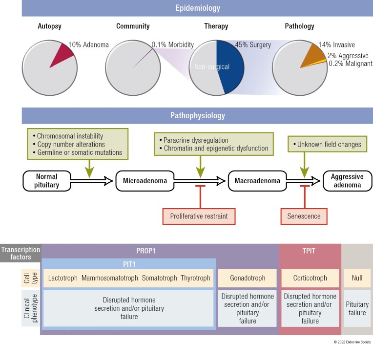
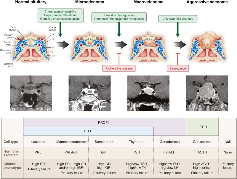
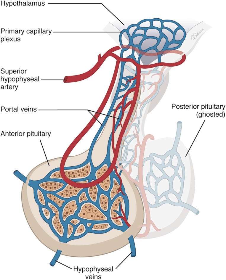
All endocrine glands are susceptible to neoplastic growth, yet the health consequences of these neoplasms differ between endocrine tissues. Pituitary neoplasms are highly prevalent and overwhelmingly benign, exhibiting a spectrum of diverse behaviors and impact on health. To understand the clinical biology of these common yet often innocuous neoplasms, we review pituitary physiology and adenoma epidemiology, pathophysiology, behavior, and clinical consequences. The anterior pituitary develops in response to a range of complex brain signals integrating with intrinsic ectodermal cell transcriptional events that together determine gland growth, cell type differentiation, and hormonal production, in turn maintaining optimal endocrine health. Pituitary adenomas occur in 10% of the population; however, the overwhelming majority remain harmless during life. Triggered by somatic or germline mutations, disease-causing adenomas manifest pathogenic mechanisms that disrupt intrapituitary signaling to promote benign cell proliferation associated with chromosomal instability. Cellular senescence acts as a mechanistic buffer protecting against malignant transformation, an extremely rare event. It is estimated that fewer than one-thousandth of all pituitary adenomas cause clinically significant disease. Adenomas variably and adversely affect morbidity and mortality depending on cell type, hormone secretory activity, and growth behavior. For most clinically apparent adenomas, multimodal therapy controlling hormone secretion and adenoma growth lead to improved quality of life and normalized mortality. The clinical biology of pituitary adenomas, and particularly their benign nature, stands in marked contrast to other tumors of the endocrine system, such as thyroid and neuroendocrine tumors.

Keywords: Cushing’s disease; acromegaly; aggressive pituitary tumor; hypothalamus; pituitary adenoma; prolactinoma.


Figures

Graphical Abstract
Graphical Abstract
Figure 1.
Figure 1.
Pathogenesis of pituitary tumors. Pituitary adenomas arise from a differentiated hormone-expressing cell or from a null cell. Clinical phenotype is determined by the cell of origin and the presence or absence of autonomous, specific hormone hypersecretion.
Figure 2.
Figure 2.
Hypothalamic-pituitary vascular and functional relationships.
Figure 3.
Figure 3.
Representative pituitary adenomas classified by immunohistochemistry for pituitary hormones and transcription factors. (A-C, Case 1) (A) A gonadotroph adenoma showing typical chromophobic cells arranged in nests, with trabecular and sinusoidal arrangements. The majority of the gonadotroph adenomas express the gonadotrophins (B) FSH-β and (C) LH-β despite being clinically silent. (A: H&E; B: FSH-β immunohistochemistry [IHC]; C: LH-β IHC; A-C: 40× original magnification). (D-F, Case 2) (D) A gonadotroph adenoma showing typical histological appearance, but (E) completely devoid of gonadotrophin (FSH-β) expression and (F) expressing the gonadotroph-lineage transcription factor SF1. (D: H&E; E: FSH-β IHC; F: SF1 IHC; D-F: 40× original magnification). (G-I, Case 3) (G) A clinically nonsecreting adenoma with chromophobic appearance on H&E, showing (H) rare ACTH-positive cells and (I) multifocal positivity for TPIT, diagnosed as corticotroph adenoma (clinically silent). (G: H&E; H: ACTH IHC; I: TPIT IHC; G-I: 40× original magnification). Note that Case 2 and Case 3 most likely would be diagnosed as null cell adenomas if transcription factors were not considered.
Figure 4.
Figure 4.
Pangenomic classification of pituitary adenomas. Multiple factor analysis of the transcriptome, miRNome, methylome, mutations, and chromosomal alterations in a series of 134 adenomas. t1: corticotroph adenomas with or without USP8 somatic mutation, t2: lactotroph adenomas, t3: silent corticotroph adenomas, t4: gonadotroph adenomas, t5: thyrotroph and plurihormonal adenomas, t6: somatotroph adenomas with or without GNAS somatic mutation. Reprinted with permission from Neou et al. (2020) (99).
Figure 5.
Figure 5.
Classification systems used to characterize pituitary adenomas. (A) Hardy classification system. Sella turcica tumors can be noninvasive (grade 0, grade I, grade II), or invasive (grade III, grade IV). Suprasellar tumors can be symmetrical (grade A, grade B, grade C), or asymmetrical (grade D, grade E). (B) Knosp classification system. Grade 0, no cavernous sinus involvement; grades 1 and 2, the tumor invades the medial wall of the cavernous sinus, but does not go beyond a hypothetical line extending between the centers of the 2 segments of the internal carotid artery (grade 1) or it goes beyond such a line, but without passing a line tangent to the lateral margins of the artery itself (grade 2); grade 3A, the tumor extends laterally to the internal carotid artery into the superior cavernous sinus compartment; grade 3B, the tumor extends laterally to the internal carotid artery into the inferior cavernous sinus compartment; grade 4, total encasement of the intracavernous carotid artery. From Di Ieva A et al. (2014) (127).
Figure 6.
Figure 6.
Epidemiology of pituitary adenomas. (A) Number of pituitary adenomas and cases plotted at age of death in 1000 unselected autopsies reported in 1936 (222). (B) Number of patients with a prolactinoma, acromegaly, Cushing’s disease, or a nonfunctioning pituitary adenoma at age of diagnosis from a nationwide study in Iceland from 1955-2012 (223). NFA, nonfunctioning adenoma. (C) Increasing prevalence of clinically significant pituitary tumors during 1972–2012 showing a clear rise since around 1990, mainly explained by the increased prevalence of prolactinomas and nonfunctioning adenomas (223). NFA, nonfunctioning adenoma.
Figure 7.
Figure 7.
Frequency of pituitary adenoma subtypes. (A) Frequency of PRL-, GH-, and ACTH-secreting and nonfunctioning adenomas in patients with clinically significant pituitary adenomas from 7 regions in Europe and the United Kingdom (149, 223, 230-234). NFA, nonfunctioning adenoma. (B) Distribution of surgically resected pituitary adenoma subtypes from the German Pituitary Tumor Registry classified by immunohistochemistry (80).
Figure 8.
Figure 8.
Cabergoline-induced shrinkage of a prolactinoma. Sequential MRIs, PRL levels, and visual fields over approximately 6 months of cabergoline treatment in a 27-year-old male with a 5 cm PRL-secreting tumor in the sphenoid with bilateral cavernous sinus extension and posterior extension to the clivus. Reprinted with permission from Dash et al. (2013) (342).
Figure 9.
Figure 9.
Clinical features of acromegaly.
Figure 10.
Figure 10.
Clinical features of Cushing’s disease.

Similar articles

Cited by

References

    1. Melmed S. Pituitary-tumor endocrinopathies. N Engl J Med. 2020;382(10):937-950. - PubMed
    1. Molitch ME. Diagnosis and treatment of pituitary adenomas: a review. JAMA. 2017;317(5):516-524. - PubMed
    1. Yavropoulou MP, Tsoli M, Barkas K, Kaltsas G, Grossman A. The natural history and treatment of non-functioning pituitary adenomas (non-functioning PitNETs). Endocr Relat Cancer. 2020;27(10):R375-R390. - PubMed
    1. Caffarini M, Orciani M, Trementino L, Di Primio R, Arnaldi G. Pituitary adenomas, stem cells, and cancer stem cells: what’s new? J Endocrinol Invest. 2018;41(7):745-753. - PubMed
    1. Kelberman D, Rizzoti K, Lovell-Badge R, Robinson IC, Dattani MT. Genetic regulation of pituitary gland development in human and mouse. Endocr Rev. 2009;30(7):790-829. - PMC - PubMed