. 2022 May:284:121490.
doi: 10.1016/j.biomaterials.2022.121490. Epub 2022 Mar 28.

A clinically relevant formulation for direct administration of nerve specific fluorophores to mitigate iatrogenic nerve injury

Affiliations

A clinically relevant formulation for direct administration of nerve specific fluorophores to mitigate iatrogenic nerve injury

Connor W Barth et al. Biomaterials. 2022 May.

Abstract

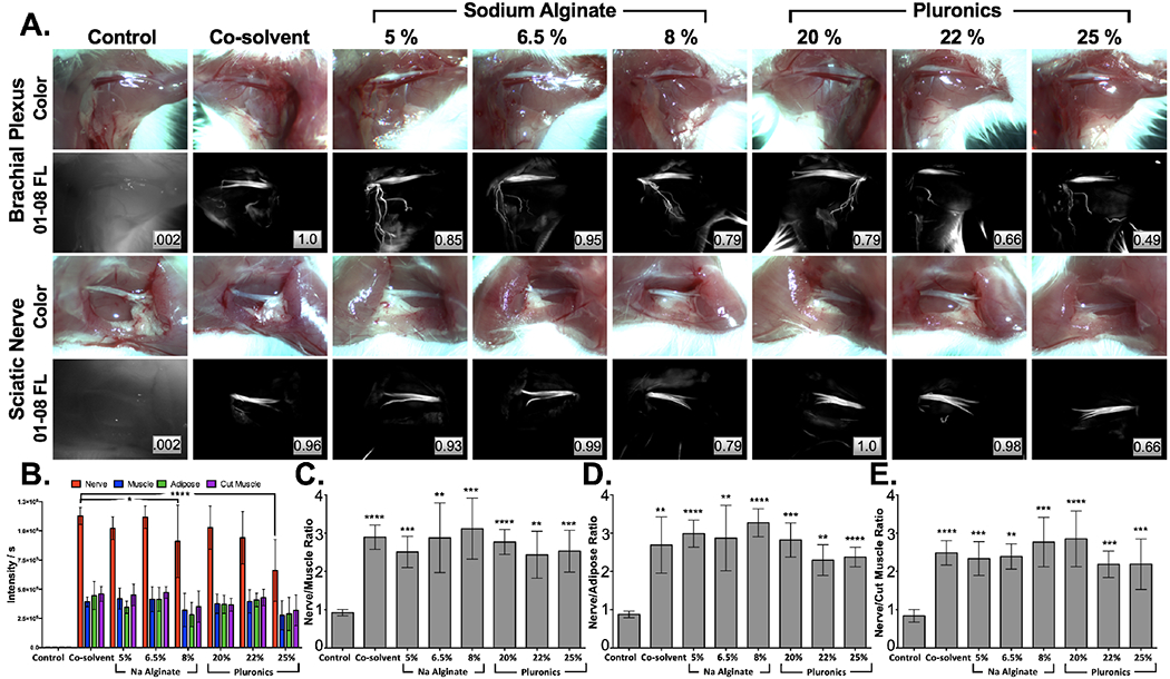
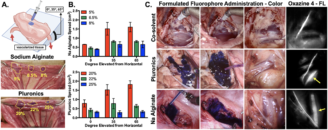
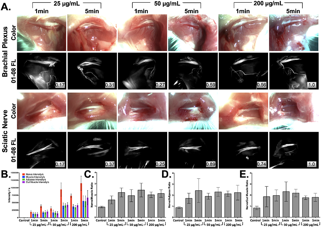
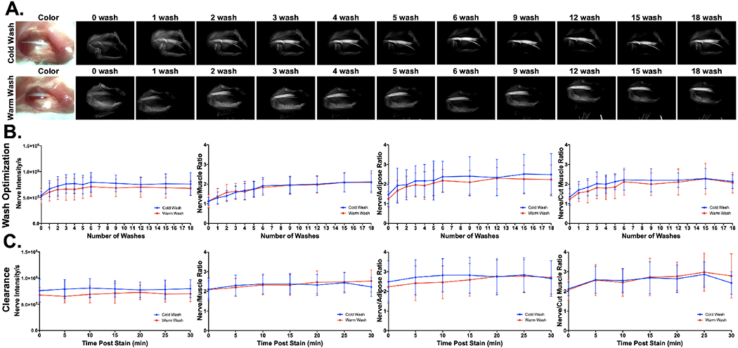
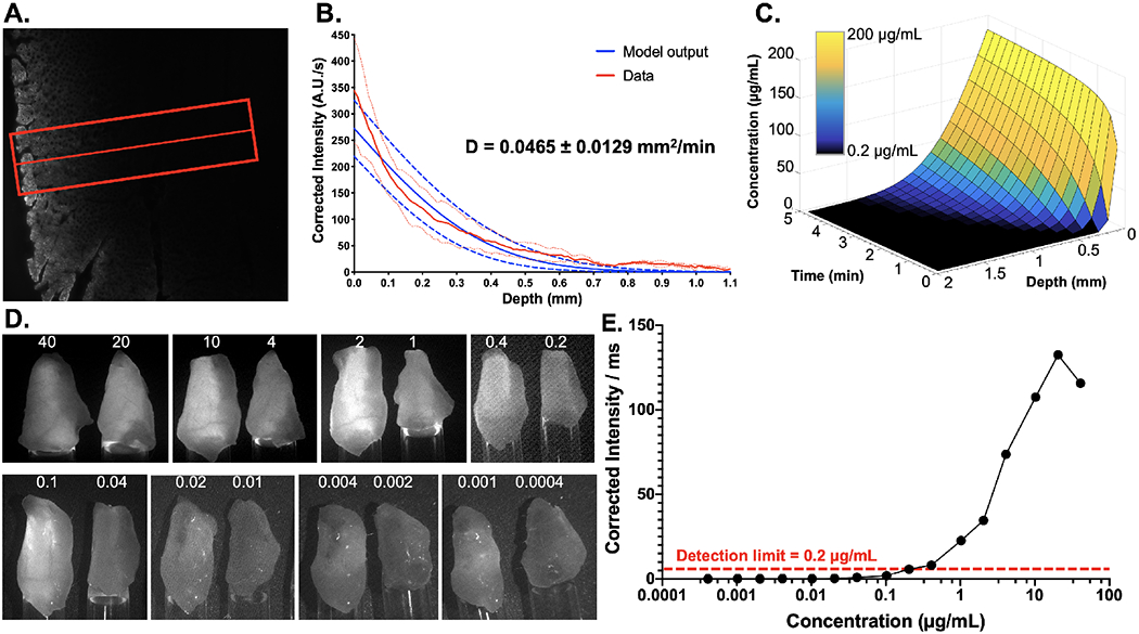
Iatrogenic nerve injury significantly affects surgical outcomes. Although intraoperative neuromonitoring is utilized, nerve identification remains challenging and the success of nerve sparing is strongly correlated with surgeon experience levels. Fluorescence guided surgery (FGS) offers a potential solution for improved nerve sparing by providing direct visualization of nerve tissue intraoperatively. However, novel probes for FGS face a long regulatory pathway to achieve clinical translation. Herein, we report on the development of a clinically-viable, gel-based formulation that enables direct administration of nerve-specific probes for nerve sparing FGS applications, facilitating clinical translation via the exploratory investigational new drug (eIND) guidance. The developed formulation possesses unique gelling characteristics, allowing it to be easily spread as a liquid followed by rapid gelling for subsequent tissue hold. Optimization of the direct administration protocol with our gel-based formulation enabled a total staining time of 1-2 min for compatibility with surgical procedures and successful clinical translation.


Conflict of interest statement

COMPETING INTERESTS

Alexander Antaris and Jonathan Sorger are employees of Intuitive Surgical. Lei Wang, Connor Barth and Summer Gibbs are inventors on patent application PCT/US19/43739 held by Oregon Health and Science University that cover the composition and methods of use of the nerve-specific oxazine compounds discussed in this manuscript. Summer Gibbs, Connor Barth, Adam Alani, Vidhi Shah, and Lei Wang are inventors on patent application PCT/US19/045347 held by Oregon Health and Science University and Oregon State University that cover the composition and methods of use of the formulations discussed in this manuscript. Lei Wang, Connor Barth and Summer Gibbs and also co-founders of Inherent Targeting, LLC.

Declaration of interests

The authors declare the following financial interests/personal relationships which may be considered as potential competing interests:

Summer Gibbs reports a relationship with Inherent Targeting, LLC that includes: equity or stocks. Connor Barth reports a relationship with Inherent Targeting, LLC that includes: equity or stocks. Lei Wang reports a relationship with Inherent Targeting, LLC that includes: equity or stocks. Jonathan Sorger reports a relationship with Intuitive Surgical Inc that includes: employment. Alexander Antaris reports a relationship with Intuitive Surgical Inc that includes: employment. Summer Gibbs has patent #PCT/US19/43739 pending to Oregon Health & Science University. Connor Barth has patent #PCT/US19/43739 pending to Oregon Health & Science University. Lei Wang has patent #PCT/US19/43739 pending to Oregon Health & Science University. Summer Gibbs has patent #PCT/US19/045347 pending to Oregon Health & Science University, Oregon State University. Connor Barth has patent #PCT/US19/045347 pending to Oregon Health & Science University, Oregon State University. Lei Wang has patent #PCT/US19/045347 pending to Oregon Health & Science University, Oregon State University. Adam Alani has patent #PCT/US19/045347 pending to Oregon Health & Science University, Oregon State University. Vidhi Shah has patent #PCT/US19/045347 pending to Oregon Health & Science University, Oregon State University.

Figures

Figure 1:
Figure 1:. Hydrogel-based formulation screening.
A. Representative murine brachial plexus (top) and sciatic nerve (bottom) images were used to B. quantify tissue intensities, enabling calculation of C. nerve to muscle D. nerve to adipose and E. nerve to cut muscle SBRs stained with 50 μg/mL LGW01-08 in each concentration of Na Alginate and Pluronics hydrogel formulations vs. the co-solvent formulation. All images are representative of data collected for n=6 nerve sites per formulation. Images are displayed without normalization. Inset boxes show intensity metrics denoting the fraction of the brightest fluorescence intensity that the image represents between images in the same row. All quantified data is presented as mean +/− standard deviation. FL = fluorescence. Nerve intensity data for each formulation was compared to the co-solvent formulation data to test for significance. Nerve SBR data for each formulation was compared to control unstained data to test for significance. * = p value < 0.05, ** = p value < 0.01, *** = p value < 0.001, **** = p value < 0.0001.
Figure 2:
Figure 2:. Hydrogel formulation viscosity assessment & large animal model staining studies.
A. Schematic, representative images, and B. quantified tissue spread surface areas for hydrogel formulation viscosity assessment, where Na Alginate (5%, 6.5% and 8%) and Pluronics (20%, 22% and 25%) were evaluated at 0°, 35° and 65° of tilt. The dotted lines indicate the area of tissue spread of each formulation in each representative image. Data is representative of n=3 replicates per formulation and tested tilt angle. C. Representative color and fluorescence images of the swine iliac plexus stained via direct administration of co-solvent, 22% Pluronics and 6.5% Na Alginate formulated oxazine 4. Yellow arrows indicate buried nerve tissue visible using fluorescence imaging up to 1 hour after direct administration. FL = fluorescence.
Figure 3:
Figure 3:. Hydrogel formulation direct administration methodology optimization.
A. Representative brachial plexus and sciatic nerve images following staining with varied concentrations (25, 50 or 200 μg/mL) of LGW01-08 in 22% Pluronics for 1- or 5-mins were B. quantified for tissue fluorescence intensity, which was used to calculate C. nerve to muscle, D. nerve to adipose and E. nerve to cut muscle SBR ratios for each tested concentration and incubation time. Images are representative of n=6 nerve sites without normalization. Inset boxes show intensity metrics denoting the fraction of the brightest fluorescence intensity that the image represents between images in the same row. FL = Fluorescence.
Figure 4:
Figure 4:. Hydrogel formulation direct administration washing protocol optimization.
A. Representative images and B. quantified nerve intensities and SBRs (i.e., nerve to muscle, nerve to adipose and nerve to cut muscle ratios) for wash optimization studies. Room temperature and cold PBS were tested to assess if Pluronics solubility was affected by temperature. All images are representative of data collected for n=6 nerve sites per wash condition, with images collected at 0, 1, 2, 3, 4, 5, 6, 9, 12, 16, and 18 washes. Each wash consisted of a quick flush of the stained tissue region with PBS. C. Nerve intensity and SBRs were calculated from images captured for 30 minutes following the final 18th wash step to visualize the effects of clearance on nerve fluorescence intensity and SBRs. All quantified data is presented as mean +/− standard deviation.
Figure 5:
Figure 5:. Hydrogel formulation tissue penetration quantification.
A. Representative fluorescence microscopy image of murine muscle tissue cross-section to visualize Pluronics formulated LGW01-08 diffusion after direct administration. The red line and box represent the center line and width used to obtain fluorescence intensity line profiles from the tissue. B. Quantified line profiles enabled the calculation of the diffusion coefficient (D) using a mathematical model, which facilitated C. prediction of tissue penetration profiles as fluorophore incubation time and concentration were varied. The graph colormap represents the fluorescence visibility at depth, as measured in LGW01-08 reference standards, where black squares represent concentrations at which the fluorescence signal is below the limit of detection at 1 mm of depth. Data is representative of n=9 line profiles and presented as mean +/− standard deviation. D. LGW01-08 tissue penetration depth was further estimated using images of LGW01-08 fluorophore in PBS titrated across a range of concentrations (0.0004-40 μg/mL). The varied LGW01-08 concentrations were placed in Eppendorf tubes that were covered with 1 mm of mouse peritoneal muscle tissue and imaged. E. Fluorescence intensities were quantified through the overlaying muscle tissue demonstrating that the concentration at which detection of fluorescence over background autofluorescence was 0.2 μg/mL.
Figure 6:
Figure 6:. Pluronics formulated LGW01-08 toxicology testing.
A. Blood marker, B. electrolyte, and C. hematological analysis results represented as concentrations (Units per liter [U/L], milligram per deciliter [mg/dL], millimole per liter [mmol/L], mole per microliter [M/μL], kilo per microliter [K/μL]) or percentages 24 hours following subcutaneous administration of 22% Pluronics either loaded with LGW01-08 for a final dose of 1.8 mg/kg or control unloaded. The dashed line boxes on each graph represent the normal expected range of values. D. The measured mean weights for mice up to 14 days after administration of the loaded and unloaded Pluronics gel vs. control un-administered animals. All data are representative of data collected for n=5 mice per group. All quantified data is presented as mean +/− standard deviation.
Figure 7:
Figure 7:. Rodent vagus nerve (VN) and recurrent laryngeal nerve (RLN) staining using the optimized direct administration methodology.
A. Representative images of murine vagus nerve tissue stained following dissection of the trachea with Pluronics formulated LGW01-08 using the optimized direct administration methodology. The displayed images are representative of n=4-stained nerve sites. The red dashed lines indicate where B. line profiles were quantified. The full-width-half-maximum (FWHM) were calculated to quantify nerve width, demonstrating visualization of sub-100 μm diameter nerves. C. Representative images of rat VN (arrows) and RLN (arrowhead) alongside thyroid (asterisk), trachea, muscle, and vasculature tissue stained following exposure with Pluronics formulated LGW01-08 using the optimized direct administration methodology. The displayed images are representative of n=4 stained nerve sites. D. Nerve signal to background ratios (SBRs) were quantified vs. muscle and thyroid tissues. Data is representative of n=4 nerve sites and presented as mean +/− standard deviation. RLN vs. muscle SBR data was compared to RLN vs. thyroid SBR data to test for significance. * = p value < 0.05. VN = vagus nerve, RLN = recurrent laryngeal nerve.

References

    1. Hall MJ, DeFrances CJ, Williams SN, Golosinskiy A & Schwartzman A National Hospital Discharge Survey: 2007 summary. Natl Health Stat Report, 1-20, 24 (2010). - PubMed
    1. Walsh PC & Donker PJ Impotence following radical prostatectomy: insight into etiology and prevention. J Urol 128, 492–497 (1982). - PubMed
    1. Ficarra V et al. Systematic review and meta-analysis of studies reporting urinary continence recovery after robot-assisted radical prostatectomy. Eur Urol 62, 405–417 (2012). - PubMed
    1. Damber JE & Khatami A Surgical treatment of localized prostate cancer. Acta Oncol 44, 599–604 (2005). - PubMed
    1. Rawla P Epidemiology of Prostate Cancer. World J Oncol 10, 63–89 (2019). - PMC - PubMed

Publication types