. 2022 May:129:102816.
doi: 10.1016/j.jaut.2022.102816. Epub 2022 Apr 5.

Autoreactive memory Th17 cells are principally derived from T-bet+RORγt+ Th17/1 effectors

Affiliations

Autoreactive memory Th17 cells are principally derived from T-bet+RORγt+ Th17/1 effectors

Nai-Wen Fan et al. J Autoimmun. 2022 May.

Abstract

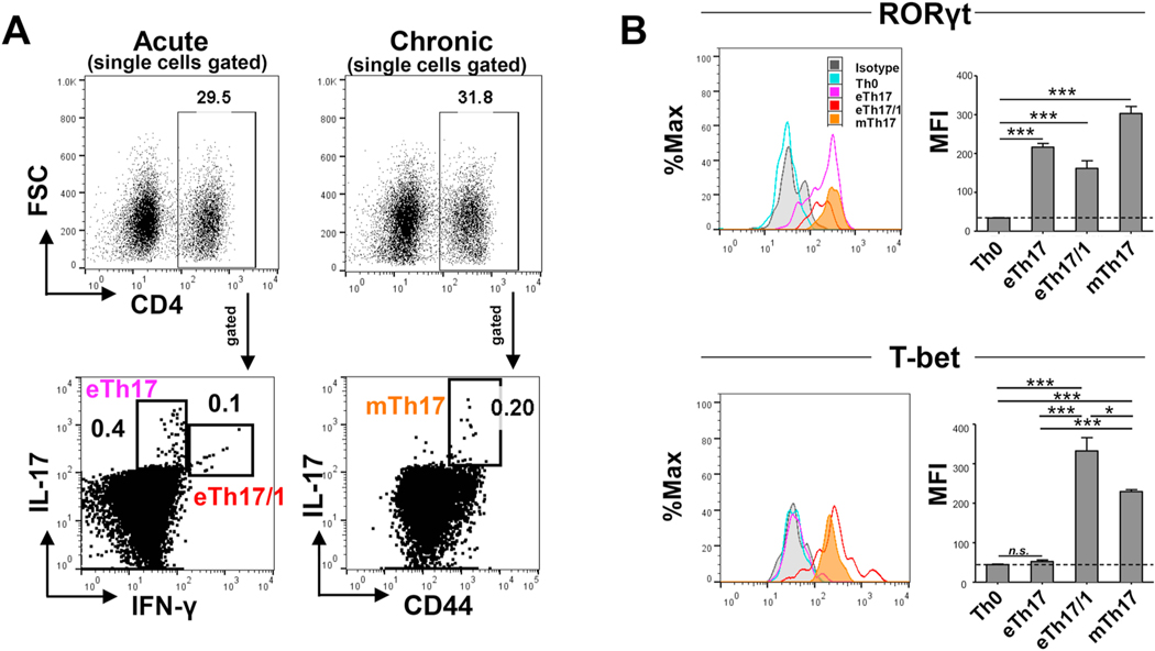
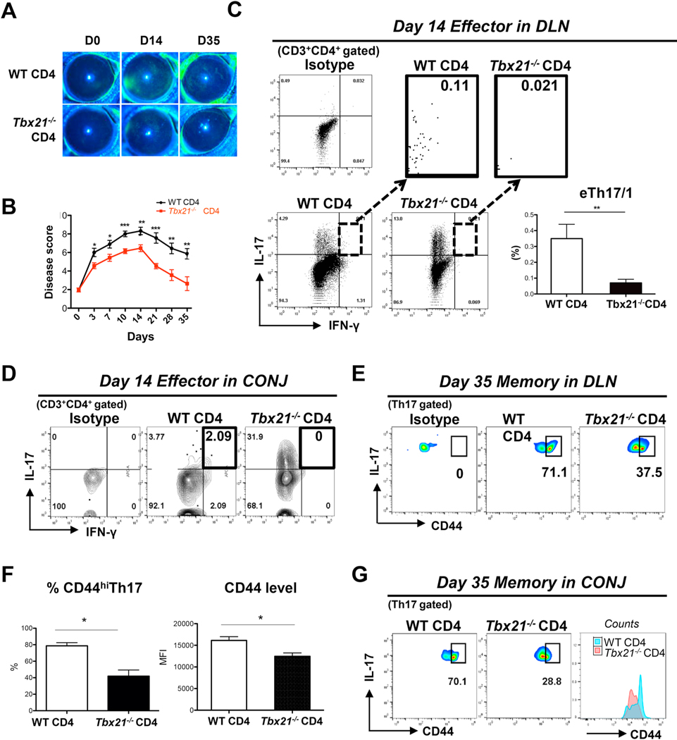
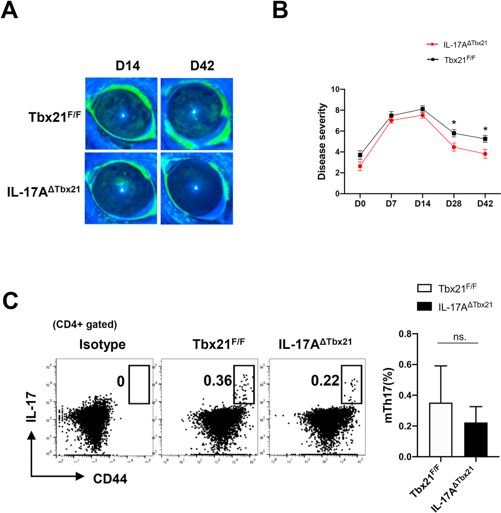
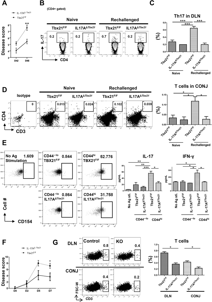
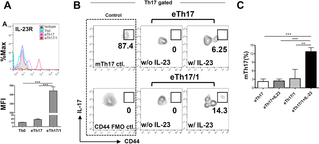
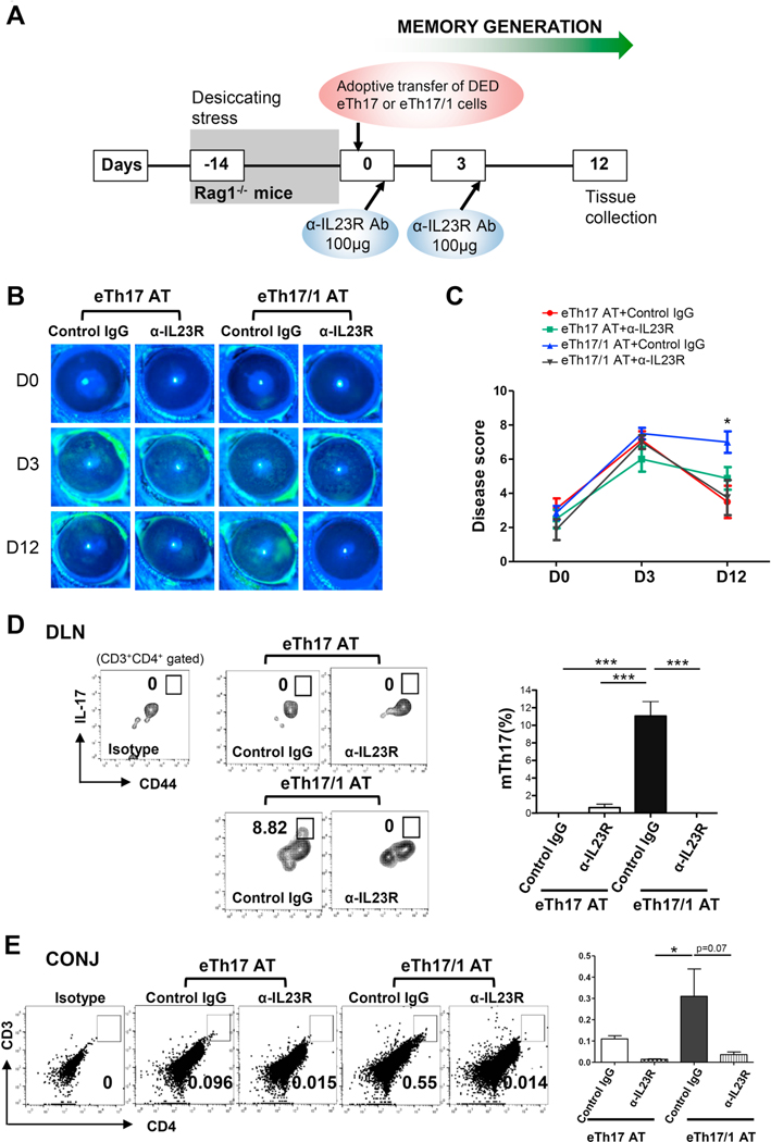
Effector Th17 cells, including IFN-γ-IL-17+ (eTh17) and IFN-γ+IL-17+ (eTh17/1) subsets, play critical pathogenic functions in the induction of autoimmunity. As acute inflammation subsides, a small proportion of the effectors survive and convert to memory Th17 cells (mTh17), which sustain chronic inflammation in autoimmune diseases. Herein, we investigated the differential contributions of eTh17 versus eTh17/1 to the memory pool using an experimental model of ocular autoimmune disease. Our results show that adoptive transfer of Tbx21-/- CD4+ T cells or conditional deletion of Tbx21 in Th17 cells leads to diminished eTh17/1 in acute phase and functionally compromised mTh17 in chronic phase. Further, adoptive transfer of disease-specific eTh17/1, but not eTh17, leads to generation of mTh17 and sustained ocular inflammation. Collectively, our data demonstrate that T-bet-dependent eTh17/1 cells generated during the acute inflammation are the principal effector precursors of pathogenic mTh17 cells that sustain the chronicity of autoimmune inflammation.

Keywords: Effector Th17/1; Memory Th17; T-bet.


Conflict of interest statement

Conflict of interest: R.D. and S.K.C. hold equity in Aramis Biosciences which owns intellectual property related to targeting IL-17 in ocular surface diseases. R.D., Y.C. and S.K.C. are inventors of a patent related to targeting memory Th17 cells in ocular immunoinflammatory diseases (owned by Massachusetts Eye and Ear).

Figures

Figure 1.
Figure 1.. Expression of transcription factors by effector Th17 (including eTh17 and eTh17/1, CD44lowIL-17+CD4+) and memory Th17 (mTh17, CD44hiIL-17+CD4+) cells in autoimmune inflammation.
(A) Mice were exposed to desiccating stress for 14 d to induce acute dry eye disease (DED), and eye-draining lymph nodes (DLNs) were collected and analyzed for eTh17 (IFN-γ-IL-17+) and eTh17/1 (IFN-γ+IL-17+) cells. In addition, another group of mice subjected to 14 d desiccating stress were returned to normal vivarium for additional 14 d to induce chronic DED, in which DLNs were collected and analyzed for mTh17 (CD44hiIL-17+). (B) Further analysis of DED-derived eTh17, eTh17/1 and mTh17 cells in (A) for their expression levels of Th17 and Th1 master transcription factors RORγt and T-bet, respectively. Representative histograms shown on the left and summary graphs of mean fluorescein intensities (MFI) are exhibited on the right. Naïve Th0 cells serve as the baseline control. Data shown are mean ± SEM from one representative experiment (n = 4–5/group) out of two performed. *, p<0.05; ***, p<0.001. n.s., not significant.
Figure 2.
Figure 2.. Adoptive transfer of naïve CD4+ T cells isolated from Tbx21−/− mice to Rag1−/− mice fails to induce chronic DED with impaired generation of mTh17 cells.
Naïve CD4+ T cells were isolated from wild-type (WT) or Tbx21−/− mice and then i.v. injected into Rag1−/− mice. After adoptive transfer, these Rag1−/− recipients were immediately subjected to desiccating stress for 14 d and then relocated to normal non-desiccating environment where they were maintained until day 35. (A) Clinical disease severity was evaluated by corneal fluorescein staining (CFS) and representative images show baseline, acute (day 14) and chronic (day 35) stage of DED. (B) Kinetic DED scores in each group are summarized as mean ± SEM in linear graphs (n = 28 eyes per group at acute stage, 16 eyes per group at chronic stage, pooled from two independent experiments). (C) Representative flow cytometry plots show frequencies of eTh17/1 cells in draining lymph nodes (DLN) of Rag1−/− recipients in acute DED (day 14). The frequencies of eTh17/1 cells (mean ± SEM) are summarized in the bar graphs (n = 6 per group). (D) Flow cytometry plots show frequencies of eTh17/1 cells in conjunctiva (CONJ) of Rag1−/− recipients in acute DED (day 14). Ocular tissues were pooled from 6 animals in each group for the analysis, and data shown are one representative out of two performed. (E) Representative flow cytometry plots show frequencies of CD44hi mTh17 cells in DLNs of Rag1−/− recipients in chronic DED (day 35). (F) The proportion of CD44hi mTh17 cells among Th17 population and the expression intensity of CD44 (MFI) by Th17 cells in DLNs of chronic DED from one representative experiment out of two independent experiments performed are summarized as mean ± SEM in the bar graphs (n = 3–6 per group). (G) Flow cytometry plots show frequencies of CD44hi mTh17 in conjunctiva of Rag1−/− recipients in chronic DED (day 35). Ocular tissues were pooled from 4 animals in each group for the analysis, and data shown are one representative out of two performed.*, p<0.05; **, p<0.01; ***, p<0.001.
Figure 3.
Figure 3.. Conditional deletion of Tbx21 in Th17 cells suppresses development of chronic ocular inflammation.
(A) IL-17AΔTbx21 and control (Tbx21F/F) mice were exposed to desiccating stress for 14 d and then transfer to normal non-desiccating vivarium. Clinical disease severity was evaluated by corneal fluorescein staining (CFS) scores and representative images at day14 (acute disease) and day 42 (chronic disease) are shown. (B) Kinetic disease scores in each group are summarized as mean ± SEM in linear graphs (n = 44 eyes in IL-17AΔTbx21, 36 eyes in Tbx21F/F group at acute stage, 28 eyes per group at chronic stage, pooled from three independent experiments). (C) Representative flow cytometry plots show frequencies of CD44hi mTh17 cells in DLNs at day 42 (chronic disease), and summarized as mean ± SEM in the bar graph (n = 14 per group, pooled from 3 independent experiments). *, p<0.05; ***, p<0.001; n.s., not significant.
Figure 4.
Figure 4.. Conditional deletion of Tbx21 in Th17 cells inhibits the generation of functional mTh17.
(A) Chronic DED mice (desiccating stress from day 1 to 14, followed by normal non-desiccating environment until day 42) were re-challenged with another short-term desiccating stress (6 days in duration until day 48). Kinetic scores summarized the disease scores immediately before re-challenge and after 6-day re-challenge. (n=20 per group, pooled from three independent experiments). (B) Representative flow cytometry plots show frequencies of Th17 effector cells (CD4+IL17+) in draining lymph nodes (DLN) after re-challenge. (C) The frequencies of effector cells (mean ± SEM) from one representative experiment out of two performed are summarized in the bar graphs (n = 4 per group). (D) Representative flow cytometry plots showing CD4+ T cells infiltration in conjunctiva (CONJ) upon re-challenge. Six eye tissues were pooled together. The frequencies of CD4+ T cells are summarized in the bar graphs (n = 3 per group). (E) CD4+CD44hi T cells were isolated from chronic DED mice (day 42) and in vitro stimulated with corneal tissue homogenates. Cells were then evaluated for CD154 expression by flow cytometry, and the culture supernatants were assayed for IL-17 and IFN-γ by ELISA (n = 3–4 per group). (F) mTh17 cells isolated from chronic DED mice were adoptively transferred to normal Rag1−/− mice which were subsequently exposed to desiccating stress, and the kinetic disease scores are summarized (n = 6 per group). (G) Representative flow cytometric plots showing CD4+ T cells infiltration in DLN and the CONJ, and the frequencies of T cells are summarized in the bar graph (n = 3 per group). *, p< 0.05; **, p<0.01, ***, p<0.001.
Figure 5.
Figure 5.. IL-23 promotes the conversion of effector Th17/1 cells to memory Th17 cells ex vivo.
(A) Representative flow cytometry histograms show the expression of IL-23R in naive T cells (Th0), and eTh17 and eTh17/1 cells from autoimmune DED mice (same gating strategy as Fig. 1A). The expression levels of IL-23R by each T cell subset are summarized as mean ± SEM of mean fluorescein intensities (MFIs) in the bar graph. (B) Enriched fresh eTh17 and eTh17/1 cells from DLNs of acute DED mice were cultured in the presence or absence of IL-23 for 48 h. Representative flow cytometry plots from four independent experiments show the population of CD44hi mTh17 cells at the end of culture. The mTh17 cells analyzed in wild-type chronic DED mice serve as a positive control for the staining (mTh17 ctl.). (C) The frequencies of mTh17 cells in each group are summarized as mean ± SEM. Bar graphs summarize data from 4 independent experiments (n = 4–6 in eTh17/1 groups, n = 12–13 in eTh17 groups). **, p<0.01; ***, p<0.001
Figure 6.
Figure 6.. Adoptive transfer of eTh17/1, but not eTh17, induces and maintains DED, along with the generation of mTh17 cells in vivo.
(A) Experimental design. Rag1−/− mice was exposed to desiccating stress for 14 d, followed by adoptive transfer of wild-type DED mice-derived eTh17 or eTh17/1 cells via i.v. injection (2×10⁴). Immediately after adoptive transfer (day 0), the Rag1−/− recipients were transferred to normal vivarium for additional 12 d. Some of the recipients also received anti-IL23 receptor (IL-23R) antibody treatment intraperitoneally at day 0 and 3. (B) Clinical disease severity was evaluated by corneal fluorescein staining scores and representative images show baseline (day 0, the day of adoptive transfer) and day 3, 12 post-adoptive transfer. (C) Clinical disease scores in each group at day 12, pooled from three independent experiments, are summarized as mean ± SEM (n = 6–8 eyes per group). *, p<0.05 for eTh17/1 AT vs eTh17 AT, eTh17/1 AT vs eTh17/1 AT+anti-IL23R, and eTh17/1 AT vs eTh17 AT+anti-IL23R. (D) Representative flow cytometry plots from three independent experiments show generation of CD44hi mTh17 cells in eye-draining lymph nodes (DLN) of mice with adoptive transfer of eTh17 or eTh17/1 cells at day 12 post-transfer. The frequencies of mTh17 cells (mean ± SEM) are summarized in the bar graphs (n = 3–4 per group). (E) Conjunctival tissues were pooled from 3 animals in each group for flow cytometric analysis of infiltration of CD4+ T cells at day 12 post-transfer, and data shown are one representative out of two performed. The frequencies of T cells (mean ± SEM) are summarized in the bar graphs (n = 3 per group). *p<0.05; ***, p<0.001

References

    1. Ahmed R, Bevan MJ, Reiner SL, Fearon DT. The precursors of memory: models and controversies. Nat Rev Immunol. 9 (2009) 662–668. 10.1038/nri2619. - DOI - PubMed
    1. Raphael I, Joern RR, Forsthuber TG. Memory CD4(+) T Cells in Immunity and Autoimmune Diseases. Cells. 9 (2020). 10.3390/cells9030531. - DOI - PMC - PubMed
    1. Yang J, Sundrud MS, Skepner J, Yamagata T. Targeting Th17 cells in autoimmune diseases. Trends Pharmacol Sci. 35 (2014) 493–500. 10.1016/j.tips.2014.07.006. - DOI - PubMed
    1. Chen Y, Chauhan SK, Lee HS, Saban DR, Dana R. Chronic dry eye disease is principally mediated by effector memory Th17 cells. Mucosal Immunol. 7 (2014) 38–45. 10.1038/mi.2013.20. - DOI - PMC - PubMed
    1. Deng Q, Luo Y, Chang C, Wu H, Ding Y, Xiao R. The Emerging Epigenetic Role of CD8+T Cells in Autoimmune Diseases: A Systematic Review. Front Immunol. 10 (2019) 856. 10.3389/fimmu.2019.00856. - DOI - PMC - PubMed

Publication types

Substances