

Skeletal and alveolar changes in conventional rapid palatal expansion (RPE) and miniscrew-assisted RPE (MARPE): a prospective randomized clinical trial using low-dose CBCT
- PMID: 35395801
- PMCID: PMC8994336
- DOI: 10.1186/s12903-022-02138-w
Skeletal and alveolar changes in conventional rapid palatal expansion (RPE) and miniscrew-assisted RPE (MARPE): a prospective randomized clinical trial using low-dose CBCT
Abstract
Background: This prospective randomized clinical trial aimed to evaluate the immediate and short-term skeletal, dentoalveolar, and periodontal effects of rapid palatal expansion (RPE) and miniscrew-assisted RPE (MARPE) in adolescent and young adult patients.
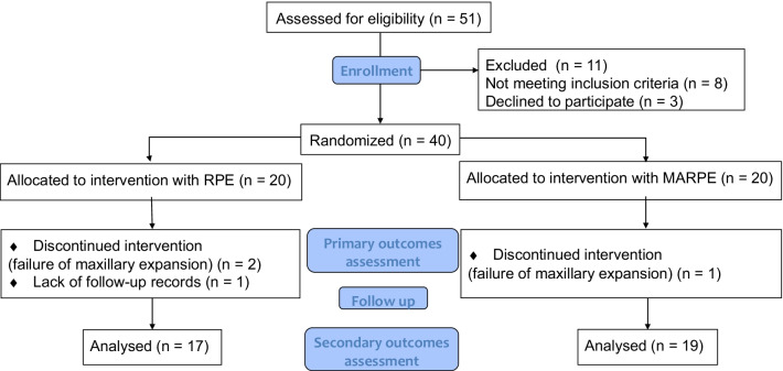
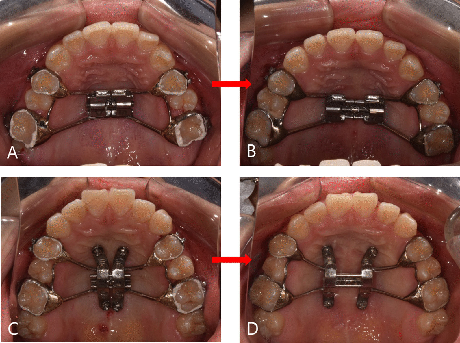
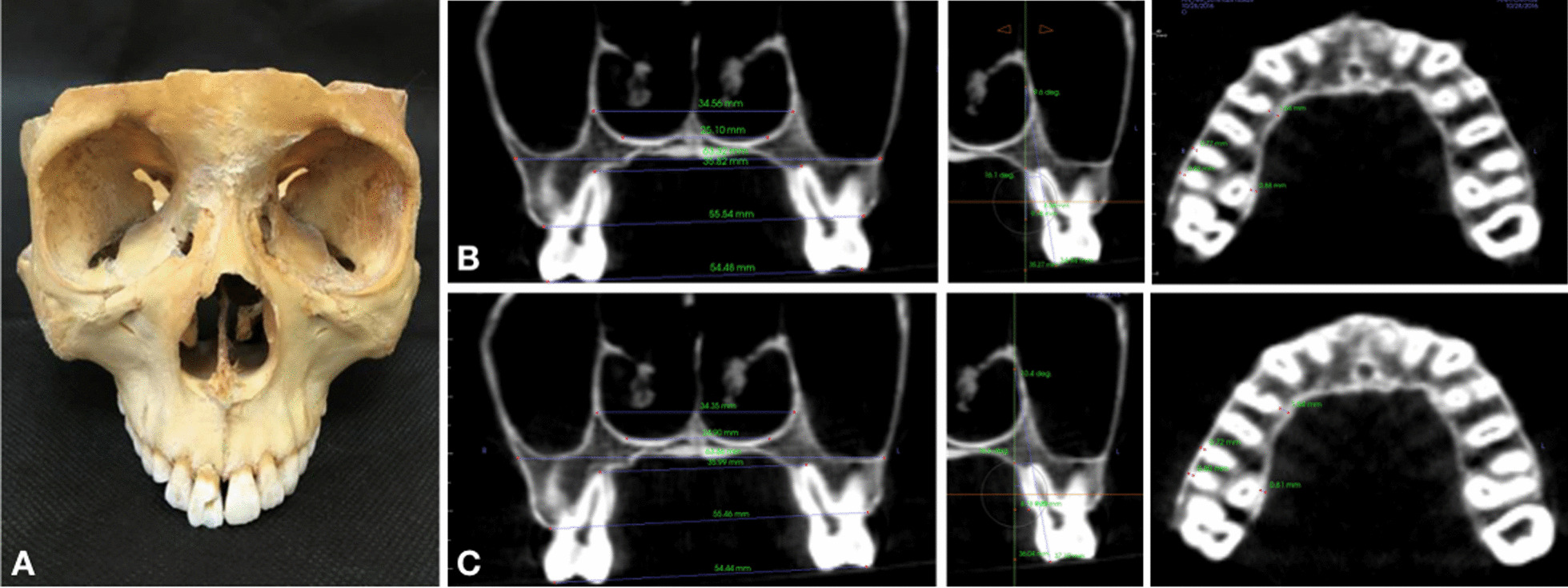
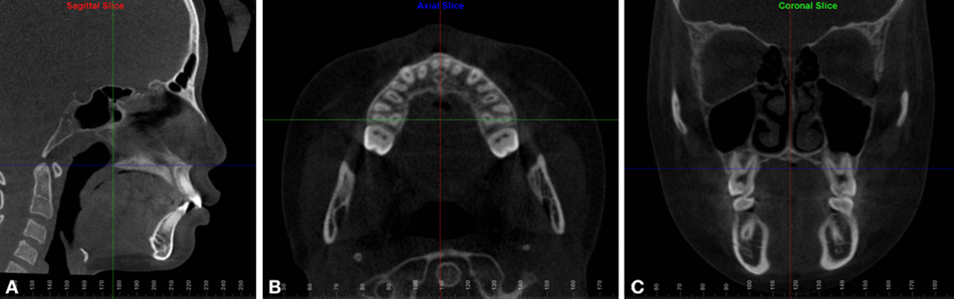
Methods: This study followed a two-arm, parallel, randomized clinical trial design that recruited patients with transverse maxillary deficiency in a 1:1 allocation ratio. Forty patients (14 men and 26 women) requiring maxillary expansion were randomly allocated to the RPE (n = 20, age = 14.0 ± 4.5) or MARPE (n = 20, age = 14.1 ± 4.2) groups. The assignment was performed via computer-generated block randomization, with a block size of four. Upon identical (35 turns) amount of expansion, low-dose cone-beam computed tomography images were taken before treatment (T0), immediately after expansion (T1), and after a 3-month consolidation period (T2). The primary outcome of this study comprised the assessment of midpalatal suture separation. Secondary outcomes included, skeletal, dentoalveolar, and periodontal measurements, which were performed at each time point.
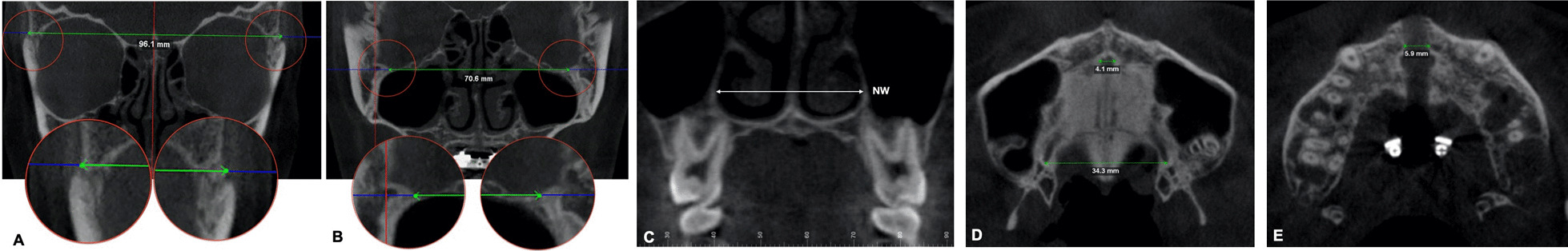
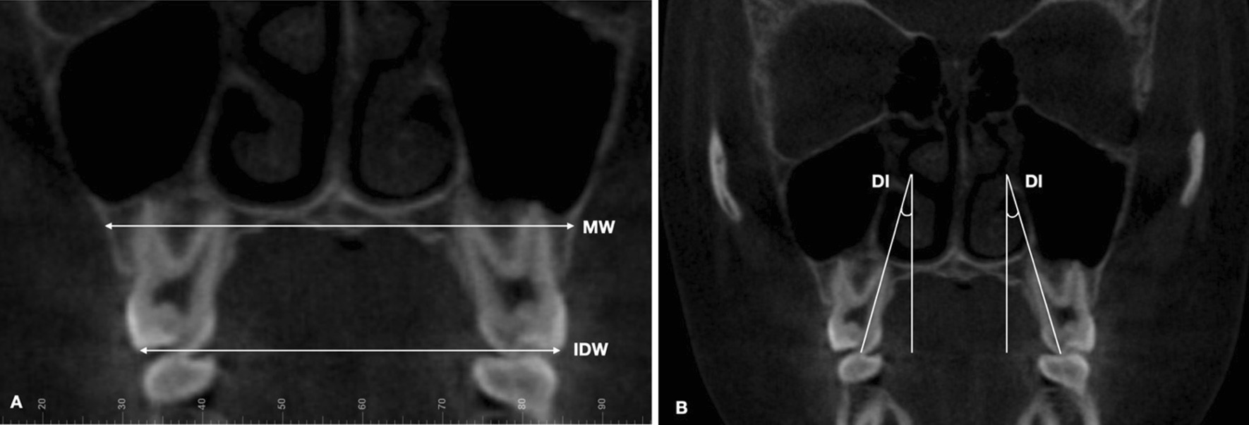
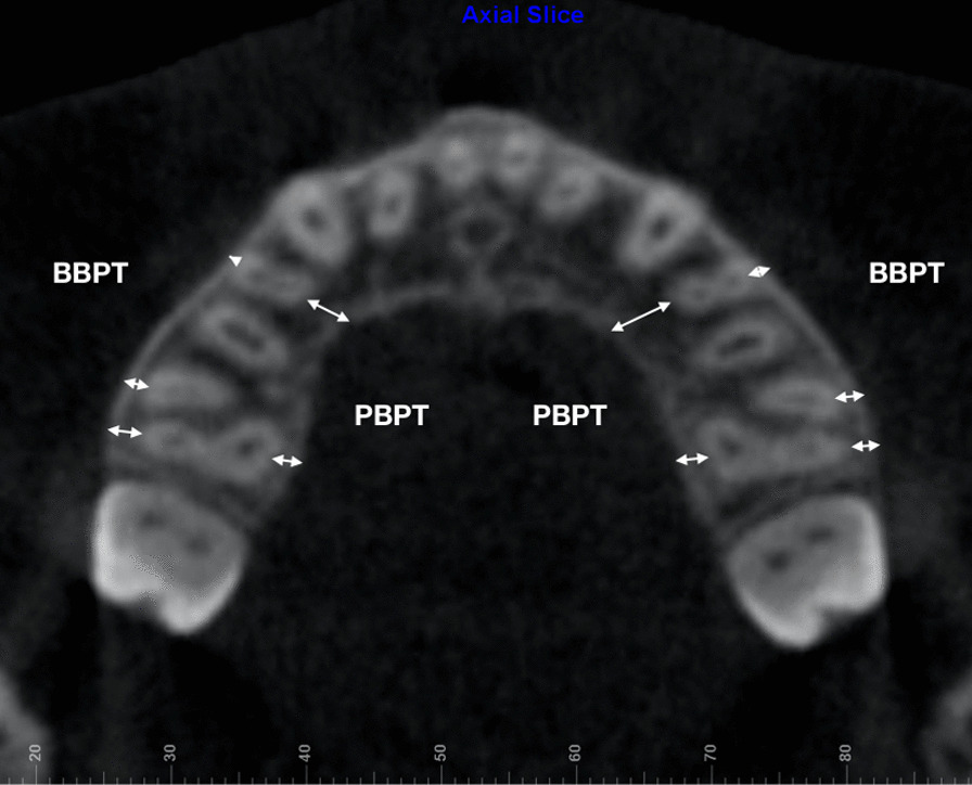
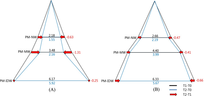
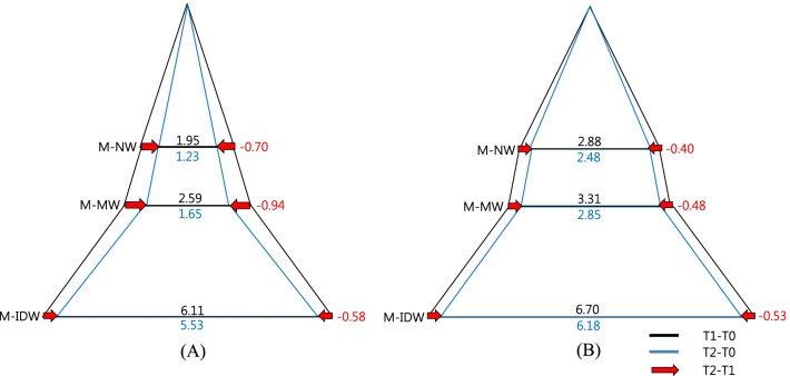
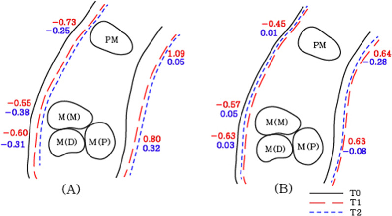
Results: The frequency of midpalatal suture separation was 90% (18/20) and 95% (19/20) for the RPE and MARPE groups, respectively. A greater increase in nasal width in the molar region (M-NW) and greater palatine foramen (GPF) was observed immediately after the expansion (T1-T0) and consolidation periods (T2-T0) in the MARPE group compared to the RPE group (P < 0.05). The MARPE and RPE groups showed similar dentoalveolar changes except for the maxillary width (PM-MW, M-MW). The MARPE group presented greater bilateral first premolar (PM-MW) and molar (M-MW) maxillary width in relation to the RPE group (P < 0.05). Through the expansion and consolidation periods (T2-T0), lesser buccal displacement of the anchor teeth was observed in the MARPE group (PM-BBPT, PM-PBPT, M-BBPT [mesial and distal roots], and M-PBPT)( P < 0.05).
Conclusions: Midpalatal suture separation was observed in 90% and 95% of patients in the RPE and MARPE groups, respectively. Both RPE and MARPE groups exhibited significant triangular basal bone expansion and skeletal relapse during consolidation. Under identical amounts of expansion, the MARPE group showed lower decrease in the skeletal, dentoalveolar and periodontal variables after consolidation. The reinforcement of RPE with miniscrews contributes to the maintenance of the basal bone during consolidation period. Trial registration WHO Institutional Clinical Trials Registry Platform (IRB No. KCT0006871 / Registration date 27/12/2021).
Keywords: Alveolar bone loss; Cone-beam computed tomography; Cranial sutures; Orthodontic anchorage procedures; Palatal expansion technique.
© 2022. The Author(s).
Conflict of interest statement
The authors declare that they have no competing interests.
Figures
References
-
- Angell EH. Treatment of irregularities of the permanent or adult tooth. Dent Cosmos. 1860;1:540–544.
-
- Haas AJ. The treatment of maxillary deficiency by opening the midpalatal suture. Angle Orthod. 1965;35:200–217. - PubMed
-
- Davis WM, Kronman JH. Anatomical changes induced by splitting of the midpalatal suture. Angle Orthod. 1969;39(2):126–132. - PubMed
Publication types
MeSH terms
LinkOut - more resources
Full Text Sources
