. 2022 May 3;9(3):ENEURO.0482-21.2022.
doi: 10.1523/ENEURO.0482-21.2022. Print 2022 May-Jun.

SMART: An Open-Source Extension of WholeBrain for Intact Mouse Brain Registration and Segmentation

Affiliations

SMART: An Open-Source Extension of WholeBrain for Intact Mouse Brain Registration and Segmentation

Michelle Jin et al. eNeuro. .

Abstract

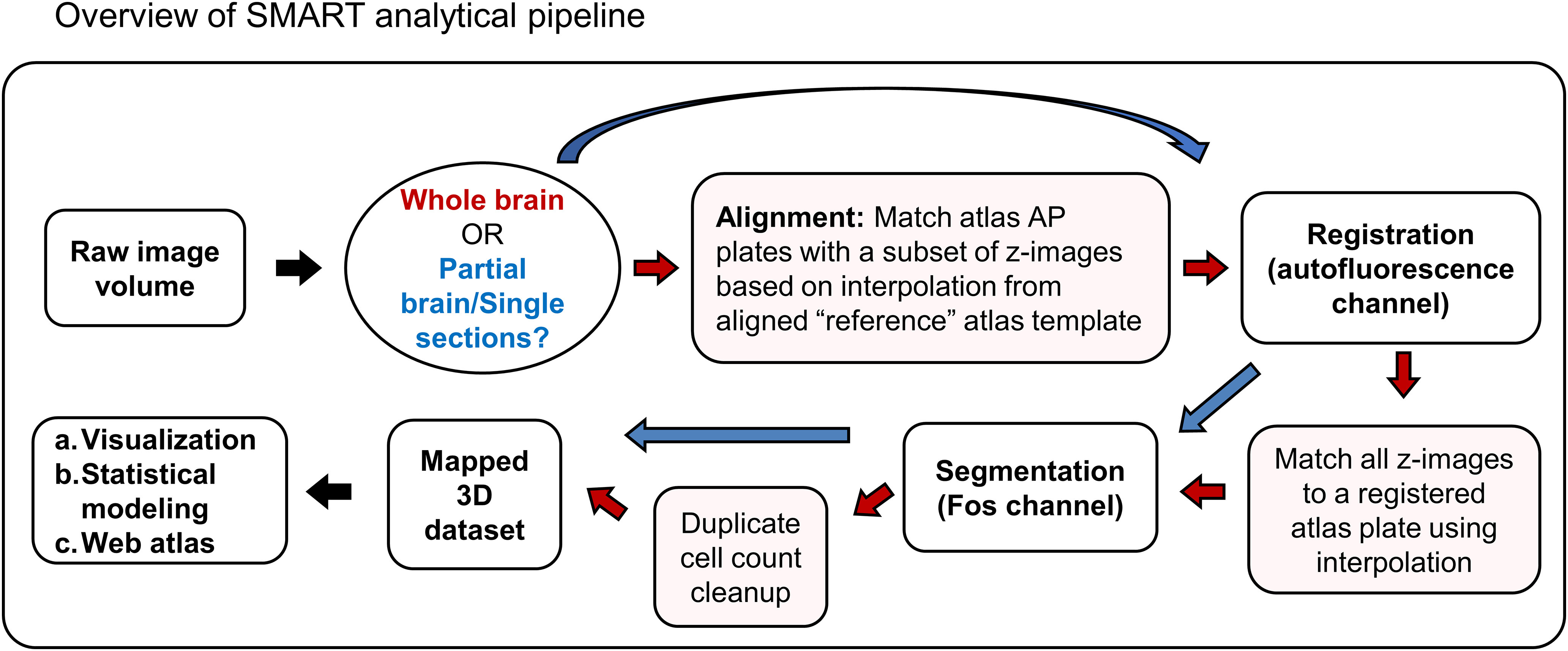
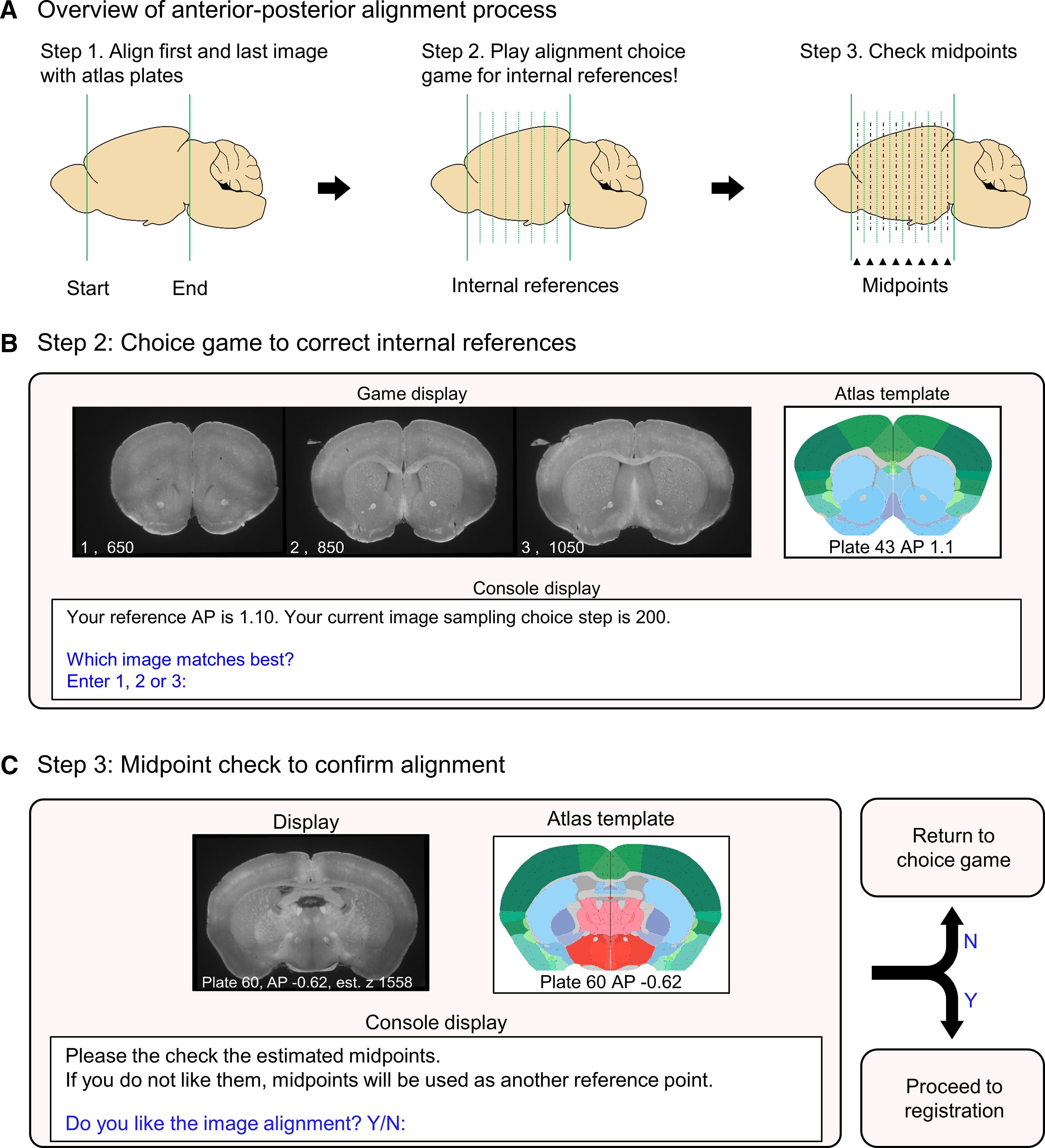
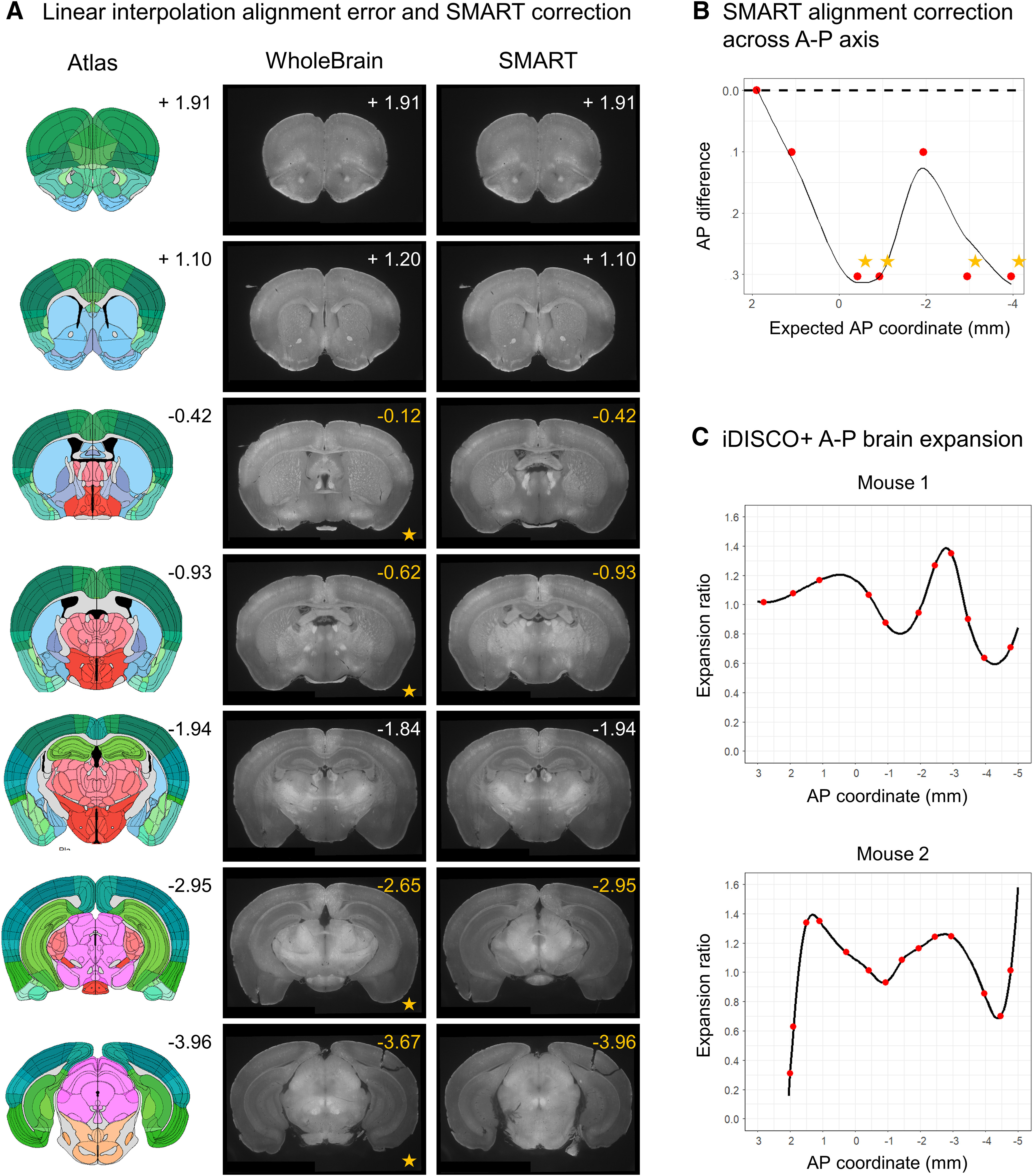
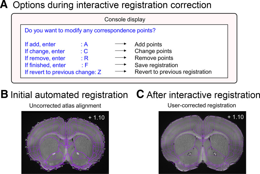
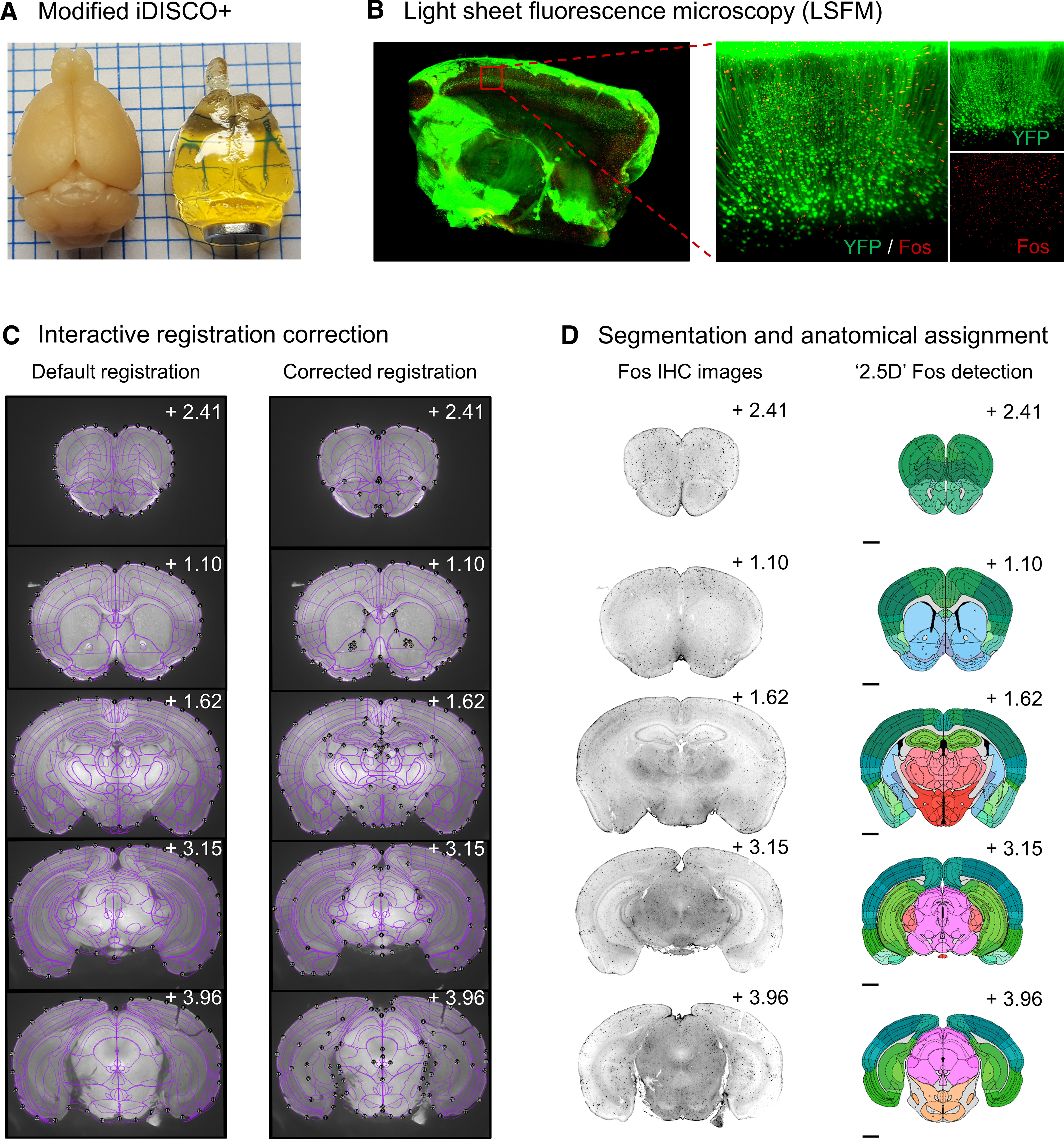
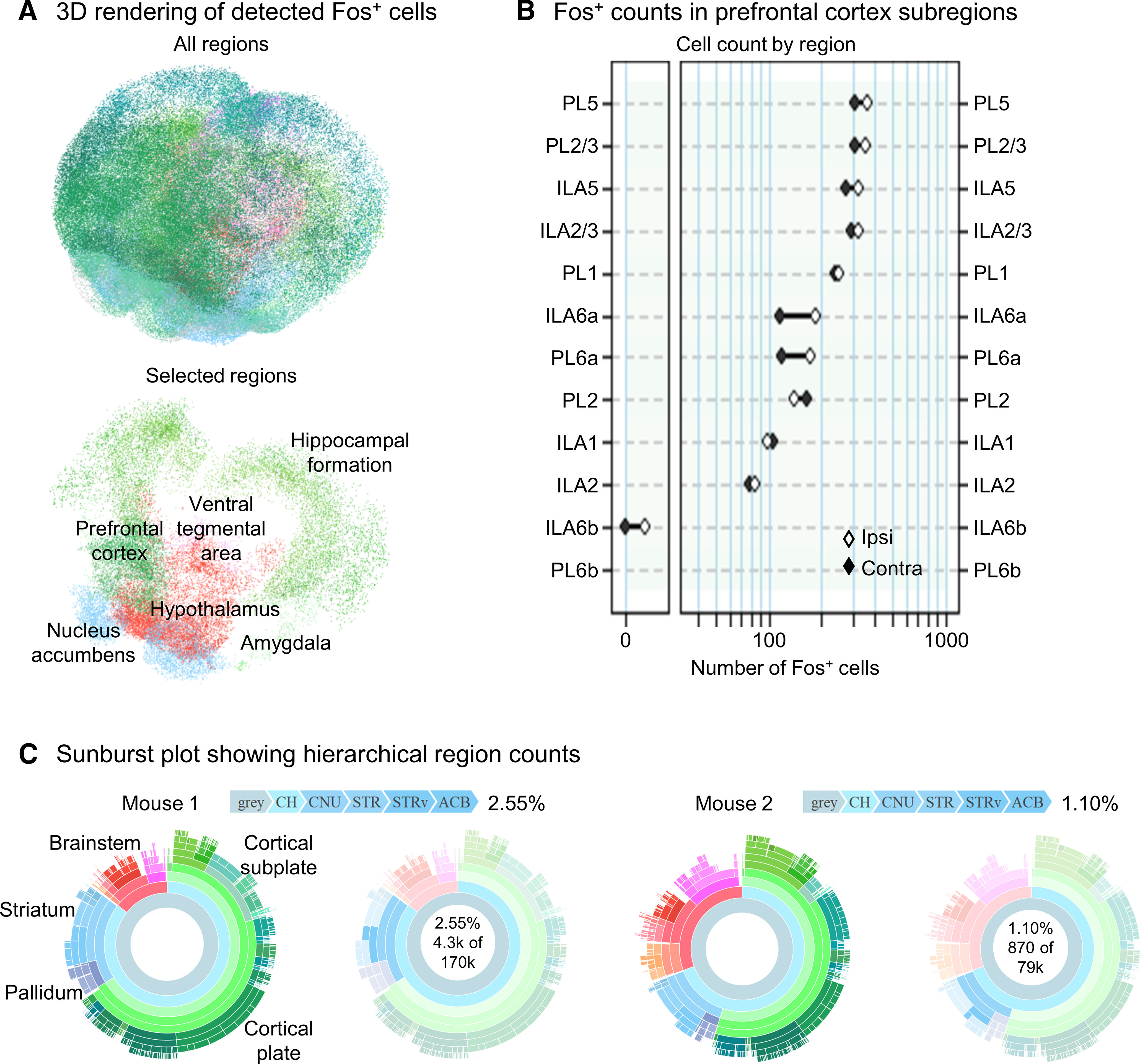
Mapping immediate early gene (IEG) expression across intact mouse brains allows for unbiased identification of brain-wide activity patterns underlying complex behaviors. Accurate registration of sample brains to a common anatomic reference is critical for precise assignment of IEG-positive ("active") neurons to known brain regions of interest (ROIs). While existing automated voxel-based registration methods provide a high-throughput solution, they require substantial computing power, can be difficult to implement and fail when brains are damaged or only partially imaged. Additionally, it is challenging to cross-validate these approaches or compare them to any preexisting literature based on serial coronal sectioning. Here, we present the open-source R package SMART (Semi-Manual Alignment to Reference Templates) that extends the WholeBrain R package framework to automated segmentation and semi-automated registration of intact mouse brain light-sheet fluorescence microscopy (LSFM) datasets. The SMART package was created for novice programmers and introduces a streamlined pipeline for aligning, registering, and segmenting LSFM volumetric datasets across the anterior-posterior (AP) axis, using a simple "choice game" and interactive menus. SMART provides the flexibility to register whole brains, partial brains or discrete user-chosen images, and is fully compatible with traditional sectioned coronal slice-based analyses. We demonstrate SMART's core functions using example datasets and provide step-by-step video tutorials for installation and implementation of the package. We also present a modified iDISCO+ tissue clearing procedure for uniform immunohistochemical labeling of the activity marker Fos across intact mouse brains. The SMART pipeline, in conjunction with the modified iDISCO+ Fos procedure, is ideally suited for examination and orthogonal cross-validation of brain-wide neuronal activation datasets.

Keywords: activity mapping; brain-wide activity mapping; immediate early gene; light sheet microscopy; neuronal ensembles; tissue clearing.


Figures

Figure 1.
Figure 1.
Overview of the SMART analytical pipeline. Red arrows indicate the appropriate trajectory of an intact brain imaging dataset through steps in the pipeline, while blue arrows indicate the appropriate trajectory for a partial brain dataset, consisting of coronal sections chosen by the user. Video tutorials for each step outlined in the schematic are provided in Extended Data Table 1-1.
Figure 2.
Figure 2.
Diagram of the alignment process to reference templates. A, A schematic illustrating the process of qualitative alignment and inspection of midpoints of reference templates. B, A visual representation of the graphical windows displayed during the choice game and the user options allowed in the R console window. The choice game is cycled through each internal reference template. During the midpoint check, the choice game is automatically played again for midpoints that are unsatisfactorily aligned; these midpoints become additional reference templates. C, A visual representation of the graphical windows displayed during the qualitative midpoint check and the user options in the R console.
Figure 3.
Figure 3.
Comparison of linearly interpolated versus choice game-aligned images to reference templates. A, A qualitative panel showing seven default internal atlas templates with their corresponding AP coordinates (left), predicted images based on linear interpolation (middle, WholeBrain), and aligned images following user alignment with the choice game (right, SMART). The actual coordinates of the images following the choice game are printed in the top-right corner of the images. B, A plot of the difference between the reference coordinates and the actual AP coordinates of the images based predicated based off linear interpolation. Starred are four AP coordinates with the greatest absolute difference; they correspond to the starred images in A for qualitive comparison. C, Normalized brain morph ratio across the AP axis for two example datasets following the choice game and a midpoint-check. Red dots indicate positions of the aligned reference templates, and the black line indicates interpolated morph ratio for AP positions between aligned reference templates.
Figure 4.
Figure 4.
The interactive registration correction process. A, A visual representation of user options in the R console display during manual correction of registrations. B, An example image showing the initial registration of the atlas template to the tissue based on correspondence points around the contours of the tissue in the autofluorescence channel. C, The atlas-tissue alignment is improved following interactive registration correction.
Figure 5.
Figure 5.
Representative images of registration correction and Fos segmentation from an example dataset. A, Transparency of intact mouse brain samples pre-iDISCO+ and post-iDISCO+ immunolabeling and clearing. B, Example LSFM tissue section of an intact cleared brain from a Thy-1 GFP transgenic mouse (left); an enlarged cortical image in the YFP and Fos imaging channels (right). C, Representative images of initial and corrected atlas-tissue registrations in the autofluorescence channel (488 nm) of the example dataset from Mouse 1 at various AP coordinates. Note the improved alignment of internal structures such as ventricles and white matter tracts. D, Images of Fos-IHC (647 nm) and segmented Fos-positive cells following automated 2.5D segmentation, cleanup of duplicate cell counts and forward warping onto atlas space.
Figure 6.
Figure 6.
Graphical representations of whole-brain Fos datasets following SMART registration, and segmentation. A, 3D rendering of the entire mapped dataset (Mouse 1) from Figure 4, left, and a 3D rendering of the indicated ROIs (right). B, Region plot quantifying segmented Fos-positive cell bodies in the PFC (Mouse 1, PL and IL). C, Sunburst plots showing region cell counts and hierarchical structural relationships for two example datasets. The innermost ring represents all cell counts, with each subsequent outer ring representing child structures of the adjacent inner ring. Regions colors are based on the Allen Mouse Brain Atlas and arc length is proportional to total cell counts. Plots are interactive and hovering over an arc reveals region identity, hierarchical path, and region cell count (right). ACB, nucleus accumbens; IL, infralimbic cortex; PL, prelimbic cortex.

References

    1. Adler D, Murdoch D, Nenadic O, Urbanek S, Chen M, Gebhardt A, Bolker B, Csardi G, Strzelecki A, Senger A, Team TRC, Eddelbuettel D (2019) rgl: 3D visualization using OpenGL, 0.99.16 edition. CRAN.
    1. Bossert JM, Stern AL, Theberge FRM, Cifani C, Koya E, Hope BT, Shaham Y (2011) Ventral medial prefrontal cortex neuronal ensembles mediate context-induced relapse to heroin. Nat Neurosci 14:420–422. 10.1038/nn.2758 - DOI - PMC - PubMed
    1. Bossert JM, Stern AL, Theberge FRM, Marchant NJ, Wang HL, Morales M, Shaham Y (2012) Role of projections from ventral medial prefrontal cortex to nucleus accumbens shell in context-induced reinstatement of heroin seeking. J Neurosci 32:4982–4991. 10.1523/JNEUROSCI.0005-12.2012 - DOI - PMC - PubMed
    1. Bostoc M, Rodden K, Warne K, Russell K, Breitwieser F, Yetman C (2019) sunburstR: 'Htmlwidget' for 'Kerry Rodden' 'd3.js' sequence and 'd2b' sunburst, 2.1.0 edition. CRAN.
    1. Bostock M, Russell K, Aisch G, Pearce A, Ortiz B (2019) d3r: 'd3.js' utilities for R, 0.8.5 edition. CRAN.

Publication types

LinkOut - more resources