

Isolating the Neural Substrates of Visually Guided Attention Orienting in Humans
- PMID: 35396326
- PMCID: PMC9121836
- DOI: 10.1523/JNEUROSCI.0205-22.2022
Isolating the Neural Substrates of Visually Guided Attention Orienting in Humans
Abstract
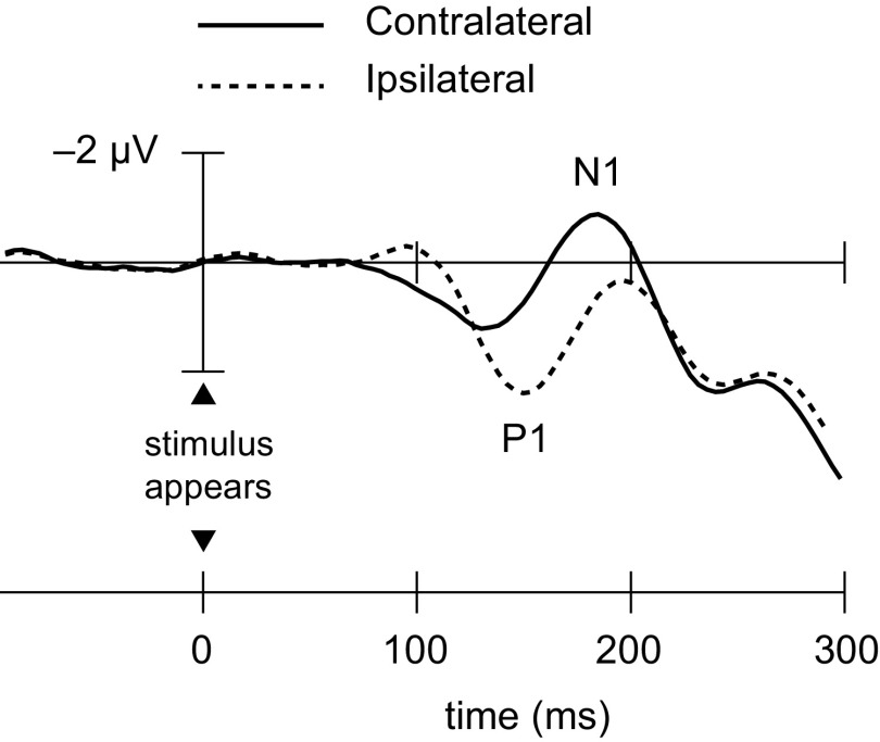
The neural processes that enable healthy humans to orient attention to sudden visual events are poorly understood because they are tightly intertwined with purely sensory processes. Here we isolated visually guided orienting activity from sensory activity using event-related potentials (ERPs). By recording ERPs to a lateral stimulus and comparing waveforms obtained under conditions of attention and inattention, we identified an early positive deflection over the ipsilateral visual cortex that was associated with the covert orienting of visual attention to the stimulus. Across five experiments with male and female adult participants, this ipsilateral visual orienting activity (VOA) could be distinguished from purely sensory-evoked activity and from other top-down spatial attention effects. The VOA was linked with behavioral measures of orienting, being significantly larger when the stimulus was detected rapidly than when it was detected more slowly, and its presence was independent of saccadic eye movements toward the targets. The VOA appears to be a specific neural index of the visually guided orienting of attention to a stimulus that appears abruptly in an otherwise uncluttered visual field.SIGNIFICANCE STATEMENT The study of visual attention orienting has been an important impetus for the field of cognitive neuroscience. Seminal reaction-time studies demonstrated that a suddenly appearing visual stimulus attracts attention involuntarily, but the neural processes associated with visually guided attention orienting have been difficult to isolate because they are intertwined with sensory processes that trigger the orienting. Here, we disentangled orienting activity from sensory activity using scalp recordings of event-related electrical activity in the human brain. A specific neural index of visually guided attention orienting was identified. Surprisingly, whereas peripheral sensory stimulation is processed initially and predominantly by the contralateral visual cortex, this electrophysiological index of visual orienting was recorded over the cerebral hemisphere that was ipsilateral to the attention-capturing stimulus.
Keywords: attention; attention capture; covert orienting; event-related potentials; visual orienting activity; visually guided orienting.
Copyright © 2022 the authors.
Figures


Similar articles
-
Shifting visual attention in space: an electrophysiological analysis using high spatial resolution mapping.Clin Neurophysiol. 2000 Jul;111(7):1241-57. doi: 10.1016/s1388-2457(00)00313-8. Clin Neurophysiol. 2000. PMID: 10880800 Clinical Trial.
-
Orienting and maintenance of spatial attention in audition and vision: an event-related brain potential study.Eur J Neurosci. 2007 Jun;25(12):3725-33. doi: 10.1111/j.1460-9568.2007.05616.x. Eur J Neurosci. 2007. PMID: 17610592
-
Event-related power modulations of brain activity preceding visually guided saccades.Brain Res. 2007 Mar 9;1136(1):122-31. doi: 10.1016/j.brainres.2006.12.018. Epub 2007 Jan 2. Brain Res. 2007. PMID: 17196943
-
Cross-modal orienting of visual attention.Neuropsychologia. 2016 Mar;83:170-178. doi: 10.1016/j.neuropsychologia.2015.06.003. Epub 2015 Jun 11. Neuropsychologia. 2016. PMID: 26072092 Review.
-
Attention and predictions: control of spatial attention beyond the endogenous-exogenous dichotomy.Front Hum Neurosci. 2013 Oct 21;7:685. doi: 10.3389/fnhum.2013.00685. eCollection 2013. Front Hum Neurosci. 2013. PMID: 24155707 Free PMC article. Review.
Cited by
-
Behavioural and electrophysiological modulations of onset primacy in visual change detection.Atten Percept Psychophys. 2025 Aug 15. doi: 10.3758/s13414-025-03142-2. Online ahead of print. Atten Percept Psychophys. 2025. PMID: 40817180
-
Novel tests of capture by irrelevant abrupt onsets: No evidence for a mediating role of search task difficulty during color search.Atten Percept Psychophys. 2023 Apr;85(3):667-684. doi: 10.3758/s13414-022-02623-y. Epub 2022 Dec 2. Atten Percept Psychophys. 2023. PMID: 36460927 Free PMC article.
-
Fast perceptual learning induces location-specific facilitation and suppression at early stages of visual cortical processing.Front Hum Neurosci. 2025 Jan 17;18:1473644. doi: 10.3389/fnhum.2024.1473644. eCollection 2024. Front Hum Neurosci. 2025. PMID: 39897083 Free PMC article.
-
The strength of confidence is involved in controlling the intensity of attentional allocation.Sci Rep. 2025 Jan 21;15(1):2688. doi: 10.1038/s41598-025-86160-2. Sci Rep. 2025. PMID: 39837927 Free PMC article.
-
Anticipatory and reactive mechanisms of habituation to visual distractors.Sci Rep. 2025 Jul 2;15(1):22953. doi: 10.1038/s41598-025-04082-5. Sci Rep. 2025. PMID: 40594143 Free PMC article.
References
Publication types
MeSH terms
LinkOut - more resources
Full Text Sources