Review
. 2022 May;29(5):961-971.
doi: 10.1038/s41418-022-00996-z. Epub 2022 Apr 8.

Of the many cellular responses activated by TP53, which ones are critical for tumour suppression?

Affiliations
Review

Of the many cellular responses activated by TP53, which ones are critical for tumour suppression?

Annabella F Thomas et al. Cell Death Differ. 2022 May.

Abstract

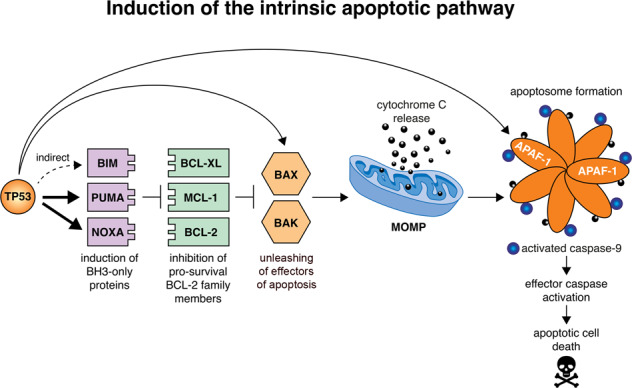
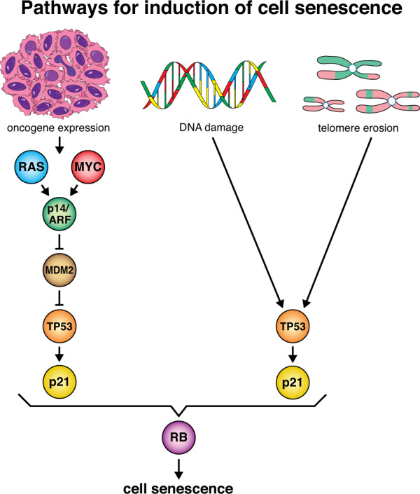
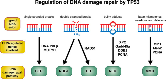
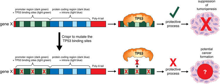
The tumour suppressor TP53 is a master regulator of several cellular processes that collectively suppress tumorigenesis. The TP53 gene is mutated in ~50% of human cancers and these defects usually confer poor responses to therapy. The TP53 protein functions as a homo-tetrameric transcription factor, directly regulating the expression of ~500 target genes, some of them involved in cell death, cell cycling, cell senescence, DNA repair and metabolism. Originally, it was thought that the induction of apoptotic cell death was the principal mechanism by which TP53 prevents the development of tumours. However, gene targeted mice lacking the critical effectors of TP53-induced apoptosis (PUMA and NOXA) do not spontaneously develop tumours. Indeed, even mice lacking the critical mediators for TP53-induced apoptosis, G1/S cell cycle arrest and cell senescence, namely PUMA, NOXA and p21, do not spontaneously develop tumours. This suggests that TP53 must activate additional cellular responses to mediate tumour suppression. In this review, we will discuss the processes by which TP53 regulates cell death, cell cycling/cell senescence, DNA damage repair and metabolic adaptation, and place this in context of current understanding of TP53-mediated tumour suppression.


Conflict of interest statement

The authors declare no competing interests.

Figures

Fig. 1
Fig. 1. Induction of the BCL-2-regulated apoptotic pathway by TP53.
This model depicts the mechanism by which activated TP53 induces the BCL-2-regulated apoptotic pathway. When TP53 is activated, it directly transcriptionally activates the genes encoding the pro-apoptotic BH3-only proteins, PUMA and NOXA, and indirectly induces expression of the gene encoding the BH3-only protein BIM. The pro-survival BCL-2 family members (e.g., BCL-XL, MCL-1 and BCL-2) are repressed by binding of the pro-apoptotic BH3-only proteins. This permits the activation of the apoptosis effectors, BAX and BAK, which in healthy cells are kept in check by binding to the pro-survival BCL-2 proteins. Activated BAX and BAK can oligomerise at the mitochondrial outer membrane, forming pores, thereby causing mitochondrial outer membrane permeabilisation (MOMP) which releases cytochrome c and other apoptogenic factors. Following MOMP, the apoptosome (composed of the adaptor protein APAF-1, dATP, pro-caspase-9 and cytochrome c) forms, which leads to the activation of caspase-9 that triggers activation of downstream effector caspases (e.g., caspases -3, -6 and -7). This caspase cascade causes proteolysis of hundreds of cellular proteins to orchestrate the ordered dismantling of the dying cells. Thick arrows indicate the direct upregulation of target genes by TP53. The thin arrow represents the indirect activation of BIM by TP53. Of note, in the absence of TP53, BAX and APAF-1 are still expressed at levels that are sufficient for effective induction of apoptosis, for example after treatment of lymphoid cells with glucocorticoids, which does not require TP53 for apoptosis induction. Upregulation of BAX and APAF-1 by TP53 may serve to enhance the efficiency of apoptosis but the TP53-mediated upregulation of their genes is not essential for induction of apoptosis, at least in haematopoietic cells.
Fig. 2
Fig. 2. Induction of cell senescence by TP53.
A variety of cellular stresses, such as oxidative damage, DNA damage, telomere shortening and downstream effects of oncogene activation, such as replication stress, can induce cellular senescence via the activation of TP53. This is in part due to the ability of TP53 to directly transcriptionally upregulate the gene for p21, although other effectors of cell senescence are likely to also play critical roles. During cellular senescence, the retinoblastoma (RB) tumour suppressor protein is activated and represses the transcription of genes encoding proteins that are critical for cell cycle progression.
Fig. 3
Fig. 3. Regulation of DNA damage repair by TP53.
Activated TP53 can transcriptionally upregulate an array of direct target genes involved in a range of DNA damage repair pathways, including base excision repair, non-homologous end joining, homologous recombination, nucleotide excision repair and mismatch repair. Thick arrows indicate the type of DNA damage repair pathway that is needed for the repair of a specific type of DNA lesion. The genes to the side of the arrows are genes that are upregulated by TP53 to orchestrate that particular damage repair pathway.
Fig. 4
Fig. 4. Proposed gene-targeting experiments to determine whether TP53-mediated upregulation of a specific direct TP53 target gene is responsible for tumour suppression.
TP53 acts as a tumour suppressor through transcriptional induction of target genes. Binding of TP53 to TP53 binding sites in the promoter and intronic regions induces upregulation of a TP53 target gene. This allows the protein encoded by this TP53 target gene to be upregulated and exert its function that contributes to TP53-mediated tumour suppression. Mutation of TP53 binding sites in target genes can identify the critical genes important for TP53-mediated tumour suppression. Gene targeting can be used to mutate the TP53 binding site(s) in a gene of interest. In cells derived from such genetically modified mice, the TP53 target gene of interest cannot be upregulated by activated TP53, but it can still be regulated by other transcription factors that bind to other sequences in its promoter or intronic regions. If these genetically modified mice, for example after introducing a potent oncogene, such as c-MYC or mutant RAS, will show accelerated tumour development, this would demonstrate that TP53-mediated upregulation of this gene is critical for tumour suppression. If these genetically modified mice are not tumour prone, this would indicate that transcription factors in addition to TP53 can also induce the expression of this protein at levels sufficient for effective tumour suppression.

Similar articles

Cited by

References

    1. Aubrey BJ, Kelly GL, Janic A, Herold MJ, Strasser A. How does p53 induce apoptosis and how does this relate to p53-mediated tumour suppression? Cell Death Differ. 2018;25:104–13. doi: 10.1038/cdd.2017.169. - DOI - PMC - PubMed
    1. Tafvizi A, Huang F, Fersht AR, Mirny LA, van Oijen AM. A single-molecule characterization of p53 search on DNA. Proc Natl Acad Sci. 2011;108:563–8. doi: 10.1073/pnas.1016020107. - DOI - PMC - PubMed
    1. Boutelle AM, Attardi LD. p53 and tumor suppression: it takes a network. Trends Cell Biol. 2021;41:298–310. doi: 10.1016/j.tcb.2020.12.011. - DOI - PMC - PubMed
    1. Haupt Y, Maya R, Kazaz A, Oren M. Mdm2 promotes the rapid degradation of p53. Nature. 1997;387:296–9. doi: 10.1038/387296a0. - DOI - PubMed
    1. Shieh S-Y, Ikeda M, Taya Y, Prives C. DNA damage-induced phosphorylation of p53 alleviates inhibition by MDM2. Cell. 1997;91:325–34. doi: 10.1016/S0092-8674(00)80416-X. - DOI - PubMed

Publication types

Substances