HIF-1α-regulated lncRNA-TUG1 promotes mitochondrial dysfunction and pyroptosis by directly binding to FUS in myocardial infarction
- PMID: 35396503
- PMCID: PMC8993815
- DOI: 10.1038/s41420-022-00969-8
HIF-1α-regulated lncRNA-TUG1 promotes mitochondrial dysfunction and pyroptosis by directly binding to FUS in myocardial infarction
Abstract
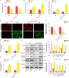
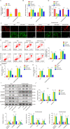
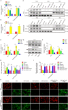
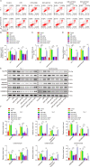
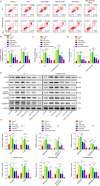
Myocardial infarction (MI) is a fatal heart disease that affects millions of lives worldwide each year. This study investigated the roles of HIF-1α/lncRNA-TUG1 in mitochondrial dysfunction and pyroptosis in MI. CCK-8, DHE, lactate dehydrogenase (LDH) assays, and JC-1 staining were performed to measure proliferation, reactive oxygen species (ROS), LDH leakage, and mitochondrial damage in hypoxia/reoxygenation (H/R)-treated cardiomyocytes. Enzyme-linked immunoassay (ELISA) and flow cytometry were used to detect LDH, creatine kinase (CK), and its isoenzyme (CK-MB) levels and caspase-1 activity. Chromatin immunoprecipitation (ChIP), luciferase assay, and RNA-immunoprecipitation (RIP) were used to assess the interaction between HIF-1α, TUG1, and FUS. Quantitative real-time polymerase chain reaction (qRT-PCR), Western blotting, and immunohistochemistry were used to measure HIF-1α, TUG1 and pyroptosis-related molecules. Hematoxylin and eosin (HE), 2,3,5-triphenyltetrazolium chloride (TTC), and terminal deoxynucleotidyl transferase dUTP risk end labelling (TUNEL) staining were employed to examine the morphology, infarction area, and myocardial injury in the MI mouse model. Mitochondrial dysfunction and pyroptosis were induced in H/R-treated cardiomyocytes, accompanied by an increase in the expression of HIF-α and TUG1. HIF-1α promoted TUG1 expression by directly binding to the TUG1 promoter. TUG1 silencing inhibited H/R-induced ROS production, mitochondrial injury and the expression of the pyroptosis-related proteins NLRP3, caspase-1 and GSDMD. Additionally, H/R elevated FUS levels in cardiomyocytes, which were directly inhibited by TUG1 silencing. Fused in sarcoma (FUS) overexpression reversed the effect of TUG1 silencing on mitochondrial damage and caspase-1 activation. However, the ROS inhibitor N-acetylcysteine (NAC) promoted the protective effect of TUG1 knockdown on H/R-induced cardiomyocyte damage. The in vivo MI model showed increased infarction, myocardial injury, ROS levels and pyroptosis, which were inhibited by TUG1 silencing. HIF-1α targeting upregulated TUG1 promotes mitochondrial damage and cardiomyocyte pyroptosis by combining with FUS, thereby promoting the occurrence of MI. HIF-1α/TUG1/FUS may serve as a potential treatment target for MI.
© 2022. The Author(s).
Conflict of interest statement
The authors declare no competing interests.
Figures



Similar articles
-
Uric acid aggravates myocardial ischemia-reperfusion injury via ROS/NLRP3 pyroptosis pathway.Biomed Pharmacother. 2021 Jan;133:110990. doi: 10.1016/j.biopha.2020.110990. Epub 2020 Nov 21. Biomed Pharmacother. 2021. PMID: 33232925
-
Effects of PGC1α on myocardial ischemia reperfusion injury and the underlying mechanisms.Zhong Nan Da Xue Xue Bao Yi Xue Ban. 2020 Oct 28;45(10):1155-1163. doi: 10.11817/j.issn.1672-7347.2020.190215. Zhong Nan Da Xue Xue Bao Yi Xue Ban. 2020. PMID: 33268575 Chinese, English.
-
IRF2 contributes to myocardial infarction via regulation of GSDMD induced pyroptosis.Mol Med Rep. 2022 Feb;25(2):40. doi: 10.3892/mmr.2021.12556. Epub 2021 Dec 8. Mol Med Rep. 2022. PMID: 34878155 Free PMC article.
-
Gasdermin D-mediated pyroptosis in myocardial ischemia and reperfusion injury: Cumulative evidence for future cardioprotective strategies.Acta Pharm Sin B. 2023 Jan;13(1):29-53. doi: 10.1016/j.apsb.2022.08.007. Epub 2022 Aug 13. Acta Pharm Sin B. 2023. PMID: 36815034 Free PMC article. Review.
-
Biological function of RNA-binding proteins in myocardial infarction: a potential emerging therapeutic limelight.Cell Biosci. 2025 May 24;15(1):65. doi: 10.1186/s13578-025-01408-8. Cell Biosci. 2025. PMID: 40413549 Free PMC article. Review.
Cited by
-
Mechanically induced pyroptosis enhances cardiosphere oxidative stress resistance and metabolism for myocardial infarction therapy.Nat Commun. 2023 Oct 2;14(1):6148. doi: 10.1038/s41467-023-41700-0. Nat Commun. 2023. PMID: 37783697 Free PMC article.
-
Anti-angiogenic effect of exo-LncRNA TUG1 in myocardial infarction and modulation by remote ischemic conditioning.Basic Res Cardiol. 2023 Jan 12;118(1):1. doi: 10.1007/s00395-022-00975-y. Basic Res Cardiol. 2023. PMID: 36635484 Clinical Trial.
-
ELF5-Regulated lncRNA-TTN-AS1 Alleviates Myocardial Cell Injury via Recruiting PCBP2 to Increase CDK6 Stability in Myocardial Infarction.Mol Cell Biol. 2024;44(8):303-315. doi: 10.1080/10985549.2024.2374083. Epub 2024 Jul 21. Mol Cell Biol. 2024. PMID: 39034459 Free PMC article.
-
Therapeutic Potential of Gasdermin D-Mediated Myocardial Pyroptosis in Ischaemic Heart Disease: Expanding the Paradigm From Bench to Clinical Insights.J Cell Mol Med. 2025 Feb;29(3):e70357. doi: 10.1111/jcmm.70357. J Cell Mol Med. 2025. PMID: 39929748 Free PMC article. Review.
-
Non-coding RNAs in the pathophysiology of heart failure with preserved ejection fraction.Front Cardiovasc Med. 2024 Jan 8;10:1300375. doi: 10.3389/fcvm.2023.1300375. eCollection 2023. Front Cardiovasc Med. 2024. PMID: 38259314 Free PMC article. Review.
References
-
- Mechanic OJ, Grossman SA. Acute Myocardial Infarction. StatPearls. Treasure Island (FL); 2020.
LinkOut - more resources
Full Text Sources
Research Materials
Miscellaneous

