. 2022 Apr 8;5(1):335.
doi: 10.1038/s42003-022-03238-7.

Rearrangement processes and structural variations show evidence of selection in oesophageal adenocarcinomas

Collaborators, Affiliations

Rearrangement processes and structural variations show evidence of selection in oesophageal adenocarcinomas

Alvin Wei Tian Ng et al. Commun Biol. .

Abstract

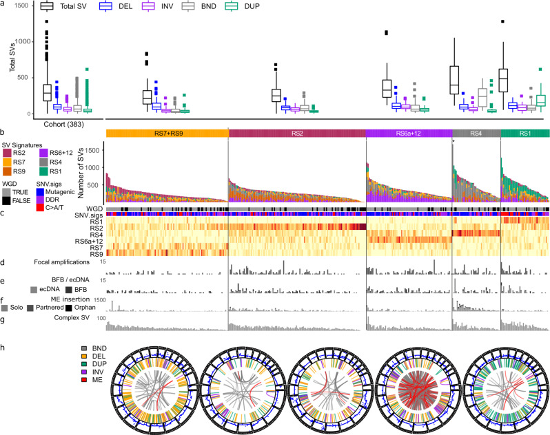
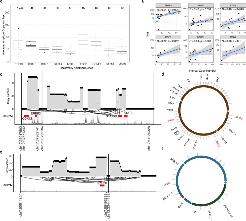
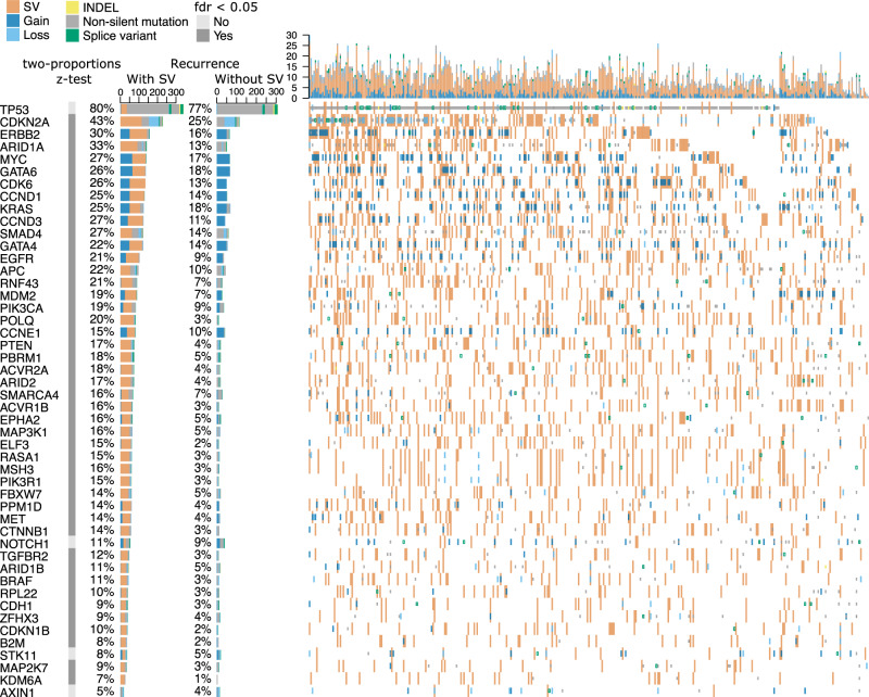
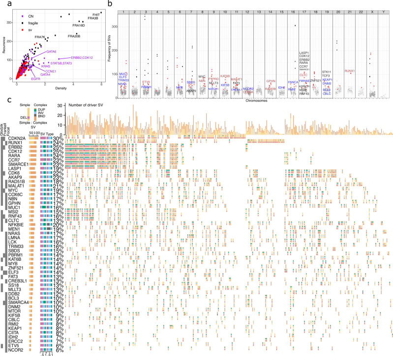
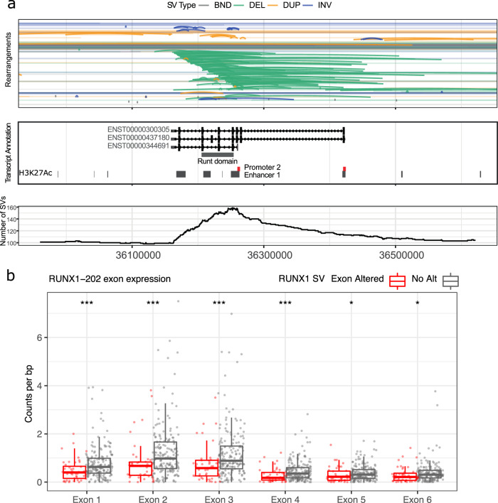
Oesophageal adenocarcinoma (OAC) provides an ideal case study to characterize large-scale rearrangements. Using whole genome short-read sequencing of 383 cases, for which 214 had matched whole transcriptomes, we observed structural variations (SV) with a predominance of deletions, tandem duplications and inter-chromosome junctions that could be identified as LINE-1 mobile element (ME) insertions. Complex clusters of rearrangements resembling breakage-fusion-bridge cycles or extrachromosomal circular DNA accounted for 22% of complex SVs affecting known oncogenes. Counting SV events affecting known driver genes substantially increased the recurrence rates of these drivers. After excluding fragile sites, we identified 51 candidate new drivers in genomic regions disrupted by SVs, including ETV5, KAT6B and CLTC. RUNX1 was the most recurrently altered gene (24%), with many deletions inactivating the RUNT domain but preserved the reading frame, suggesting an altered protein product. These findings underscore the importance of identification of SV events in OAC with implications for targeted therapies.


Conflict of interest statement

R.C.F. has devised an early detection technology called Cytosponge, this device technology and the associated TFF3 biomarker are licensed to Covidien GI solutions (now owned by Medtronic) by the Medical Research Council. R.C.F. and M.O. are named inventors on patents pertaining to the Cytosponge and associated technology. R.C.F. is a shareholder of Cyted Ltd., a company working on early detection technology. R.C.F. has received consulting and/or speaker fees from Medtronic, Roche and Bristol Myers Squibb. The other authors declare no competing interests.

Figures

Fig. 1
Fig. 1. Classification of OACs according to the proportions of SV types and signatures.
Tumours are shown classified into groups according to their predominant SV signature defined by Nik-Zainal et al. (2016). a Box plot showing numbers of SVs by SV type for the entire cohort and in each group (named after the simplest rearrangement that could generate such a junction, DEL: deletion, INV: inversion, BND: ‘breakend’, i.e. an inter-chromosome junction or translocation, DUP: tandem duplication). b Bar plots of rearrangements associated to each rearrangement signatures in OAC. c Heatmap showing proportions of SVs associated to each signature and a comparison with related variables: whole genome doubling (WGD), SNV signature classification (Mutagenic, DDR and C > A/T) described by Secrier (2016), d focal amplifications, e number of BFB and ecDNA cycles, f number of mobile element insertions and g complex SV clusters. h Circos plots of representative tumours from each signature group with ME insertions highlighted in red. *Denotes tumour with >2500 SVs excluded from plot.
Fig. 2
Fig. 2. Complex SVs leading to amplification of oncogenes.
a Recurrent amplicons detected by Amplicon Architect associated with known OAC oncogenes. The number of tumours with detected amplicon is shown above. Y-axis showing copy number of segments spanning each gene, averaged along the length of segment. b Correlation of gene expression (TPM) and copy number of amplicons. c Example of an amplified region spanning CDK12, ERBB2, STAT3 and STAT5B, resembling ecDNA and d Reconstructed amplicon as an extrachromosomal circle containing ERBB2 and a CDK12-STAT5B fusion. e An amplified region spanning EGFR and joining chromosomes 7 and 13, forming an ecDNA and reconstructed as a circle (f).
Fig. 3
Fig. 3. Estimates of recurrence in known driver alterations with and without SVs.
Oncoplot showing recurrence of known OAC driver gene mutations (taken from Frankell et al., 2019 and Campbell et al., 2020) with and without SV. Estimates of recurrence without SVs includes copy number gains and losses, INDELs and SNVs. Recurrence with SVs are counted when the interval between two breakpoints overlaps with exon or exons of the gene. Two-proportions z-test with multiple hypothesis testing (FDR) used to test if recurrence is significantly higher with SVs included.
Fig. 4
Fig. 4. Recurrence and density of SVs in 1 Mb genomic bins.
a Scatter plot showing recurrence, the number of patients with an SV break in each 1 Mb bin (y-axis) and density, the average number of SV breaks in the bin over all tumours (x-axis). Bins are labelled with genes or fragile sites that they overlap: black, fragile sites; purple, intervals of amplification and deletion; red, putative genes under selection. b Manhattan plot showing 1MB bins containing putative drivers (red) and fragile sites (black) and genes coloured by methods discovered: Glodzik model adjusting for genomic context (Black), Focal (F, blue) and both methods (brown: FG). c Oncoplot showing candidate driver genes identified using focal and Glodzik methods and annotated if each gene was found in Frankell et al. (2019). Horizontal bar plots show total number of simple (light orange) and complex (dark orange) SVs found in the given gene; proportions of SVs classified as simple that are of the various SV types; and similarly for SVs classified as complex. Each oncoplot cell shows if each patient has a simple or complex SV and the combination of SV types.
Fig. 5
Fig. 5. Deletions and duplications in RUNX1 affecting RUNT domain exons.
a Genomic regions with SVs at the RUNX1 locus (arcs) with cumulative numbers of SV intervals at each position (bottom). RUNX1 is transcribed from the negative strand. RUNT domain and enhancers, from H3K27Ac data, in grey, and promoters in red. b Exon expression of RUNX1-202 (ENST00000344691) for with tumours with alterations in RUNX1 (red) and no alterations (grey). Read counts were normalized to length of exons. *, *** denotes p ≤ 0.05 and p ≤ 0.001 respectively. Gene structure for RUNX1-202 shown as it was determined to be highest expressed transcript by GTEx and in the cohort of 214 tumours. No transcription from promoter 1 was detected.

Similar articles

Cited by

References

    1. Li, Y. et al. Patterns of somatic structural variation in human cancer genomes. Nature10.1038/s41586-019-1913-9 (2020). - PMC - PubMed
    1. Nik-Zainal S, et al. Landscape of somatic mutations in 560 breast cancer whole-genome sequences. Nature. 2016;534:47–54. - PMC - PubMed
    1. Campbell PJ, et al. Pan-cancer analysis of whole genomes. Nature. 2020;578:82–93. - PMC - PubMed
    1. Sabarinathan, R. et al. The whole-genome panorama of cancer drivers. Preprint at bioRxiv10.1101/190330 (2017).
    1. Ciriello G, et al. Emerging landscape of oncogenic signatures across human cancers. Nat. Genet. 2013;45:1127–1133. - PMC - PubMed

Publication types

Substances

Supplementary concepts