

Hippocampal interneurons are direct targets for circulating glucocorticoids
- PMID: 35397117
- PMCID: PMC9232959
- DOI: 10.1002/cne.25322
Hippocampal interneurons are direct targets for circulating glucocorticoids
Abstract
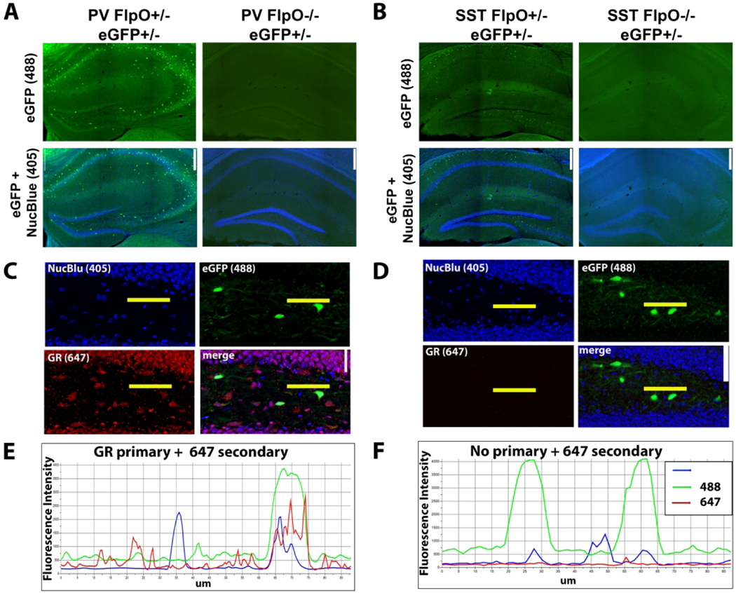
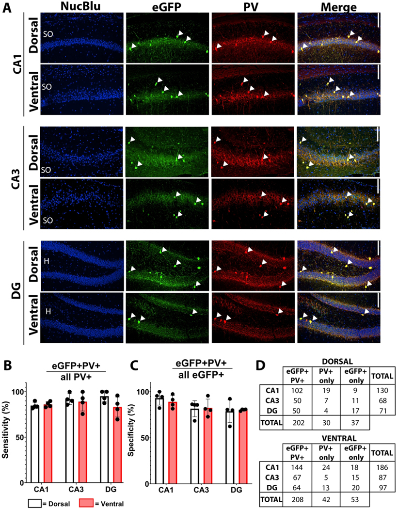
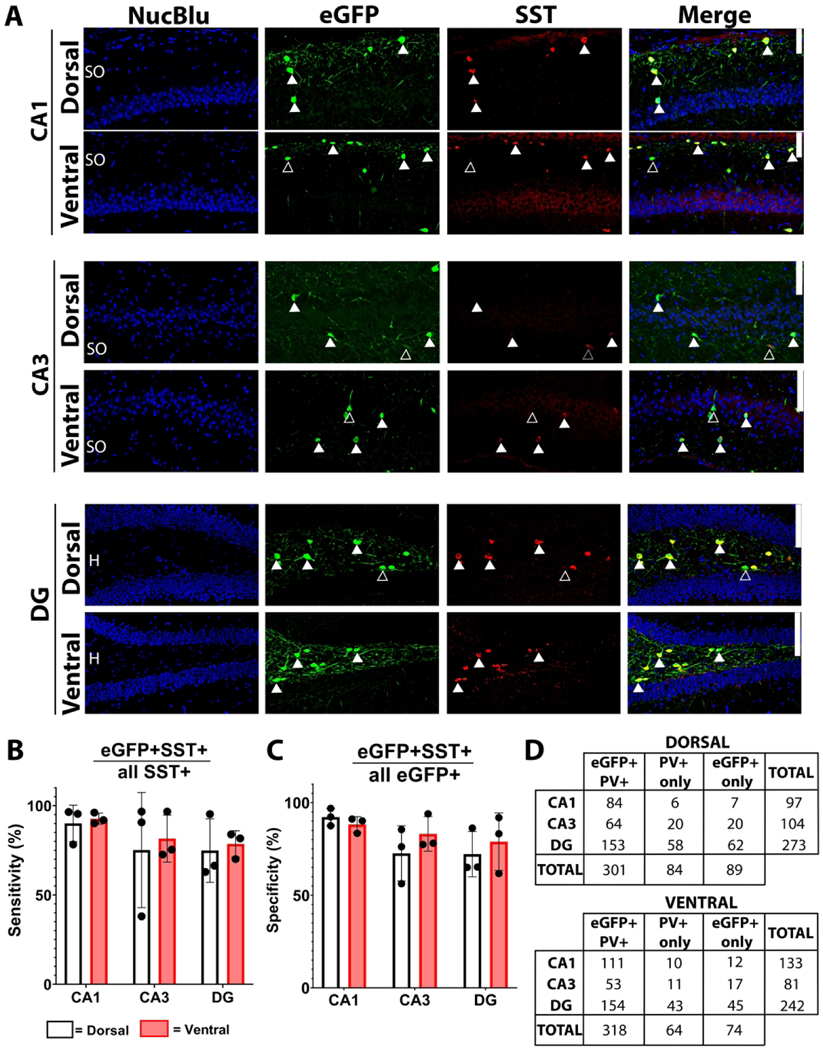
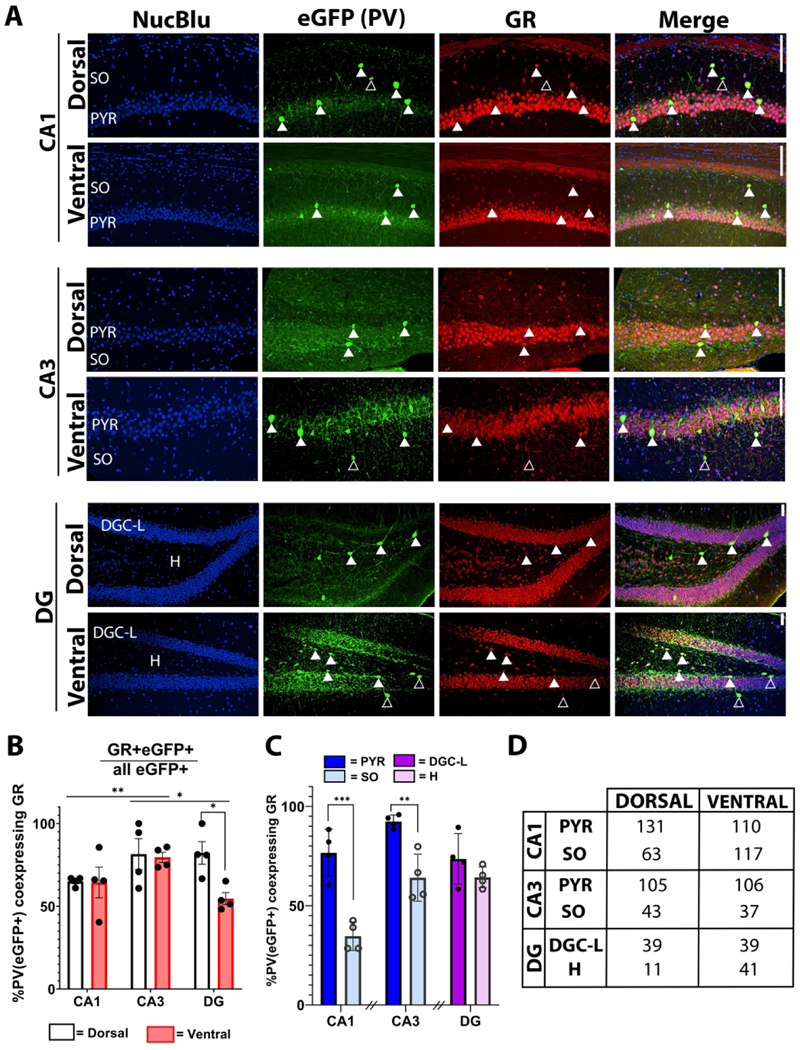
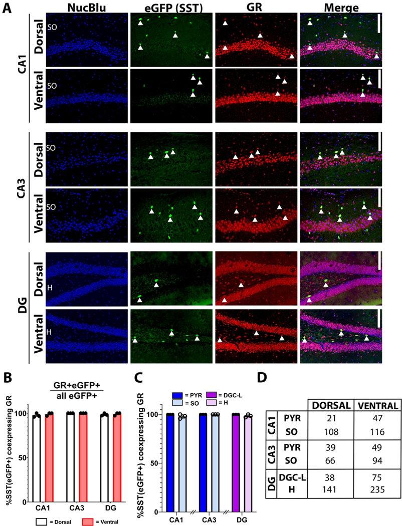
The hippocampus has become a significant target of stress research in recent years because of its role in cognitive functioning, neuropathology, and regulation of the hypothalamic-pituitary-adrenal (HPA) axis. Despite the pervasive impact of stress on psychiatric and neurological disease, many of the circuit- and cell-dependent mechanisms giving rise to the limbic regulation of the stress response remain unknown. Hippocampal excitatory neurons generally express high levels of glucocorticoid receptors (GRs) and are therefore positioned to respond directly to serum glucocorticoids. These neurons are, in turn, regulated by neighboring interneurons, subtypes of which have been shown to respond to stress exposure. However, GR expression among hippocampal interneurons is not well characterized. To determine whether key interneuron populations are direct targets for glucocorticoid action, we used two transgenic mouse lines to label parvalbumin-positive (PV+) and somatostatin-positive (SST+) interneurons. GR immunostaining of labeled interneurons was characterized within the dorsal and ventral dentate hilus, dentate cell body layer, and CA1 and CA3 stratum oriens and stratum pyramidale. While nearly all hippocampal SST+ interneurons expressed GR across all regions, GR labeling of PV+ interneurons showed considerable subregion variability. The percentage of PV+, GR+ cells was highest in the CA3 stratum pyramidale and lowest in the CA1 stratum oriens, with other regions showing intermediate levels of expression. Together, these findings indicate that, under baseline conditions, hippocampal SST+ interneurons are a ubiquitous glucocorticoid target, while only distinct populations of PV+ interneurons are direct targets. This anatomical diversity suggests functional differences in the regulation of stress-dependent hippocampal responses.
Keywords: glucocorticoid receptor (GR); hippocampus; interneurons; parvalbumin (PV); somatostatin (SST); stress.
© 2022 Wiley Periodicals LLC.
Conflict of interest statement
CONFLICT OF INTEREST
The authors have no conflicts of interest to disclose.
Figures
Similar articles
-
Excitatory Inputs Determine Phase-Locking Strength and Spike-Timing of CA1 Stratum Oriens/Alveus Parvalbumin and Somatostatin Interneurons during Intrinsically Generated Hippocampal Theta Rhythm.J Neurosci. 2016 Jun 22;36(25):6605-22. doi: 10.1523/JNEUROSCI.3951-13.2016. J Neurosci. 2016. PMID: 27335395 Free PMC article.
-
Calcium-binding protein (calbindin-D28k) and parvalbumin immunocytochemistry: localization in the rat hippocampus with specific reference to the selective vulnerability of hippocampal neurons to seizure activity.J Comp Neurol. 1989 Feb 8;280(2):183-96. doi: 10.1002/cne.902800203. J Comp Neurol. 1989. PMID: 2925892
-
HIV-1 Tat causes cognitive deficits and selective loss of parvalbumin, somatostatin, and neuronal nitric oxide synthase expressing hippocampal CA1 interneuron subpopulations.J Neurovirol. 2016 Dec;22(6):747-762. doi: 10.1007/s13365-016-0447-2. Epub 2016 May 13. J Neurovirol. 2016. PMID: 27178324 Free PMC article.
-
Chronic stress, hippocampus and parvalbumin-positive interneurons: what do we know so far?Rev Neurosci. 2016 Jun 1;27(4):397-409. doi: 10.1515/revneuro-2015-0042. Rev Neurosci. 2016. PMID: 26751865 Review.
-
Brain corticosteroid receptor balance in health and disease.Endocr Rev. 1998 Jun;19(3):269-301. doi: 10.1210/edrv.19.3.0331. Endocr Rev. 1998. PMID: 9626555 Review.
Cited by
-
Chronic stress causes striatal disinhibition mediated by SOM-interneurons in male mice.Nat Commun. 2022 Nov 29;13(1):7355. doi: 10.1038/s41467-022-35028-4. Nat Commun. 2022. PMID: 36446783 Free PMC article.
-
Hippocampal glucocorticoid receptors modulate status epilepticus severity.Neurobiol Dis. 2023 Mar;178:106014. doi: 10.1016/j.nbd.2023.106014. Epub 2023 Jan 23. Neurobiol Dis. 2023. PMID: 36702319 Free PMC article.
-
Altered regulation of oligodendrocytes associated with parvalbumin neurons in the ventral hippocampus underlies fear generalization in male mice.Neuropsychopharmacology. 2023 Oct;48(11):1668-1679. doi: 10.1038/s41386-023-01611-6. Epub 2023 Jun 5. Neuropsychopharmacology. 2023. PMID: 37277574 Free PMC article.
-
Serum cortisol and insulin-like growth factor 1 levels in major depressive disorder and schizophrenia.Sci Rep. 2023 Jan 20;13(1):1148. doi: 10.1038/s41598-023-28449-8. Sci Rep. 2023. PMID: 36670169 Free PMC article.
-
Somatostatin interneuron fate-mapping and structure in a Pten knockout model of epilepsy.Front Cell Neurosci. 2024 Oct 21;18:1474613. doi: 10.3389/fncel.2024.1474613. eCollection 2024. Front Cell Neurosci. 2024. PMID: 39497922 Free PMC article.
References
-
- Allen Reference Atlas - Mouse Brain [coronal]. Available from atlas.brain-map.org.
-
- Castro OW, Santos VR, Pun RYK, McKlveen JM, Batie M, Holland KD, Gardner M, Garcia-Cairasco N, Herman JP, & Danzer SC (2012). Impact of corticosterone treatment on spontaneous seizure frequency and epileptiform activity in mice with chronic epilepsy. PLoS ONE, 7(9), e46044. 10.1371/journal.pone.0046044 - DOI - PMC - PubMed
MeSH terms
Substances
Grants and funding
LinkOut - more resources
Full Text Sources
Medical
Molecular Biology Databases
Miscellaneous
