. 2022 Jun:130:105156.
doi: 10.1016/j.jmbbm.2022.105156. Epub 2022 Mar 18.

Assessing the aneurysm occlusion efficacy of a shear-thinning biomaterial in a 3D-printed model

Affiliations

Assessing the aneurysm occlusion efficacy of a shear-thinning biomaterial in a 3D-printed model

Grant Schroeder et al. J Mech Behav Biomed Mater. 2022 Jun.

Abstract

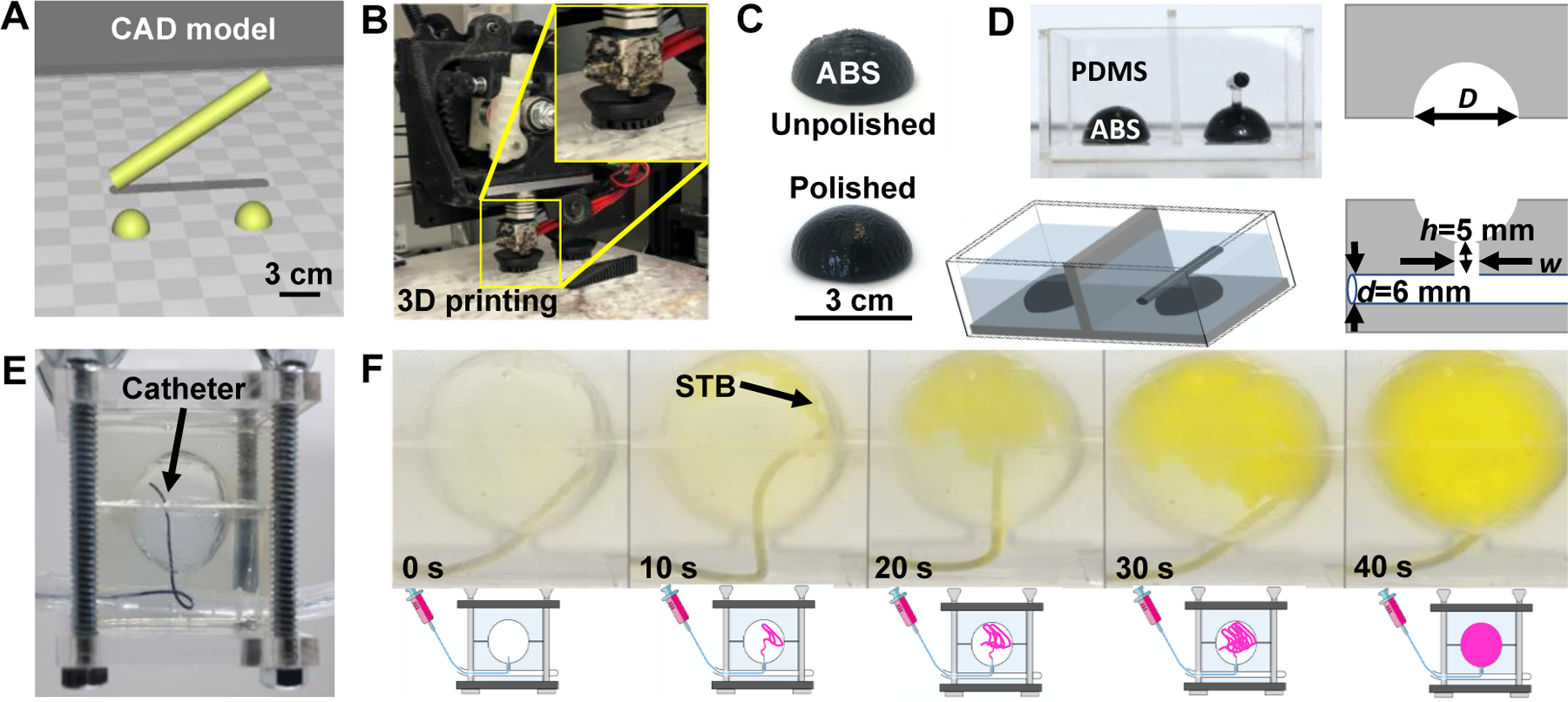
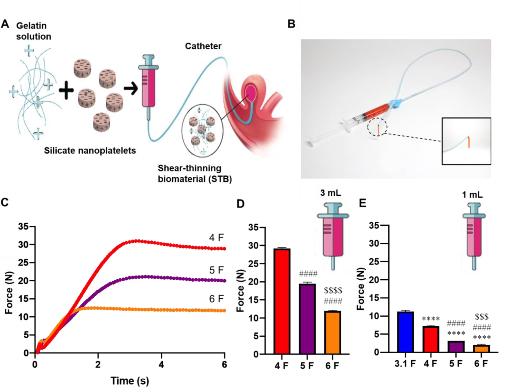
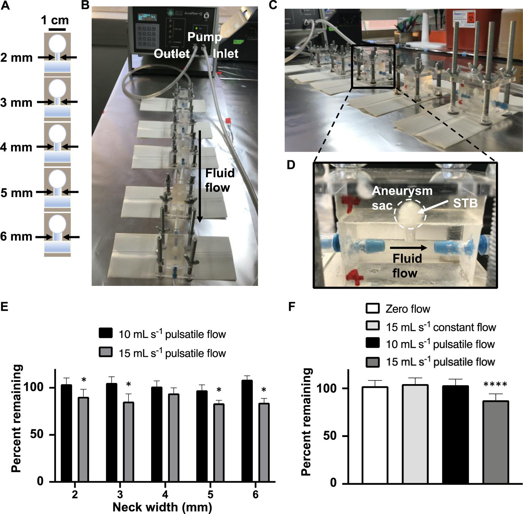
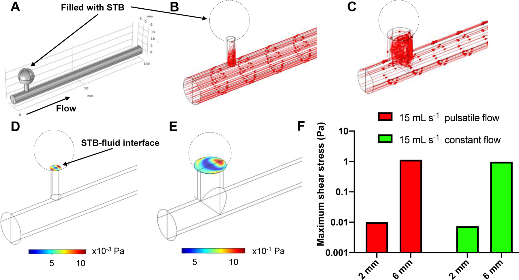
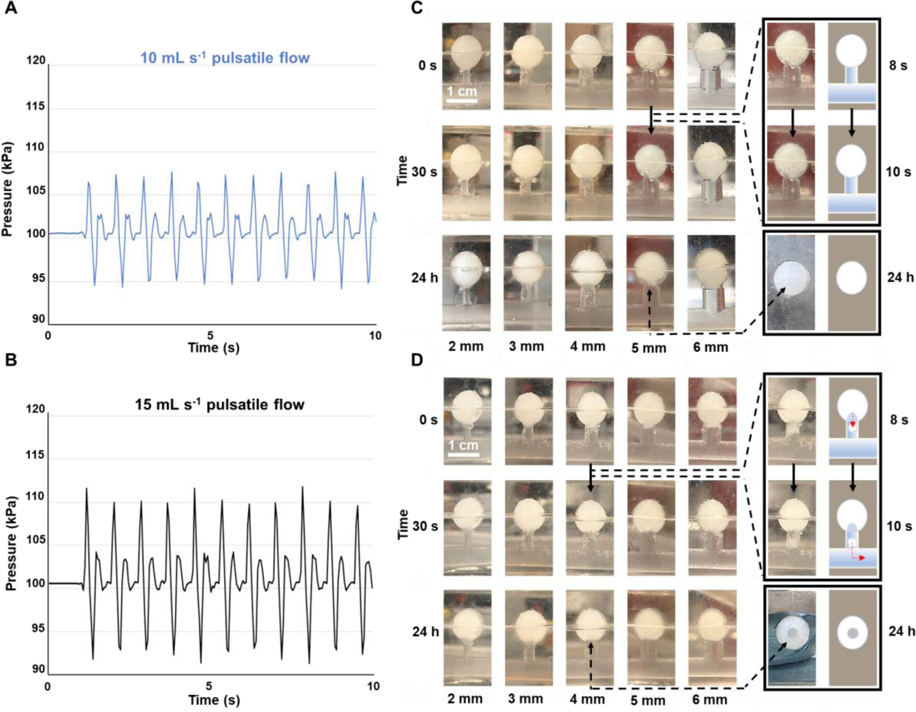
Metallic coil embolization is a common method for the endovascular treatment of visceral artery aneurysms (VAA) and visceral artery pseudoaneurysms (VAPA); however, this treatment is suboptimal due to the high cost of coils, incomplete volume occlusion, poor reendothelialization, aneurysm puncture, and coil migration. Several alternative treatment strategies are available, including stent flow diverters, glue embolics, gelfoam slurries, and vascular mesh plugs-each of which have their own disadvantages. Here, we investigated the in vitro capability of a shear-thinning biomaterial (STB), a nanocomposite hydrogel composed of gelatin and silicate nanoplatelets, for the minimally-invasive occlusion of simple necked aneurysm models. We demonstrated the injectability of STB through various clinical catheters, engineered an in vitro testing apparatus to independently manipulate aneurysm neck diameter, fluid flow rate, and flow waveform, and tested the stability of STB within the models under various conditions. Our experiments show that STB is able to withstand at least 1.89 Pa of wall shear stress, as estimated by computational fluid dynamics. STB is also able to withstand up to 10 mL s-1 pulsatile flow with a waveform mimicking blood flow in the human femoral artery and tolerate greater pressure changes than those in the human aorta. We ultimately found that our in vitro system was limited by supraphysiologic pressure changes caused by aneurysm models with low compliance.

Keywords: Catheters; Hydrogels; Minimally invasive; Pseudoaneurysms; Shear-thinning biomaterials; Silicate nanoplatelets; Visceral artery aneurysms.


Conflict of interest statement

Conflict of Interest

A. K. is a co-founder of a start‐up company, Obsidio, Inc., which is based on shear‐thinning embolic materials.

Declaration of interests

The authors declare that they have no known competing financial interests or personal relationships that could have appeared to influence the work reported in this paper.

Ali Khademhosseini reports financial support was provided by National Institutes of Health. Ali Khademhosseini reports a relationship with Obsidio, Inc that includes: board membership and equity or stocks. Ali Khademhosseini has patent Shear-thinning compositions as an intravascular embolic agent issued to Obsidio, Inc.

Figures

Fig. 1.
Fig. 1.. Fabrication of in vitro aneurysm models and filling them with STB.
(A) CAD-based fabrication of aneurysm model components, including a parent artery and the saccular aneurysm pieces. (B) 3D-printing of aneurysm model components using ABS. (C) Polishing the surface of 3D printed ABS pieces to render the surfaces smooth. (D) Formation of negative PDMS mold using the 3D printed ABS pieces, and the removal of ABS yielding aneurysm models with the following dimensions: blood vessel diameter, d: 6 mm, neck height, h: 5 mm, neck diameter (width), w: variable (2, 3, 4, 5, or 6 mm), and aneurysm sac diameter, D: variable (1 cm or 3 cm). (E) Navigation of a catheter into a 3 cm diameter aneurysm model. (F) Time course of STB filling of the 3 cm aneurysm model using a microcatheter under pulsatile flow in less than 1 min. The STB is stained with commercial yellow food dye.
Fig. 2.
Fig. 2.. Formulation and properties of shear-thinning biomaterial (STB).
(A) Gelatin and LAPONITE® XLG-XR (silicate nanoplatelets) are mixed to form STB, which is injected into an aneurysm using a catheter. (B) Picture of colored STB loaded into a syringe and injected via a catheter. (C) Injection force versus injection time and (D) injection force plateaus for STB injected through catheters connected to a 3 mL syringe at a syringe depression rate of 33.96 mm min-1. (E) Injection force plateaus for STB injected through catheters connected to a 1 mL syringe at the same rate of syringe depression as in panel D. One-way ANOVA with multiple comparisons (Tukey post-test) were performed: * indicates comparison with 3.1 F; # shows comparison with 4 F; and $ is for the comparison with 5 F (***, ###, or $ $ $ indicate p < 0.001; **** ####, or $ $ $ $ show p < 0.0001).
Fig. 3.
Fig. 3.. Stability of STB in aneurysm models over 24 h.
(A) Schematic of 1 cm aneurysm models used in 24-h flow experiments, with the neck width ranging from 2 to 6 mm. (B and C) Experimental setup showing the AccuFlow-Q Physiological Flow System connected to aneurysm models in series. (D) Close-up image of a 1 cm aneurysm model with a 4 mm neck width, showing the sac filled with STB. (E) Comparison of the percent STB recovered from models with neck widths ranging from 2 to 6 mm exposed to 10 mL s−1 and 15 mL s−1 pulsatile flows after 24 h, showing significant differences (One-way ANOVA with multiple comparisons (Fisher’s post-test); * p ≤ 0.05) for all aneurysm neck sizes except the 4 mm neck. (F) STB percent remaining after 24 h from all neck sizes for zero flow, 15 mL s−1 constant flow, 10 mL s−1 pulsatile flow, and 15 mL s−1 pulsatile flow, showing significant differences (One-way ANOVA with multiple comparisons (Tukey post-test); **** p ≤ 0.0001) for 15 mL s−1 pulsatile flow compared with all other flow patterns.
Fig. 4.
Fig. 4.. CFD simulation of fluid flow in aneurysm models.
(A) Simulation computational domain, comprising a finite length 3D conduit that is 100 mm long with an inner diameter of 6 mm, connected to an aneurysm sac through a 5 mm long cylindrical neck of a diameter varying from 2 mm to 6 mm. For the simulation, the neck was capped at the STB-fluid interface to reflect the in vitro experiments in which STB filled the aneurysm sac. (B and C) Flow streamlines determined by CFD for 15 mL s−1 pulsatile flow, demonstrating more fluid flow into the aneurysm neck for the 6 mm neck compared with the 2 mm neck. (D) Shear stress as determined from CFD modeling at the STB-fluid interface for the 2 mm neck aneurysm model experiencing 15 mL s−1 pulsatile flow. (E) Shear stress as determined from CFD modeling at the STB-fluid interface for the 6 mm neck aneurysm model experiencing 15 mL s−1 pulsatile flow. (F) Magnitude of maximum shear stress at the STB-fluid interface for the 2 mm and 6 mm neck-size aneurysm models under 15 mL s−1 pulsatile flow and 15 mL s−1 constant flow, demonstrating a discordance between maximal shear stress and material stability (STB was stable for 24 h under 15 mL s−1 constant flow regardless of aneurysm neck size, whereas it was lost within 24 h under 15 mL s−1 pulsatile flow regardless of aneurysm neck size).
Fig. 5.
Fig. 5.. Supraphysiologic pressure changes resulting in STB loss.
Pressure versus time for 10 mL s−1 and (B) 15 mL s−1 pulsatile flow. (C) Images of 1 cm aneurysm models ranging from 2 mm to 6 mm neck widths under 10 mL s−1 pulsatile flow at varying time points, demonstrating the stability of STB for 24 h. (D) Images of 1 cm aneurysm models ranging from 2 mm to 6 mm neck widths under 15 mL s−1 pulsatile flow at varying time points, showing STB loss within the first 30 s, followed by no additional material loss for the remainder of the 24 h.

Similar articles

Cited by

References

    1. Hosn MA, Xu J, Sharafuddin M, Corson JD. Visceral Artery Aneurysms: Decision Making and Treatment Options in the New Era of Minimally Invasive and Endovascular Surgery. Int J Angiol 2019;28(1):11–16. doi:10.1055/s-0038-1676958 - DOI - PMC - PubMed
    1. Tipaldi MA, Krokidis M, Orgera G, et al. Endovascular management of giant visceral artery aneurysms. Sci Rep 2021;11(1):1–6. doi:10.1038/s41598-020-80150-2 - DOI - PMC - PubMed
    1. Loffroy R, Favelier S, Pottecher P, et al. Endovascular management of visceral artery aneurysms: When to watch, when to intervene? World J Radiol 2015;7(7):143. doi:10.4329/wjr.v7.i7.143 - DOI - PMC - PubMed
    1. Chiaradia M, Novelli L, Deux J-F, et al. Ruptured visceral artery aneurysms. Diagn Interv Imaging 2015;96(7–8):797–806. doi:10.1016/j.diii.2015.03.012 - DOI - PubMed
    1. Sachdev U, Baril DT, Ellozy SH, et al. Management of aneurysms involving branches of the celiac and superior mesenteric arteries: A comparison of surgical and endovascular therapy. J Vasc Surg 2006;44(4):718–724. doi:10.1016/j.jvs.2006.06.027 - DOI - PubMed

Publication types

Substances