Review
. 2022 Jul 6;12(7):1642-1655.
doi: 10.1158/2159-8290.CD-22-0145.

Somatic Mutation: What Shapes the Mutational Landscape of Normal Epithelia?

Affiliations
Review

Somatic Mutation: What Shapes the Mutational Landscape of Normal Epithelia?

Joanna C Fowler et al. Cancer Discov. .

Abstract

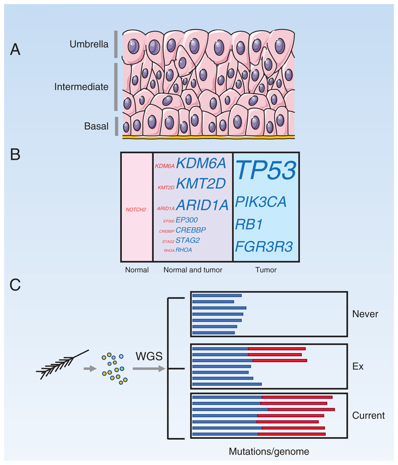
Epithelial stem cells accumulate mutations throughout life. Some of these mutants increase competitive fitness and may form clones that colonize the stem cell niche and persist to acquire further genome alterations. After a transient expansion, mutant stem cells must revert to homeostatic behavior so normal tissue architecture is maintained. Some positively selected mutants may promote cancer development, whereas others inhibit carcinogenesis. Factors that shape the mutational landscape include wild-type and mutant stem cell dynamics, competition for the niche, and environmental exposures. Understanding these processes may give new insight into the basis of cancer risk and opportunities for cancer prevention.

Significance: Recent advances in sequencing have found somatic mutations in all epithelial tissues studied to date. Here we review how the mutational landscape of normal epithelia is shaped by clonal competition within the stem cell niche combined with environmental exposures. Some of the selected mutant genes are oncogenic, whereas others may be inhibitory of transformation. Discoveries in this area leave many open questions, such as the definition of cancer driver genes, the mechanisms by which tissues constrain a high proportion of oncogenic mutant cells, and whether clonal fitness can be modulated to decrease cancer risk.


Conflict of interest statement

Conflict of interest Disclosure

The authors declare no competing interests.

Figures

Figure 1
Figure 1. Detecting somatic mutants in normal epithelia
A: Clonality and somatic mutant detection in tumors and normal epithelia. Tumors are clonal yellow) with sub-clonal mutations (green, blue and red). Normal epithelia harbor scattered somatic mutant clones (colored circles) in a wild type background). When sequenced at standard depth, the founder clone and common subclones in a tumor are readily identified, whereas only the largest mutant clones in normal tissue exceed the lower limit of detection. B-D: Methods to detect somatic mutant clones in normal tissues B: A single cell suspension is generated from normal epithelium, cultured at clonal density and individual colonies whole genome sequenced (WGS). C: Laser capture microdissection (LCM) is used to isolate areas of tissue such as individual stem cell niches like the colonic crypt. Special protocols are used to perform WGS, whole exome or targeted sequencing and mutations can then be located within the tissue section from which they came. D: Larger areas of epithelia can be dissected into a gridded array of typically 2mm2 samples, deep targeted sequencing (DTS) performed, and a statistical approach used to call rare somatic mutants in each sample, which can then be mapped (colored circles) within the sample grid.
Figure 2
Figure 2. Colon and endometrium
A: Colonic crypt: Stem cells (red) reside in a niche at the base of the crypt. Differentiation of stem cells generates short lived proliferating cells (green) which migrate up into the crypt and differentiate into post-mitotic differentiated cells (blue). Right hand panels show top-down views of stem cells in the crypt base. When a stem cell differentiates and exits the niche (red arrow), it is replaced by division of one of its neighboring stem cells with equal probability (0.5). B: Possible outcomes of a neutral mutation in a crypt stem cell: A new mutation that does not alter stem cell fate (yellow) may generate a clone that is outcompeted by wild type cells and lost or replaces other stem cells so the mutant becomes fixed in the crypt as no non-mutant cells remain. Positively selected mutations confer a greater than 50% chance of replacing a wild type neighbor and a high likelihood of taking over a crypt. C: Crypt Fission: Crypt fission occurs rarely in wild type crypts, but the rate is greatly accelerated by activating KRAS and nonsense STAG2 mutations, which spread to colonize the crypt and then induce the crypt to split, creating two independent crypts. The process may repeat, allowing a mutation to spread widely across the epithelium. D: Structure of human endometrial epithelium: The basalis layer (blue) contains a branching network of interconnected glands through which clonal mutations may spread. The overlying functionalis layer (pink) contains glands connected with the basalis layer and undergoes cyclical growth, apoptosis, and shedding between menarche and menopause.
Figure 3
Figure 3. Epidermis
A: Structure of the epidermis. Proliferating cells are confined to the basal cell layer. On commitment to differentiation, cells exit the basal layer and migrate to the surface. Division of a basal stem cell (dark pink) is coupled to the exit of an adjacent cell (blue) from the basal layer. Inset: Stem cell division results in two stem cells (pink), two differentiating cells (blue), or one cell of each type. The likelihoods of each division outcome (P) are balanced so that an average division generates 50% stem cells and 50% differentiating cells. B: Typical outcomes of a neutral mutation in a stem cell (yellow). Most clones are lost by differentiation and shedding, after all basal layer cells differentiate (left hand panel). By chance, a few clones will become large and are likely to persist (right hand panel). C: Mutant genes in normal epidermis and keratinocyte tumors Percentage by area occupied by cells carrying mutant genes in epidermis of normal typical sun-exposed skin (pink) compared with percentage of tumors carrying mutants (blue). Mutations that appear in both normal skin and tumor are also indicated (purple). Font size indicates percentage. PTCH1, underlined, is only commonly selected in basal cell carcinoma. D: Dynamics of Trp53 mutant clones in mouse epidermis Induction of a missense dominant negative Trp53 mutation in single cells in mouse epidermis tilts the normal balance of stem cell fate towards proliferation, with the probability of divisions resulting in two stem cell daughters increasing by Δ above the likelihood of divisions producing two differentiating daughters. This increases the odds of mutant clones persisting rather than being lost by differentiation and produces an exponential increase in the proportion of Trp53 mutant cells in the epidermis. However, once areas of the epidermis have been colonized, the mutant cells within them revert towards balanced cell production (green curve, Δ decreases), so the tissue remains histologically normal and functional.
Figure 4
Figure 4
A: Top down view of mutant clones in 1cm2 of normal esophagus from a 75-year old non-smoker mapped by DTS. Clones containing mutant genes under positive selection are represented by colored circles. From Martincorena I, Fowler JC, Wabik A, Lawson ARJ, Abascal F, Hall MWJ, et al. Somatic mutant clones colonize the human esophagus with age. Science (New York, NY) 2018;362(6417):911-7 doi 10.1126/science.aau3879. Reprinted with permission from AAAS B: Clonal competition in the mouse esophagus. A stem cell carrying a positively selected mutant (cyan) grows into a mutant clone due to a proliferative advantage conferred by the mutation. The clone expands laterally until it encounters neighboring clones of similar fitness (red and green), at which point the mutant cells revert to neutral competition and balanced production of stem and differentiated daughters (79). C: Elimination of intra-epithelial tumor by expanding clones. In a mouse model, highly competitive mutant clones (green and blue) in normal wild type epithelium (pink) have been shown to remove microscopic tumors (orange) from the esophagus as they expand by displacing them from the proliferating cell layer (magenta) (80). D: Mutant genes in normal esophagus and squamous cell carcinoma Percentage by area occupied by cells carrying mutant genes in the middle third of the esophagus (pink) compared with percentage of esophageal squamous cell carcinoma cells carrying mutations (blue). Mutations that appear in both normal esophagus and tumor cells are also indicated (purple). Font size indicates percentage (19,81).
Figure 5
Figure 5. Bladder and Bronchus
A: Urothelium. Proliferation is very rare. The basal layer is thought to contain stem cells, but following injury, cells in the intermediate layer may be recruited into cycle. The upper layer of ‘umbrella’ cells is binucleate and post mitotic. B: Mutant genes in normal urothelium and bladder cancer. Proportion by area occupied by cells carrying mutant genes in normal urothelium (pink) compared with percentage of bladder cancer cells carrying mutations (blue). Mutations that appear in both normal urothelium and tumor cells are also indicated (purple). Font size indicates percentage. C: Analysis of mutant cells in bronchial epithelium. Single cells from a small area were sampled via a brush biopsy, cultured, and WGS of single colonies performed. Bar chart depicts mutational burden, with colors showing age-correlated mutational signatures (blue) and tobacco-linked signatures (red), in subjects who have never smoked, ex-smokers, and current smokers. The data implies that the epithelium in ex-smokers becomes colonized by lightly mutated cells protected from the effects of smoke exposure.

References

    1. Blokzijl F, de Ligt J, Jager M, Sasselli V, Roerink S, Sasaki N, et al. Tissue-specific mutation accumulation in human adult stem cells during life. Nature. 2016;538(7624):260–4. doi: 10.1038/nature19768. - DOI - PMC - PubMed
    1. Ju YS, Martincorena I, Gerstung M, Petljak M, Alexandrov LB, Rahbari R, et al. Somatic mutations reveal asymmetric cellular dynamics in the early human embryo. Nature. 2017;543(7647):714–8. doi: 10.1038/nature21703. - DOI - PMC - PubMed
    1. Park S, Mali NM, Kim R, Choi JW, Lee J, Lim J, et al. Clonal dynamics in early human embryogenesis inferred from somatic mutation. Nature. 2021;597(7876):393–7. doi: 10.1038/s41586-021-03786-8. - DOI - PubMed
    1. Coorens THH, Moore L, Robinson PS, Sanghvi R, Christopher J, Hewinson J, et al. Extensive phylogenies of human development inferred from somatic mutations. Nature. 2021;597(7876):387–92. doi: 10.1038/s41586-021-03790-y. - DOI - PubMed
    1. Gerstung M, Jolly C, Leshchiner I, Dentro SC, Gonzalez S, Rosebrock D, et al. The evolutionary history of 2,658 cancers. Nature. 2020;578(7793):122–8. doi: 10.1038/s41586-019-1907-7. - DOI - PMC - PubMed

Publication types