

Pulmonary valve perforation with multiple cardiac anomalies: a case report
- PMID: 35397515
- PMCID: PMC8994286
- DOI: 10.1186/s12872-022-02595-9
Pulmonary valve perforation with multiple cardiac anomalies: a case report
Abstract
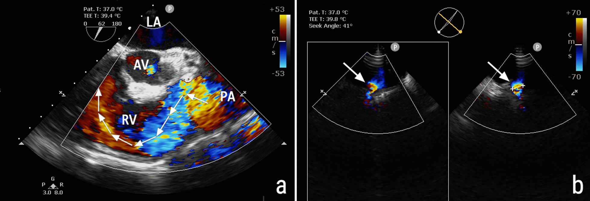
Background: Large pulmonary valve perforation, which is rarely seen with infective endocarditis, general atrophy, or congenital fenestration, often leads to potentially fatal outcomes, including heart failure.
Case presentation: Transthoracic and transesophageal echocardiographic evaluation of a 69-year-old woman revealed a severely eccentric pulmonary regurgitation with concomitant pulmonary valve stenosis, patent ductus arteriosus, patent foramen ovale, and pulmonary artery aneurysm. In the operation, a large perforation was found in the pulmonary valve leaflet. She underwent complicated surgery that involved closure of the congenital heart defects and replacement of a pulmonary valve with successful results. But the cause of her pulmonary valve perforation remained undetermined.
Conclusion: This case highlights two important points: the need for timely management of congenital heart disease and being aware of the possibility of pulmonary valve perforation, which in this case was indicated by an eccentric pulmonary regurgitant jet seen on echocardiography.
Keywords: Case report; Echocardiography; Infective endocarditis; Pulmonary valve.
© 2022. The Author(s).
Conflict of interest statement
The authors declare that they have no competing interests.
Figures

References
Publication types
MeSH terms
LinkOut - more resources
Full Text Sources
Medical
