. 2022 Apr 9;12(1):5999.
doi: 10.1038/s41598-022-10057-7.

The SARS-CoV-2 spike residues 616/644 and 1138/1169 delineate two antibody epitopes in COVID-19 mRNA COMINARTY vaccine (Pfizer/BioNTech)

Affiliations

The SARS-CoV-2 spike residues 616/644 and 1138/1169 delineate two antibody epitopes in COVID-19 mRNA COMINARTY vaccine (Pfizer/BioNTech)

Jessica Andries et al. Sci Rep. .

Erratum in

Abstract

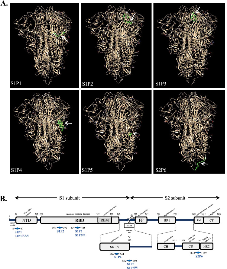
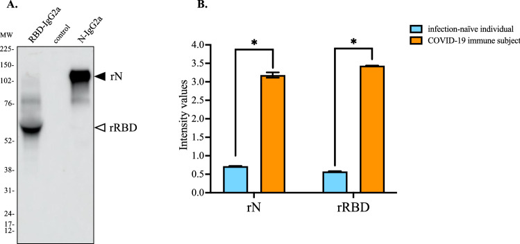
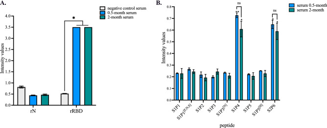
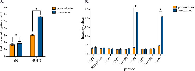
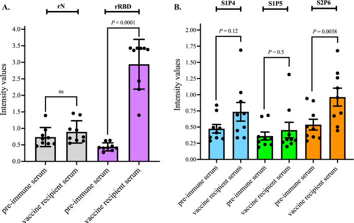
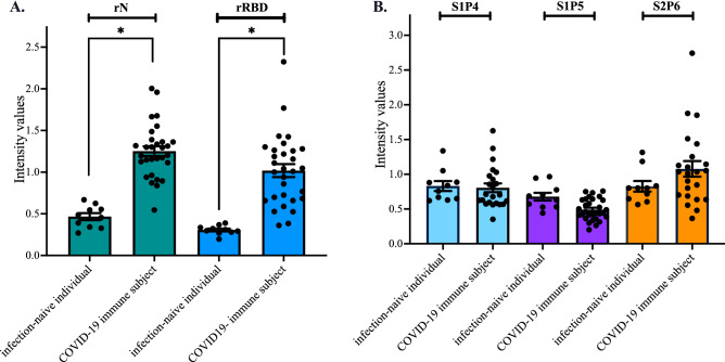
The newly identified coronavirus SARS-CoV-2 is responsible for the worldwide pandemic COVID-19. Considerable efforts have been devoted for the development of effective vaccine strategies against COVID-19. The SARS-CoV-2 spike protein has been identified as the major antigen candidate for the development of COVID-19 vaccines. The Pfizer-BioNTech COVID-19 vaccine COMIRNATY is a lipid nanoparticle-encapsulated mRNA encoding a full-length and prefusion-stabilized SARS-CoV-2 spike protein. In the present study, synthetic peptide-based ELISA assays were performed to identify linear B-cell epitopes into the spike protein that contribute to elicitation of antibody response in COMIRNATY-vaccinated individuals. The synthetic S2P6 peptide containing the spike residues 1138/1169 and to a lesser extent, the synthetic S1P4 peptide containing the spike residues 616/644 were recognized by the immune sera from COMIRNATY vaccine recipients but not COVID-19 recovered patients. We assume that the synthetic S2P6 peptide and to a lesser extent the synthetic S1P4 peptide, could be of interest to measure the dynamic of antibody response to COVID-19 mRNA vaccines. The S2P6 peptide has been identified as immunogenic in adult BALB/c mice that received protein-peptide conjugates in a prime-boost schedule. This raises the question on the role of the B-cell epitope peptide containing the SARS-CoV-2 spike residues 1138/1169 in protective efficacy of the Pfizer-BioNTech COVID-19 vaccine COMIRNATY.


Conflict of interest statement

All authors declare no conflict of interest.

Figures

Figure 1
Figure 1
Position of B-cell epitope peptides on SARS-CoV-2 spike protein. In (A), positions of the six B-cell epitope peptides colored in green on a single protomer of SARS-CoV-2 spike protein trimer (EMD data resource: EMD-2256). The blank arrow indicates their position on the protomer. In (B), schematic organization of SARS-CoV-2 spike protein with the S1 and S2 subunits and their different domains and motifs is shown. The six synthetic peptides S1P1, S1P2, S2P3, S1P4, S1P5, and S2P6 peptides and their variants S1P1[F,T,S], S1P3[N], and S1P5[H] are mapped on the SARS-CoV-2 spike protein.
Figure 2
Figure 2
Antigenic reactivity of recombinant N and RBD proteins with human serum-specific SARS-CoV-2 antibody. In (A), CHO cells were transfected 72 h with plasmids expressing RBD-IgG2A (rN) and N-IgG2A (rRBD) proteins or empty vector plasmid (control). Aliquots of clarified cell culture supernatants (C3S) were analyzed by immunoblot assay using goat anti-mouse IgG heavy chain HRP antibody. The original full-length gel is presented in Supplementary Figure S1. In (B), pools of four COVID-19 donor serum samples (Table S2, serum donors n°16, 18, 19, and 20) (COVID-19 donor serum) and four negative donor serum samples (Table S3) (negative donor serum) at dilution 1:100 were tested for anti-N and anti-rRBD antibodies by indirect ELISA using rN and rRBD for protein-based antibody capture. The intensity values of serum samples were measured at O.D. 450 nm. The results are the mean (± SEM) of three replicates. Unpaired t tests between COVID-19 immune subject and infection-naïve individual were performed and statistically significant comparisons are shown as * p < 10–4.
Figure 3
Figure 3
Antigenic reactivity of synthetic peptides with comirnaty vaccine recipient sera. Serum samples from an infection-naïve individual who received comirnaty vaccine in a 2-dose regimen were collected at 0.5 month or 2 months after the injection of a second dose. A pool of ten infection-naïve individuals collected in 2019 (Table S3) served as negative control serum. In (A), serum samples at dilution 1:100 were assessed for the detection of antibodies against SARS-CoV-2 N and S proteins by indirect ELISA using soluble rN and rRBD proteins for antigen-based antibody capture. The intensity values of serum samples were measured at O.D. 450 nm. The results are the mean (± SEM) of three replicates. Statistical comparisons were performed between serum samples. Statistically significant comparisons are shown as * p < 10–4 (ns: non-statistically significant, p > 0.05). In (B), the serum samples were assayed at dilution 1:50 on synthetic peptides and their variants (200 ng.ml−1) through synthetic peptide-based ELISA. The intensity values of vaccine serum sample were measured at O.D. 450 nm. The results are the mean (± SEM) of three replicates. Pairwise comparisons between peptides showed that the experimental points for S1P4 and S2P6 peptides are significantly different from the other peptides (** p < 10–4, * p < 10–3). The differences between 0.5 month and 2.0 month for the synthetic S1P4 and S2P6 peptides are non-statistically significant (ns, p > 0.05).
Figure 4
Figure 4
Antigenic reactivity of synthetic peptides with immune sera from a COVID-19 immune subject who received comirnaty vaccine. Serum samples from a COVID-19 patient were collected few weeks after recovery of SARS-CoV-2 infection (post-infection) and then two weeks after the injection of a single dose of comirnaty vaccine (vaccination). Vaccine administration was performed three months after COVID-19 recovery. In (A), serum samples at dilution 1:100 were assayed for the detection of antibodies against SARS-CoV-2 N and spike proteins by indirect ELISA using recombinant rN and rRBD proteins for antigen-based antibody capture. A pool of serum samples from infection-naïve individuals (Table S3) served as negative control serum. The intensity values of serum samples were measured at O.D. 450 nm and their immune reactivity was estimated as a fold increase of intensity values obtained with negative control serum. The results are the mean (± SEM) of three replicates. Statistically significant comparisons are shown as * p < 10–4 (ns: non-statistically significant, p > 0.05). In (B), serum samples at dilution 1:50 were assayed for the detection of specific antibodies through peptide-based ELISA. The intensity values of serum samples were measured at O.D. 450 nm. The results are the mean (± SEM) of three replicates. Paired t tests on experimental points between post-infection and vaccination immune were performed and statistically significant comparisons are shown as * p < 10–4. Differences between experimental points considered as non-statistically significant are not shown.
Figure 5
Figure 5
Immune reactivity of comirnaty vaccine recipient sera against the synthetic S1P4 and S2P6 peptides. Serum samples from comirnaty vaccine recipients (n = 9) (Table S4) were collected few weeks after the receipt of the second vaccine dose. The pre-immune serum of each individual was collected prior vaccination. In (A), serum samples at dilution 1:100 have been tested for SARS-CoV-2 N and S antibodies by indirect ELISA using soluble rN and rRBD proteins for antigen-based antibody capture. The intensity values of serum samples were measured at O.D. 450 nm. Paired t tests were performed between pre-immune serum and vaccine recipient serum (ns: non-statistically significant, p > 0.05). In (B), the serum samples were assayed on the synthetic S2P4 and S2P6 peptides (200 ng.ml−1) through peptide-based ELISA at serum dilution 1:50. The synthetic S1P5 peptide served as negative control serum. The intensity values of vaccine serum samples were measured at O.D. 450 nm. Paired t tests were performed between pre-immune serum and vaccine recipient serum.
Figure 6
Figure 6
Antigenic reactivity of the synthetic peptides with human serum-specific SARS-CoV-2 antibody. Serum samples from COVID-19 recovered patients (n = 30) (Table S2) and ten infection- naïve individuals (n = 10) (Table S3) were tested for the antibody reactivity against the N and RBD proteins through indirect ELISA (A), or the S1P4, S1P5, and S2P6 peptides through synthetic peptide-based ELISA (B). The intensity values of serum samples were measured at O.D. 450 nm. In (A), the serum samples at dilution 1:100 were assayed for the detection of antibodies against SARS-CoV-2 N and spike proteins by indirect ELISA using recombinant rN and rRBD proteins (1 µg.ml−1) for antigen-based antibody capture. The intensity values of serum samples were measured at O.D. 450 nm. Unpaired t tests between infection-naïve individuals and COVID-19 immune subjects were performed and statistically significant comparisons are shown as * p < 10–4. In (B), The serum samples (n = 23) among the COVID-19 recovered patients who developed higher anti-RBD antibody titers were assayed for the detection of antibodies against the S1P4, S1P5, and S2P6 peptides (200 ng.ml−1) through synthetic peptide-based ELISA at serum dilution 1:50. The results are the mean (± SEM) of three replicates. The intensity values of serum samples were measured at O.D. 450 nm. Unpaired t tests between infection-naïve individual and COVID-19 immune subject were performed and differences between experimental points considered as non-statistically significant (p > 0.05) are not shown.
Figure 7
Figure 7
Immune reactivity of mouse antibodies raised against B-cell epitope peptides. In (A), serum samples from mice (n = 5) that received the KLH-peptide conjugates were assessed for peptide-reactive antibodies through peptide-based ELISA using the synthetic S1P4, S1P5, and S2P6 peptides (200 ng.mL−1) for antigen-based antibody capture. The pre-immune serum of each individual that received the KLH-peptide conjugates was tested. The intensity values of serum samples at dilution 1:50 were measured at O.D. 450 nm. Paired t tests between pre-immune serum and KLH-peptide serum were performed (ns: non-statistically significant, p > 0.05). The results are representative of two independent experiments. In (B), synthetic peptide-based ELISA using the S2P6 and S2P6.2.0 peptides (200 ng.mL−1) for peptide-based antibody capture. At the left, pre-immune and immune serum samples of an immunized mouse with KLH-S2P6 conjugates. At the right, serum samples from a COVID-19 immune subject collected after recovery (post-infection) and after injection of a single dose of comirnaty vaccine (vaccination). Synthetic S1P5 peptide served as negative peptide control. The intensity values of serum samples in a dose-curve response were measured at O.D. 450 nm.

Similar articles

Cited by

References

    1. Gralinski LE, Menachery VD. Return of the coronavirus: 2019-nCoV. Viruses. 2020;12:135. doi: 10.3390/v12020135. - DOI - PMC - PubMed
    1. Wiersinga WJ, Rhodes A, Cheng AC, Peacock SJ, Prescott HC. Pathophysiology, transmission, diagnosis, and treatment of coronavirus disease 2019 (COVID-19: A review. JAMA. 2020;324:782–793. doi: 10.1001/jama.2020.12839. - DOI - PubMed
    1. Yuki K, Fujiogi M, Koutsiogiannaki S. COVID-19 pathophysiology: a review. Clin. Immunol. 2020;215:108427. doi: 10.1016/j.clim.2020.108427. - DOI - PMC - PubMed
    1. Parasher A. COVID-19: Current understanding of its pathophysiology, clinical presentation and treatment. Postgrad. Med. J. 2021;97:312–320. doi: 10.1136/postgradmedj-2020-138577. - DOI - PMC - PubMed
    1. Osuchowski MF, et al. The COVID-19 puzzle: Deciphering pathophysiology and phenotypes of a new disease entity. Lancet Respir. Med. 2021;9:622–642. doi: 10.1016/S2213-2600(21)00218-6. - DOI - PMC - PubMed