. 2022 May;298(5):101913.
doi: 10.1016/j.jbc.2022.101913. Epub 2022 Apr 7.

The dimerization mechanism of the N-terminal domain of spider silk proteins is conserved despite extensive sequence divergence

Affiliations

The dimerization mechanism of the N-terminal domain of spider silk proteins is conserved despite extensive sequence divergence

Médoune Sarr et al. J Biol Chem. 2022 May.

Abstract

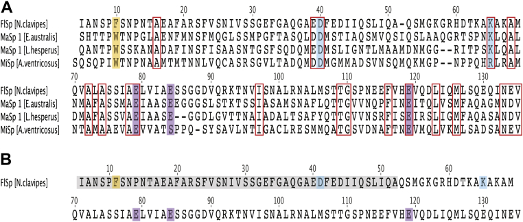
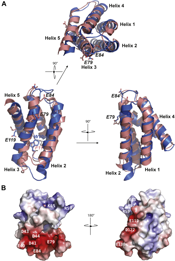
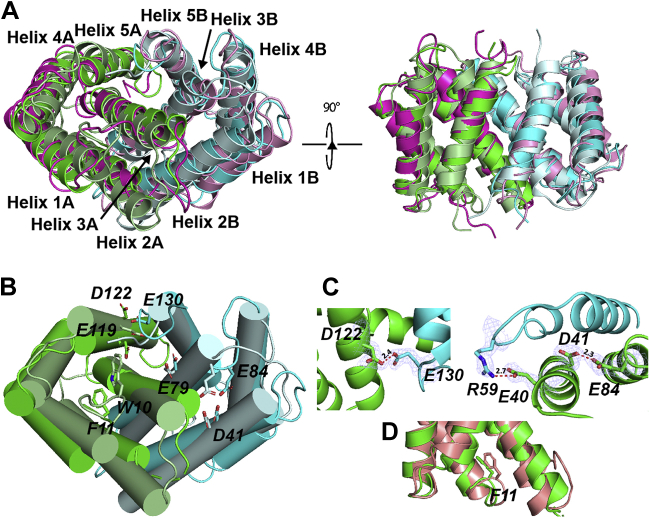
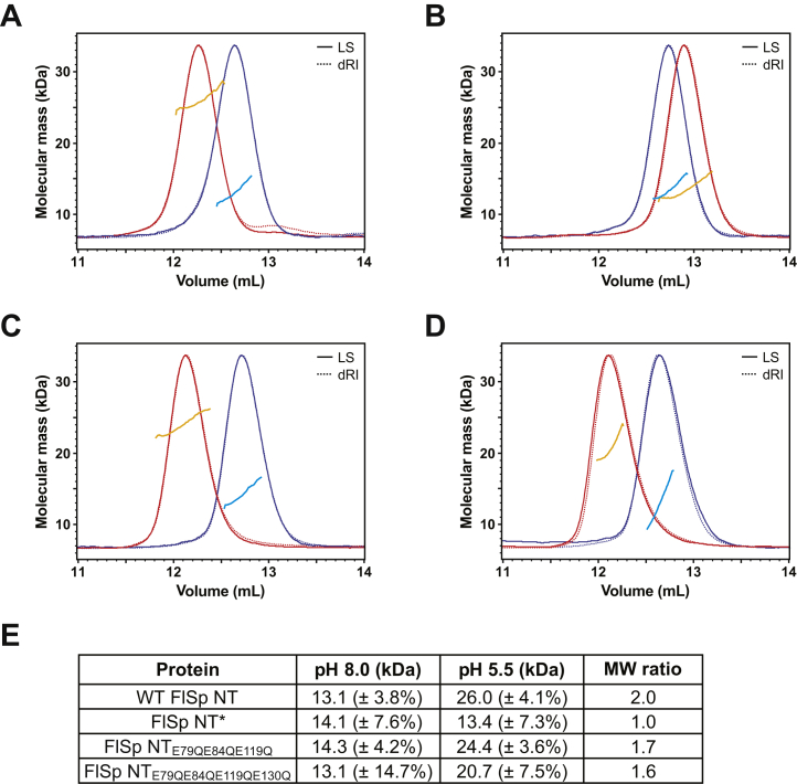
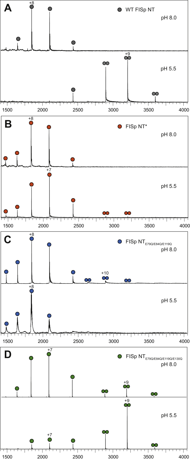
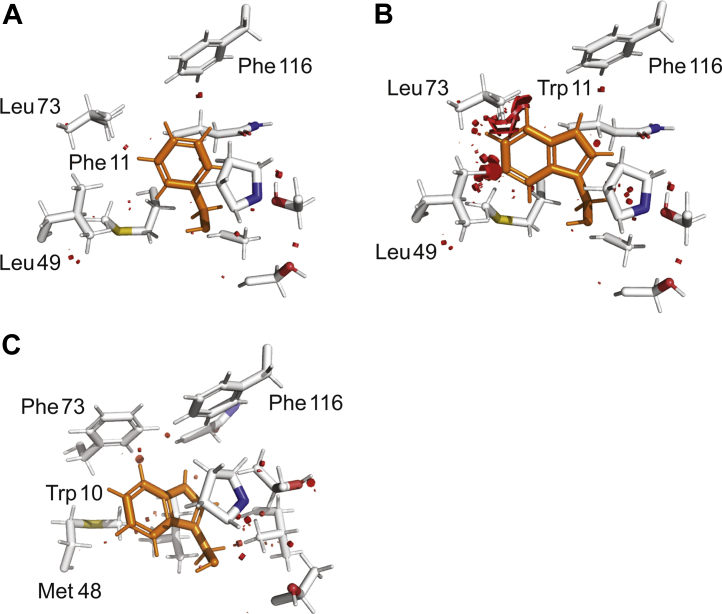
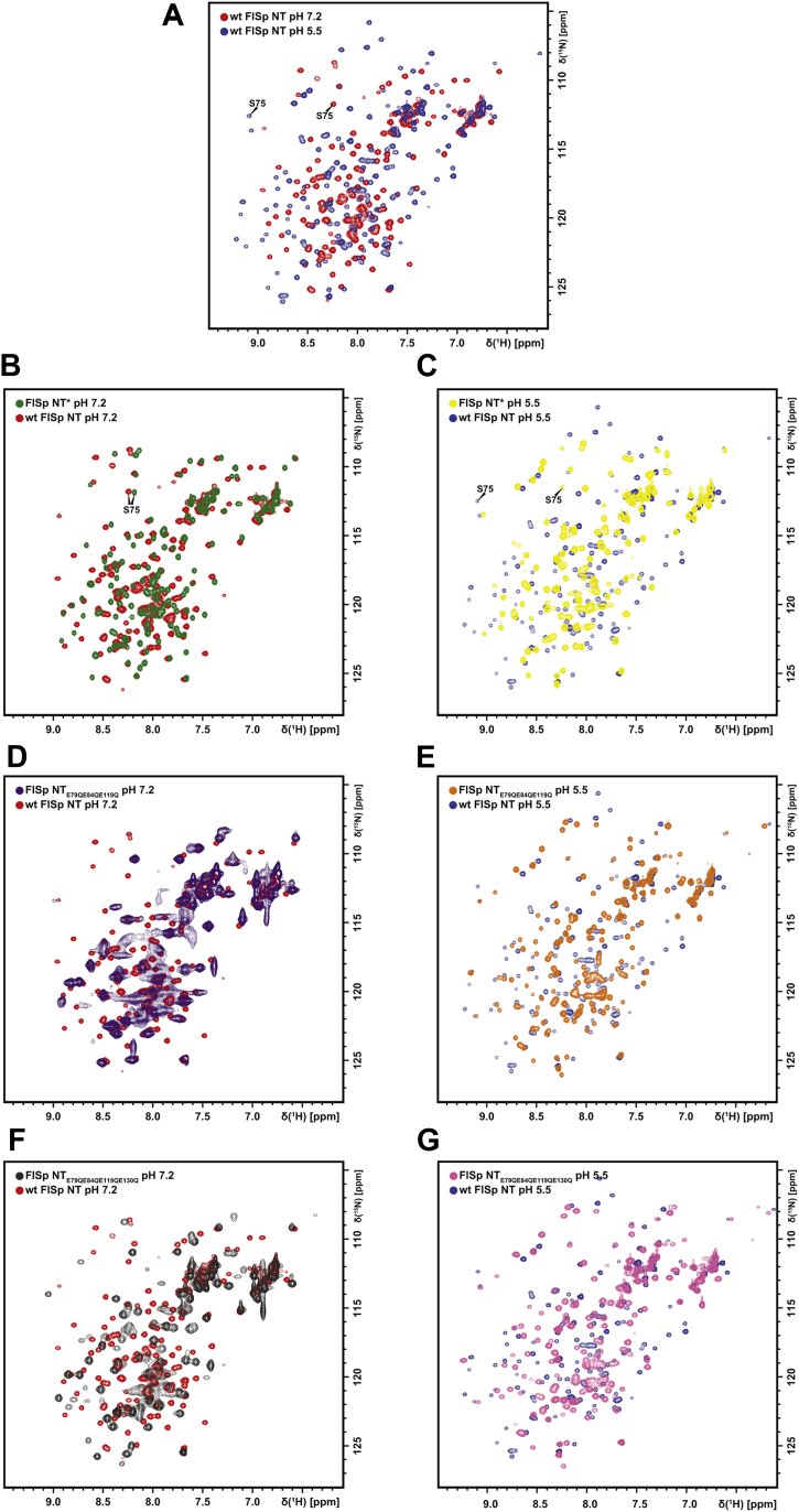
The N-terminal (NT) domain of spider silk proteins (spidroins) is crucial for their storage at high concentrations and also regulates silk assembly. NTs from the major ampullate spidroin (MaSp) and the minor ampullate spidroin are monomeric at neutral pH and confer solubility to spidroins, whereas at lower pH, they dimerize to interconnect spidroins in a fiber. This dimerization is known to result from modulation of electrostatic interactions by protonation of well-conserved glutamates, although it is undetermined if this mechanism applies to other spidroin types as well. Here, we determine the solution and crystal structures of the flagelliform spidroin NT, which shares only 35% identity with MaSp NT, and investigate the mechanisms of its dimerization. We show that flagelliform spidroin NT is structurally similar to MaSp NT and that the electrostatic intermolecular interaction between Asp 40 and Lys 65 residues is conserved. However, the protonation events involve a different set of residues than in MaSp, indicating that an overall mechanism of pH-dependent dimerization is conserved but can be mediated by different pathways in different silk types.

Keywords: NMR structure; X-ray structure; dimerization; protein domain; silk assembly; spidroin.


Conflict of interest statement

Conflict of interest The authors declare that they have no conflicts of interest with the contents of this article.

Figures

Figure 1
Figure 1
Comparison of N. clavipes FlSp NT with NT domains from other spidroins and spider species.A, sequence alignment of FlSp NT from N. clavipes with MaSp NT from E. australis and L. hesperus, and MiSp NT from A. ventricosus. Sequence alignment of all known spidroin NTs is shown in Fig. S1. The wedged Trp in MaSp and MiSp NT is substituted by a Phe in FlSp NT (orange shade). Positions with strictly conserved residues or conserved side chain charges are boxed in red. Mutated residues in MaSp NT constitutive monomer and dimer mutants are also indicated in the alignment. Asp 40 and Lys 65 are swapped in NT∗ (blue shade). E79, E84, and E119 are mutated to glutamines in NTE79QE84QE119Q (purple shade). B, the residue numbers used for FlSp NT in this paper differ by +1 at residues 5 to 51 (gray shade) compared to MaSp NT and the sequence alignment in (A). FlSp, flagelliform spidroin; MaSp, major ampullate spidroin; MiSp, minor ampullate spidroin; NT, N-terminal.
Figure 2
Figure 2
Comparison of FlSp NT and MaSp NT monomeric structures.A, superposition of structures of FlSp NT (salmon, PDB ID: 7A0I, this work) and MaSp NT (blue, PDB ID: 2LPJ) at pH 7.2. The side chains of Glu 79, Glu 84, Glu 119, and the side chain of Phe 11 in FlSp which corresponds to Trp 10 in MaSp are labeled and displayed as sticks. B, charge distribution of FlSp NT displayed in a surface representation. The negatively and positively charged residues are colored in red and blue, respectively. FlSp, flagelliform spidroin; MaSp, major ampullate spidroin; NT, N-terminal.
Figure 3
Figure 3
Comparison of FlSp NT and MaSp NT dimeric structures.A and B, superposition of structures of FlSp NT (X-ray structure in green/cyan and NMR in magenta/pink, PDB IDs: 7OOM and 7A0O, this work) and MaSp NT (pale green/pale cyan, PDB ID: 3LR2) at pH 5.5. In B, the side chains of Asp 41, Glu 79, Glu 84, Glu 119, Asp 122, Glu 130, and the side chain of Phe 11 in FlSp which corresponds to Trp 10 in MaSp are labeled and displayed as sticks. C, zoom-in on dimer formation promoting hydrogen bond interactions between protonated acidic residues (Glu 130 and Asp 122 of the other subunit, Glu 84 and Asp 41 of the same subunit, which in turn favors formation of salt bridge among Glu 40 and Arg 59 of the other subunit). Distances are shown in Å between interacting atoms. Electron density contoured at 1σ is shown for interacting residues. D, superposition of the NMR structure of FlSp NT monomer at pH 7.2 (salmon) and a subunit of FlSp NT dimer X-ray structure (green). The side chain of Phe 11 is labeled and displayed as sticks. FlSp, flagelliform spidroin; MaSp, major ampullate spidroin; NT, N-terminal.
Figure 4
Figure 4
Dimerization of FlSp NT analyzed by SEC-MALS. Proteins purified at pH 5.5 and pH 8.0 were analyzed using a Superdex 75 increase SEC column equilibrated at each pH. The chromatograms show the differential refractive index (dRI, broken lines) and light scattering signal (LS, solid lines) at pH 5.5 (red) and pH 8.0 (blue) together with the molecular mass calculated by MALS at pH 5.5 (orange) and pH 8.0 (cyan). Representative measurements are shown for (A) WT FlSp NT, (B) FlSp NT∗, (C) FlSp NTE79QE84QE119Q, and (D) FlSp NTE79QE84QE119QE130Q. MALS data for FlSp NTE79QE84QE119QE130Q were associated with higher uncertainty (%). E, the experimentally determined average molecular masses were used to calculate the molecular weight ratio between pH 5.5 and pH 8.0 for each protein. FlSp, flagelliform spidroin; MALS, multiangle light scattering; NT, N-terminal; SEC, size-exclusion chromatography.
Figure 5
Figure 5
Dimerization of FlSp NT analyzed by ESI-MS. Spectra were measured at pH 8.0 and 5.5 of (A) WT FlSp NT, (B) FlSp NT∗, (C) FlSp NTE79QE84QE119Q, and (D) FlSp NTE79QE84QE119QE130Q. ESI-MS, electrospray ionization mass spectrometry; FlSp, flagelliform spidroin; NT, N-terminal.
Figure 6
Figure 6
Close-up views of Trp/Phe pockets. Side chains surrounding (A) Phe 11 in FlSp NT, (B) modeled Trp 11 in FlSp NTTrp, and (C) Trp 10 in MaSp NT. The main unfavorable contacts are highlighted in red. FlSp, flagelliform spidroin; MaSp, major ampullate spidroin; NT, N-terminal.
Figure 7
Figure 7
HSQC NMR spectra of WT FlSp NT and mutants. Overlaid spectra of (A) WT FlSp NT at pH 7.2 (red) and pH 5.5 (blue), (B) FlSp NT∗ (green), and WT FlSp NT (red) at pH 7.2, (C) FlSp NT∗ (yellow) and WT FlSp NT (blue) at pH 5.5, (D) FlSp NTE79QE84QE119Q (purple) and WT FlSp NT (red) at pH 7.2, (E) FlSp NTE79QE84QE119Q (orange) and WT FlSp NT (blue) at pH 5.5, (F) FlSp NTE79QE84QE119QE130Q (gray) and WT FlSp NT (red) at pH 7.2, (G) FlSp NTE79QE84QE119QE130Q (pink) and WT FlSp NT (blue) at pH 5.5. FlSp, flagelliform spidroin; HSQC, heteronuclear single quantum coherence; NT, N-terminal.

References

    1. Peakall D.B. Synthesis of silk, mechanism and location. Am. Zool. 1969;9:71–79.
    1. Gosline J.M., Guerette P.A., Ortlepp C.S., Savage K.N. The mechanical design of spider silks: From fibroin sequence to mechanical function. J. Exp. Biol. 1999;202:3295–3303. - PubMed
    1. Bell A.L., Peakall D.B. Changes in fine structure during silk protein production in the ampullate gland of the spider Araneus sericatus. J. Cell Biol. 1969;42:284–295. - PMC - PubMed
    1. Casem M.L., Tran L.P., Moore A.M. Ultrastructure of the major ampullate gland of the black widow spider, Latrodectus hesperus. Tissue Cell. 2002;34:427–436. - PubMed
    1. Giesa T., Perry C.C., Buehler M.J. Secondary structure transition and critical stress for a model of spider silk assembly. Biomacromolecules. 2016;17:427–436. - PubMed

Publication types