Multicenter Study
. 2022 Jun 2;12(6):1435-1448.
doi: 10.1158/2159-8290.CD-21-0521.

PD-1 Blockade in Solid Tumors with Defects in Polymerase Epsilon

Affiliations
Multicenter Study

PD-1 Blockade in Solid Tumors with Defects in Polymerase Epsilon

Benoit Rousseau et al. Cancer Discov. .

Abstract

Missense mutations in the polymerase epsilon (POLE) gene have been reported to generate proofreading defects resulting in an ultramutated genome and to sensitize tumors to checkpoint blockade immunotherapy. However, many POLE-mutated tumors do not respond to such treatment. To better understand the link between POLE mutation variants and response to immunotherapy, we prospectively assessed the efficacy of nivolumab in a multicenter clinical trial in patients bearing advanced mismatch repair-proficient POLE-mutated solid tumors. We found that only tumors harboring selective POLE pathogenic mutations in the DNA binding or catalytic site of the exonuclease domain presented high mutational burden with a specific single-base substitution signature, high T-cell infiltrates, and a high response rate to anti-PD-1 monotherapy. This study illustrates how specific DNA repair defects sensitize to immunotherapy. POLE proofreading deficiency represents a novel agnostic biomarker for response to PD-1 checkpoint blockade therapy.

Significance: POLE proofreading deficiency leads to high tumor mutational burden with high tumor-infiltrating lymphocytes and predicts anti-PD-1 efficacy in mismatch repair-proficient tumors. Conversely, tumors harboring POLE mutations not affecting proofreading derived no benefit from PD-1 blockade. POLE proofreading deficiency is a new tissue-agnostic biomarker for cancer immunotherapy. This article is highlighted in the In This Issue feature, p. 1397.


Figures

Figure 1.
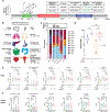
Figure 1.. Epidemiology and Immunogenomic landscape of solid tumors harboring POLE proofreading defects
A. Prevalence of POLE pathogenic missense variants leading to proofreading deficiency and other POLE molecular alterations in the MSK IMPACT Database from primary tumor samples (all stages) and from metastatic samples. POLE hotspot mutations cluster in the exonucleasic domain ranging from amino acid 268 to 471. B. Estimated prevalence of POLE proofreading deficiency in solid tumors based on results from the PanCancer TCGA and MSK-IMPACT databases. C. Prevalence of individual missense proofreading deficient variants according to microsatellite status, tumor primary in the PanCancer TCGA database. The upper panel represent the total number of samples in each group. D. Mutational burden according to microsatellite status and type of POLE alteration in the PanCancer TCGA database. POLE-pd: POLE proofreading deficientvariants;MSI-H: Microsatellite instability high E. Selected immune cell quantification and related genes according to TMB, microsatellite status and POLE alteration in the PanCancer TCGA database. Statistical comparisons are adjusted for multiple comparisons using POLE-d MSS tumors as a reference. Additional and numerical results are available in Supplementary table S4. Legend: MSI: Microsatellite Instable high tumors, TMB-H: Tumor mutational burden high (>10 mutation/Mb), POLE-pd: POLE proofreading deficient variant. From left to right: Cytotoxic T cells (MCPcounter), Th1 lymphocytes (XCELL), PDCD1: Programmed Cell Death 1 gene, CD274: Programmed Cell Death Ligand 1 gene, IFNG: Interferon Gamma gene. ns: non significant, >0.05 ; *: ≥0.05 and >0.01 ; **: ≤0.01 and >0.001, ***: ≤0.001 and >0.0001; ****≤0.0001. F. Selected immune cell quantification and related genes of CRC tumors from the COAD/READ database of the TCGA according to microsatellite and POLE-pd status. Statistical comparisons are adjusted for multiple comparisons using POLE-d MSS tumors as a reference. Additional and numerical results are available in Supplementary table S4. Legend: MSS: Microsatellite stable, MSI: Microsatellite Instable High, POLE: POLE proofreading deficient, MSS mutated tumors. From left to right: Cytotoxic T cells (MCPcounter), Th1 lymphocytes (XCELL), PDCD1: Programmed Cell Death 1 gene, CD274: Programmed Cell Death Ligand 1 gene, IFNG: Interferon Gamma gene. ns: non significant, >0.05 ; *: ≥0.05 and >0.01 ; **: ≤0.01 and >0.001, ***: ≤0.001 and >0.0001; ****≤0.0001.
Figure 2.
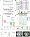
Figure 2.. Acsé Nivolumab trial : primary endpoint results for the POLE cohort
A. Flow chart of the Acsé Nivolumab POLE phase II trial showing how patients were selected from a nationwide screening strategy, through the eligibility and pathogenicity assessment of each POLE variants by an ad hoc molecular board (4 molecular biologists, 1 medical oncologist), to the two-stages phase II trial with Bayesian enrichment design. B. Summary of clinico-demographic characteristics of enrolled patients, in total and according to their proofreading category. Legend: POLE-pp: POLE proofreading proficient; VUS: Variants of Unknown significance; POLE-pd: POLE proofreading deficient. C. POLE variants and pathogenicity according to primary tumor. Legend: POLE-pd: POLE proofreading deficient variant, +: yes, -: no, ?: Variant of Unknown Significance. Of note, one variant N363K is germline. #: one patient with a colorectal cancer and S459F POLE-pd variant was included in the trial but didn’t receive Nivolumab because of rapid clinical deterioration and is further excluded from efficacy analyses. D. Best response of target lesions according to IRC RECIST 1.1 assessment presented by primary tumor, and according to molecular features. POLE-proofreading category, Tumor mutational burden and COSMIC POLE Single base substitution signature. Legend: POLE-pd: POLE proofreading deficient variant, +: yes, -: no, ?: Variant of Unknown Significance. TMB high: Tumor mutational Burden high +: TMB ≥10 mt/Mb; -: TMB <10 mt/Mb; NA: Non Available. POLE SBS: proportion of POLE related single base substitution assessed by whole exome sequencing, +: POLE SBS signature >60%; ±: POLE SBS signature >1% and ≤60%; -: POLE SBS signature of 0%; #: low cellularity of the sample; NA: Non Available. CR : Complete Response; PR : Partial Response; SD : Stable Disease; PD : Progressive Disease. Three patients experienced early clinical progression and one withdrew consent after cycle 1 of nivolumab and didn’t perform a CT Scan, so the total number of patients displayed is 16. E. Volume change of target lesions at each radiological assessment according to IRC RECIST 1.1 presented by primary tumor. Three patients experienced early clinical progression and one withdrew consent after cycle 1 of nivolumab and didn’t perform a CT Scan, so the total number of patients displayed is 16. F. Radiologic responses according to IRC RECIST 1.1 assessment at 12W (Primary endpoint) and best response in the overall cohort. ORR at 12 W: Overall response rate according to IRC RECIST 1.1; DCR at 12 W : Disease Control Rate at 12 W according to IRC RECIST 1.1; PD: Progressive Disease. One patient withdrew consent after one cycle of Nivolumab and was not assessable for response. * three patients who experienced early clinical progression have been classified as progressors. G. Progression-free survival curve from initiation of Nivolumab in the overall POLE cohort (N=20). H. Overall survival curve from initiation of Nivolumab in the overall POLE cohort (N=20). I. Abdominal CT scan showing pretreatment and 12 W post nivolumab nearly complete response of a patient with unresectable locally advanced transverse colon cancer with a POLE-pd P286R variant.
Figure 3.
Figure 3.. POLE proofreading deficiency drives benefit from Nivolumab
A. Swimmer plot of overall survival, time to best response and time to progression according to primary, POLE variant proofreading category and TMB. Legend: POLE-pd: POLE proofreading proficient, VUS: Variant of Unknown Significance, POLE-pp: POLE proofreading proficient. TMB: Tumor mutational Burden +: TMB ≥10 mt/Mb; -: TMB <10 mt/Mb; NA: Non Available. CR: Complete Response, PR: Partial Response, SD: Stable Disease, PD: Clinical or radiological progressive disease, SAE: Serious Adverse Event. B. Assessment of IRC RECIST 1.1 best response with molecular features. From left to right panel: TMB: tumor mutational burden, POLE SBS: POLE related single base substitution COMSIC signature, POLE signature TMB: TMB value imputable to POLE SBS signature calculated as the product of % of POLE SBS signature and TMB of a specific tumor. P-values have been calculated using Fisher’s test. The patient with the V464A VUS variant has not been considered for these analyses because of low tumor purity. ns: non significant, >0.05; *: ≥0.05 and >0.01; **: ≤0.01 and >0.001, ***: ≤0.001 and >0.0001; ****≤0.0001. C. Progression-free survival and overall survival curves from start of Nivolumab comparing outcomes of patients according to their tumor Tumor Mutational Burden. P-value has been computed by log-rank test. TMB: Tumor Mutational Burden low: <10 mt/Mb; high: ≥ 10 mt/Mb; Unknown: Not Available. D. Main oncologic outcomes according to POLE variant proofreading category assessed by the molecular tumor board. POLE-pd: POLE proofreading proficient, VUS: Variant of Unknown Significance, POLE-pp: POLE proofreading proficient.; ORR at 12 W : Overall response rate according to IRC RECIST 1.1; DCR at 12 W : Disease Control Rate at 12 W according to IRC RECIST 1.1; PD: Progressive Disease. The three patients without assessment at 12W have been classified as progressors. mPFS : median progression-free survival; mOS : median overall survival; HR (CI95%) : Hazard Ratio (95% Confidence interval) corresponding to Cox model comparison of non-pathogenic vs pathogenic/VUS group. All patients that received at least one dose of Nivolumab are considered for survival outcome assessment (N=20). *: one patient withdrew consent at 2 weeks after one cycle of Nivolumab and is not accounted for response, but is accounted for survival analyses. E. Progression-free survival and overall survival curves from start of Nivolumab comparing outcomes of patients according to their tumor POLE proofreading category. POLE-pd: POLE proofreading proficient, VUS: Variant of Unknown Significance, POLE-pp: POLE proofreading proficient. P-value has been computed by log-rank test. F. Individual responses for each POLE variant classified asPOLE-pd, VUS orPOLE-pp, and corresponding median TMB for each category. Legend: POLE-pd: POLE proofreading proficient, VUS: Variant of Unknown Significance, POLE-pp: POLE proofreading proficient; mTMB: median Tumor Mutational Burden; CR: Complete Response; PR: Partial Response; SD: Stable Disease; PD: Progressive Disease; NA: Not Assessed. $: Patient with the V464A variant had low TMB with low tumor purity.
Figure 4.
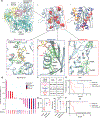
Figure 4.. In silico analyses reveal two tridimensional hotspots sites correlating with benefit from PD-1 blockade
A. In silico model of mutations located in the Exo domain. (a) Overall structure of POLE showing all the domains with the exception of the thumbs domain for clarity and zoom into the Exo domain in the same orientation. The DNA is modeled in the Editing conformation. (b) Exo Domain with mutations represented in red spheres (c) topology of the Exo domain, with the β strands numbered in square boxes and α helices in rounded boxes. The α helices contributing to the catalytic site (2 and 7) are colored orange and red. Mutations, represented in green, (d) found in the DNA binding groove (e) found in the hydrophobic core near the catalytic site (f) in surface residues mutations away from the functional sites, expected to have a neutral impact. The exonuclease domain (Figure 4A a to c) has a β1 β2 β3 β4 β5 α1 β6 α2 α3 β7 α4 α5 α6 α7 topology. The β strands form a central β-sheet, and the C-terminal α7 helix (red in Figure 4A.c) packs on the interior side of the β-sheet to form the main hydrophobic core of the protein. The metal binding residues of the catalytic site are located on β1, β2, α2 (orange in Figure 3A.c) and α7. The predicted DNA binding site is located in the groove lining the active site, with α2 and α7 on the left and right and the β1 β2 loop and α4 α5 bundle on the top and bottom in the orientation of Figure 4A.c. The P286, N363 and V411 residues, located in the β1-β2 loop, in α2 and in α4 respectively, line the DNA binding site in the Exo domain (Figure 4A.d and S7). Mutations of these residues are predicted to either interfere directly with DNA binding (N363K) or indirectly by destabilizing structural elements in contact with the DNA (P286R and V411L). The N363 residue is also near the metal binding site (in the same helix as D368), and the mutation to a basic lysine residue could also destabilize the metal binding of the acidic active site residues. The S297, G330, S459, A463 and V464 residues cluster in the hydrophobic core formed between the α7 helix and the β sheet (Figure 4A.e and S8). In addition to the general destabilization of the protein by disruption of the hydrophobic core, the close proximity of these mutations with the metal binding residues of the β1 (S459, E277) and α7 (D462) residues will have direct effect of the catalytic activity of the enzyme. This region appears as a mutation hotspot with pathogenic consequences. K425, D435 and R446 are surface residues far away from the active site (Figure 4A.f and S9). The mutations of these residues could disrupt salt bridges with neighboring residues but are not predicted to directly interfere with exonuclease function. R47 and W775 are not found in the exonuclease domain but belong to the N-terminal and thumb/fingers domains respectively (Supplementary Figure S9). None of these mutations is predicted to impact the function of the Exo Domain. B. Summary of best response observed in Acsé trial according to POLE variant tridimensional site predicted by in silico analyses: DNA binding site, Catalytic site or Neutral site. C. Radiologic best responses according to IRC RECIST 1.1 assessment for tridimensional categories of POLE variants in DNA binding, Catalytic or Neutral site. ORR: Overall Response Rate; DCR: Disease Control Rate. The three patients who experienced early clinical progression have been classified as progressors all in the neutral site category. One patient withdrew consent after one cycle of Nivolumab and was not assessable for response (catalytic site variant). D. Correlation of molecular features with tridimensional categories. From left to right panel: TMB: tumor mutational burden, POLE SBS: POLE related single base substitution COMSIC signature, POLE signature TMB: TMB value imputable to POLE SBS signature calculated as the product of % of POLE SBS signature and TMB of a specific tumor. P-values have been calculated using Fisher’s test. The patient with the V464A VUS variant has not been considered for these analyses because of low tumor purity. ns: non significant, >0.05; *: ≥0.05 and >0.01; **: ≤0.01 and >0.001, ***: ≤0.001 and >0.0001; ****≤0.0001. E. Progression-free survival observed in Acsé trial according to POLE variant site predicted by in silico analyses: DNA binding site, Catalytic site or away from these sites. P-value corresponds to a log-rank test. F. Overall survival observed in Acsé trial according to POLE variant site predicted by in silico analyses: DNA binding site, Catalytic site or away from these sites. P-value corresponds to a log-rank test.

References

    1. Rayner E, van Gool IC, Palles C, Kearsey SE, Bosse T, Tomlinson I, et al. A panoply of errors: polymerase proofreading domain mutations in cancer. Nat Rev Cancer. 2016;16:71–81. - PubMed
    1. Campbell BB, Light N, Fabrizio D, Zatzman M, Fuligni F, de Borja R, et al. Comprehensive Analysis of Hypermutation in Human Cancer. Cell. 2017;171:1042–1056.e10. - PMC - PubMed
    1. Chalmers ZR, Connelly CF, Fabrizio D, Gay L, Ali SM, Ennis R, et al. Analysis of 100,000 human cancer genomes reveals the landscape of tumor mutational burden. Genome Medicine. 2017;9:34. - PMC - PubMed
    1. Alexandrov LB, Kim J, Haradhvala NJ, Huang MN, Tian Ng AW, Wu Y, et al. The repertoire of mutational signatures in human cancer. Nature. Nature Publishing Group; 2020;578:94–101. - PMC - PubMed
    1. Hodel KP, Sun MJS, Ungerleider N, Park VS, Williams LG, Bauer DL, et al. POLE Mutation Spectra Are Shaped by the Mutant Allele Identity, Its Abundance, and Mismatch Repair Status. Molecular Cell. 2020;78:1166–1177.e6. - PMC - PubMed

Publication types

MeSH terms