

Matrix metalloproteinase-21 promotes metastasis via increasing the recruitment and M2 polarization of macrophages in HCC
- PMID: 35398966
- PMCID: PMC9899621
- DOI: 10.1111/cas.15368
Matrix metalloproteinase-21 promotes metastasis via increasing the recruitment and M2 polarization of macrophages in HCC
Abstract
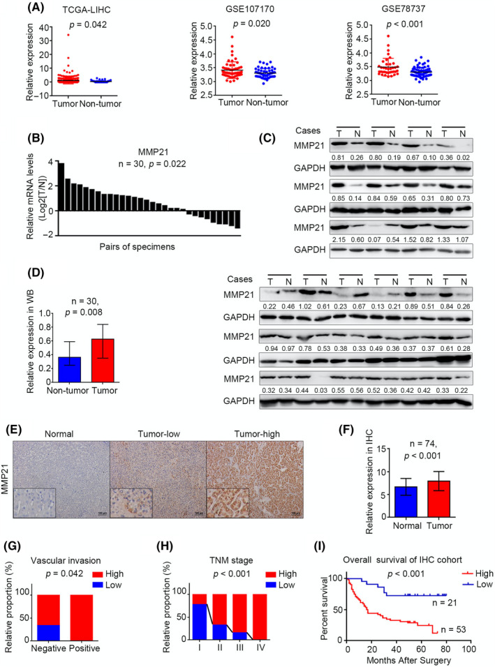
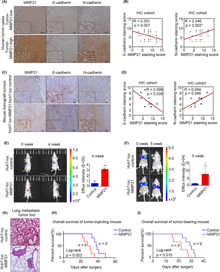
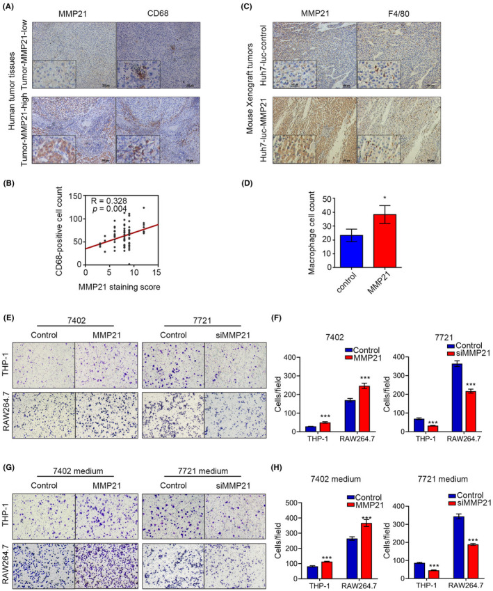
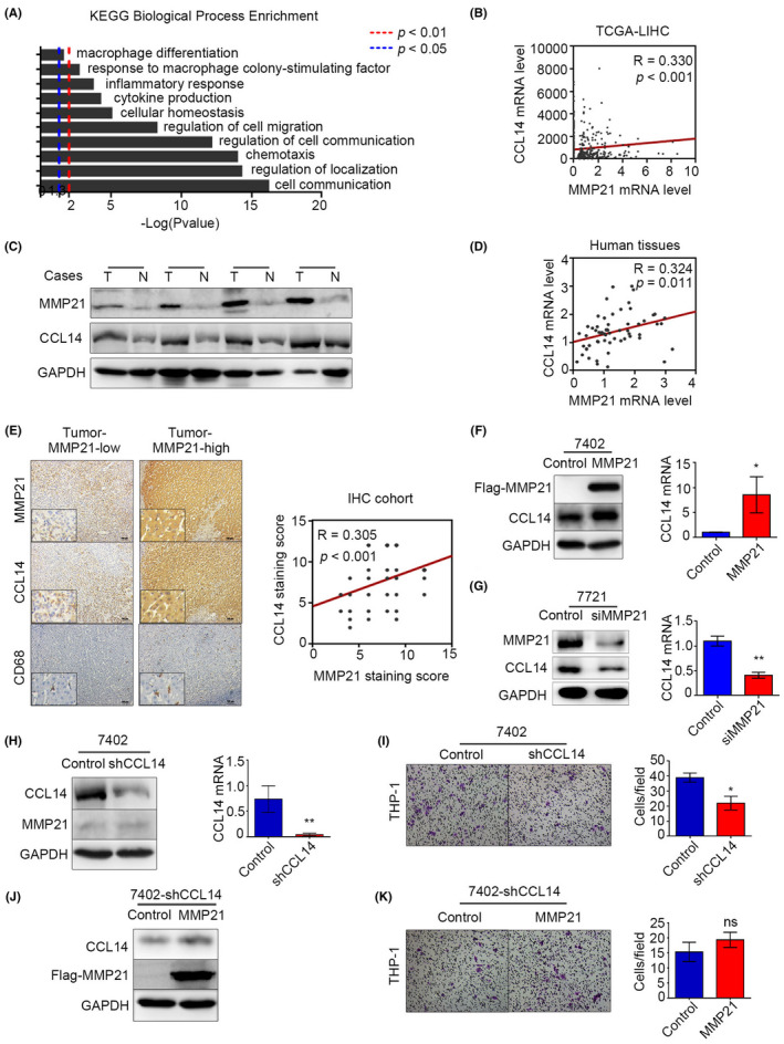
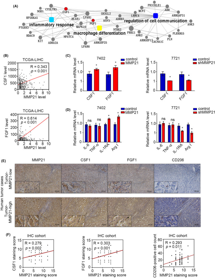
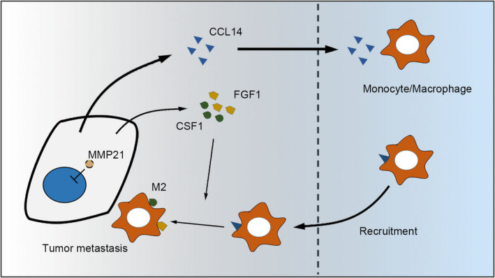
MMP-21 is a newly identified member of the matrix metalloproteinase family and has been reported to regulate both embryonic development and tumor progression. However, the roles of MMP-21 in hemofiltrate C-C chemokine (HCC) remain largely unclear. In this study, we used western blot, qPCR and immunohistochemistry (IHC) to determine the upregulation of MMP-21 in HCC tissues, and showed that the increase in MMP-21 was associated with vascular invasion and poor prognosis. Although changing levels of MMP-21 in HCC cell lines had no significant effect on cell migration or invasion abilities in in vitro transwell tests, both IHC analysis and in vivo mouse models proved that upregulated MMP-21 promoted metastasis. Functional enrichments of MMP-21 using The Cancer Genome Atlas (TCGA) data suggested that MMP-21 might regulate metastasis via macrophages. Further experiments proved that MMP-21 enhanced macrophage recruitment by increasing CCL-14 levels and promoted M2-type polarization of macrophage by elevating the expression of CSF-1 and FGF-1. Taken together, this study revealed that MMP-21 controlled the tumor microenvironment remodeling and functional regulation of macrophages to regulate HCC metastasis.
Keywords: CCL-14; CSF-1; FGF-1; HCC; MMP-21; macrophage; metastasis.
© 2022 The Authors. Cancer Science published by John Wiley & Sons Australia, Ltd on behalf of Japanese Cancer Association.
Conflict of interest statement
The authors declare that they have no competing interests.
Figures
Similar articles
-
Exosomal DLX6-AS1 from hepatocellular carcinoma cells induces M2 macrophage polarization to promote migration and invasion in hepatocellular carcinoma through microRNA-15a-5p/CXCL17 axis.J Exp Clin Cancer Res. 2021 May 26;40(1):177. doi: 10.1186/s13046-021-01973-z. J Exp Clin Cancer Res. 2021. Retraction in: J Exp Clin Cancer Res. 2022 Apr 9;41(1):134. doi: 10.1186/s13046-022-02353-x. PMID: 34039401 Free PMC article. Retracted.
-
ADAM17-regulated CX3CL1 expression produced by bone marrow endothelial cells promotes spinal metastasis from hepatocellular carcinoma.Int J Oncol. 2020 Jul;57(1):249-263. doi: 10.3892/ijo.2020.5045. Epub 2020 Apr 13. Int J Oncol. 2020. PMID: 32319605 Free PMC article.
-
Promotion of hepatocellular carcinoma metastasis through matrix metalloproteinase activation by epithelial-mesenchymal transition regulator Twist1.J Cell Mol Med. 2011 Mar;15(3):691-700. doi: 10.1111/j.1582-4934.2010.01052.x. J Cell Mol Med. 2011. PMID: 20219012 Free PMC article.
-
Suppressive effects of microRNA-16 on the proliferation, invasion and metastasis of hepatocellular carcinoma cells.Int J Mol Med. 2015 Dec;36(6):1713-9. doi: 10.3892/ijmm.2015.2379. Epub 2015 Oct 16. Int J Mol Med. 2015. PMID: 26499886
-
Fibulin-5 inhibits hepatocellular carcinoma cell migration and invasion by down-regulating matrix metalloproteinase-7 expression.BMC Cancer. 2014 Dec 12;14:938. doi: 10.1186/1471-2407-14-938. BMC Cancer. 2014. PMID: 25494879 Free PMC article.
Cited by
-
Mechanisms of tumor-associated macrophages affecting the progression of hepatocellular carcinoma.Front Pharmacol. 2023 Aug 17;14:1217400. doi: 10.3389/fphar.2023.1217400. eCollection 2023. Front Pharmacol. 2023. PMID: 37663266 Free PMC article. Review.
-
Role of exosomes in hepatocellular carcinoma and the regulation of traditional Chinese medicine.Front Pharmacol. 2023 Jan 17;14:1110922. doi: 10.3389/fphar.2023.1110922. eCollection 2023. Front Pharmacol. 2023. PMID: 36733504 Free PMC article. Review.
-
Integration of histopathological images and immunological analysis to predict M2 macrophage infiltration and prognosis in patients with serous ovarian cancer.Front Immunol. 2025 Mar 17;16:1505509. doi: 10.3389/fimmu.2025.1505509. eCollection 2025. Front Immunol. 2025. PMID: 40165975 Free PMC article.
-
Prognostic and Immune Landscape Analysis of Ubiquitination-related Genes in Hepatocellular Carcinoma: Based on Bulk and Single-cell RNA Sequencing Data.J Cancer. 2024 Mar 11;15(9):2580-2600. doi: 10.7150/jca.93425. eCollection 2024. J Cancer. 2024. PMID: 38577593 Free PMC article.
-
Cancer-associated fibroblast-derived MMP11 promotes tumor progression in pancreatic cancer.Cancer Sci. 2025 Mar;116(3):643-655. doi: 10.1111/cas.16418. Epub 2024 Dec 5. Cancer Sci. 2025. PMID: 39639765 Free PMC article.
References
-
- Bray F, Ferlay J, Soerjomataram I, Siegel RL, Torre LA, Jemal A. Global cancer statistics 2018: GLOBOCAN estimates of incidence and mortality worldwide for 36 cancers in 185 countries. CA Cancer J Clin. 2018;68(6):394‐424. - PubMed
-
- Govaere O, Roskams T. Pathogenesis and prognosis of hepatocellular carcinoma at the cellular and molecular levels. Clin Liver Dis. 2015;19(2):261‐276. - PubMed
-
- Malaguarnera G, Giordano M, Paladina I, Berretta M, Cappellani A, Malaguarnera M. Serum markers of hepatocellular carcinoma. Dig Dis Sci. 2010;55(10):2744‐2755. - PubMed
-
- Visse R, Nagase H. Matrix metalloproteinases and tissue inhibitors of metalloproteinases: structure, function, and biochemistry. Circ Res. 2003;92(8):827‐839. - PubMed
MeSH terms
Substances
LinkOut - more resources
Full Text Sources
Medical
Molecular Biology Databases
Research Materials
Miscellaneous
