. 2022 Mar 25:13:841076.
doi: 10.3389/fphys.2022.841076. eCollection 2022.

Arterial Baroreflex Inhibits Muscle Metaboreflex Induced Increases in Effective Arterial Elastance: Implications for Ventricular-Vascular Coupling

Affiliations

Arterial Baroreflex Inhibits Muscle Metaboreflex Induced Increases in Effective Arterial Elastance: Implications for Ventricular-Vascular Coupling

Joseph Mannozzi et al. Front Physiol. .

Abstract

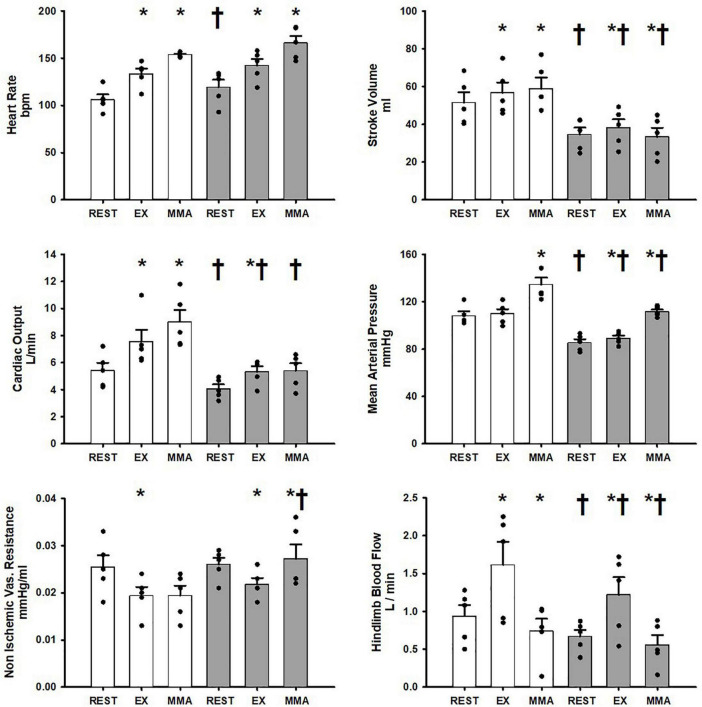
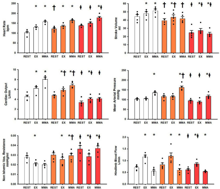
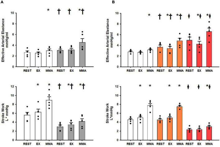
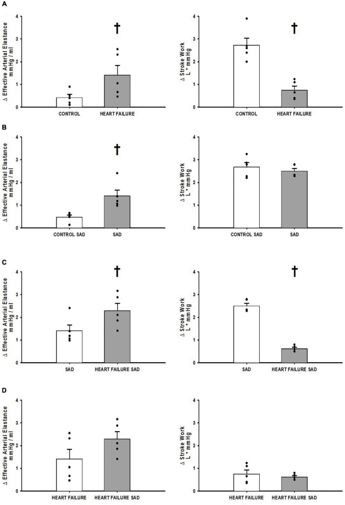
The ventricular-vascular relationship assesses the efficacy of energy transferred from the left ventricle to the systemic circulation and is quantified as the ratio of effective arterial elastance to maximal left ventricular elastance. This relationship is maintained during exercise via reflex increases in cardiovascular performance raising both arterial and ventricular elastance in parallel. These changes are, in part, due to reflexes engendered by activation of metabosensitive skeletal muscle afferents-termed the muscle metaboreflex. However, in heart failure, ventricular-vascular uncoupling is apparent and muscle metaboreflex activation worsens this relationship through enhanced systemic vasoconstriction markedly increasing effective arterial elastance which is unaccompanied by substantial increases in ventricular function. This enhanced arterial vasoconstriction is, in part, due to significant reductions in cardiac performance induced by heart failure causing over-stimulation of the metaboreflex due to under perfusion of active skeletal muscle, but also as a result of reduced baroreflex buffering of the muscle metaboreflex-induced peripheral sympatho-activation. To what extent the arterial baroreflex modifies the metaboreflex-induced changes in effective arterial elastance is unknown. We investigated in chronically instrumented conscious canines if removal of baroreflex input via sino-aortic baroreceptor denervation (SAD) would significantly enhance effective arterial elastance in normal animals and whether this would be amplified after induction of heart failure. We observed that effective arterial elastance (Ea), was significantly increased during muscle metaboreflex activation after SAD (0.4 ± 0.1 mmHg/mL to 1.4 ± 0.3 mmHg/mL). In heart failure, metaboreflex activation caused exaggerated increases in Ea and in this setting, SAD significantly increased the rise in Ea elicited by muscle metaboreflex activation (1.3 ± 0.3 mmHg/mL to 2.3 ± 0.3 mmHg/mL). Thus, we conclude that the arterial baroreflex does buffer muscle metaboreflex induced increases in Ea and this buffering likely has effects on the ventricular-vascular coupling.

Keywords: arterial baroreflex; effective arterial elastance (Ea); muscle metaboreflex activation; neural control of cardiovascular system; ventricular vascular coupling.


Conflict of interest statement

The authors declare that the research was conducted in the absence of any commercial or financial relationships that could be construed as a potential conflict of interest.

Figures

FIGURE 1
FIGURE 1
Average 1-min steady state hemodynamics from the Controlgroup at rest, exercise (EX), and exercise with muscle metaboreflex activation (MMA) before (white) and after induction of heart failure (gray). Data are reported as means with standard error. Observed data points are overlain on corresponding bar graphs. Statistical significance vs. previous workload is depicted as * and vs. previous condition as where P < 0.05. (N = 5).
FIGURE 2
FIGURE 2
Average 1-min steady state hemodynamics from the SADgroup at rest, exercise (EX), and exercise with muscle metaboreflex activation (MMA) before (white) an after SAD (orange) and after induction of heart failure post SAD (red). Data are reported as means with standard error. Observed data points are overlain on corresponding bar graphs. Statistical significance vs. previous workload depicted as * where P < 0.05. Comparisons of the condition of SAD vs. control depicted as where P < 0.05. Comparisons of the condition of heart failure vs. SAD depicted as where P < 0.05. (N = 5).
FIGURE 3
FIGURE 3
(A) Average 1-min steady state values of Effective Arterial Elastance and Stroke Work in the Controlgroup at rest, exercise (EX), and exercise with muscle metaboreflex activation (MMA) before (white) and after induction of heart failure (gray). Statistical significance vs. previous workload depicted * and vs. previous condition as where P < 0.05. (N = 5). (B) Average 1-min steady state values of Effective Arterial Elastance and Stroke Work in the SADgroup at res, exercise (EX), and exercise with muscle metaboreflex activation (MMA) before (white) and after SAD (orange), and after heart failure induction post SAD (red). Statistical significance vs. previous workload depicted as * where P < 0.05. Comparisons of the condition of SAD vs. control depicted as where P < 0.05. Comparisons of the condition of heart failure vs. SAD depicted as where P < 0.05. (N = 5) Data for (A,B) are reported as means with standard error. Observed data points are overlain on corresponding bar graphs.
FIGURE 4
FIGURE 4
(A) The relative change in Effective Arterial Elastance and Stroke Work in the transition from exercise to exercise with muscle metaboreflex activation before (white) and after induction of heart failure (gray) in the Controlgroup. (B) The relative change in Effective Arterial Elastance and Stroke Work in the transition from exercise to exercise with muscle metaboreflex activation before (white) and after SAD (gray) in the SADgroup. (C) The relative change in Effective Arterial Elastance and Stroke Work in the transition from exercise to exercise with muscle metaboreflex activation in normal animals with SAD (white) and after induction of heart failure post SAD (gray) in the SADgroup. (D) The relative change in Effective Arterial Elastance and Stroke Work in the transition from exercise to exercise with muscle metaboreflex activation in heart failure animals from the Controlgroup (white) and heart failure animals post SAD from the SADgroup (gray). For all panels data are reported as means with standard error where data points are overlain on corresponding bar graphs. Statistical significance vs. the previous state as where P < 0.05. An N = 5 was used for each bar graph in every panel.
FIGURE 5
FIGURE 5
(A) Average 1-min steady state values of central venous pressure from the Controlgroup at rest, exercise (EX), and exercise with muscle metaboreflex activation (MMA) before (white) and after induction of heart failure (gray). Data are reported as means with standard error. Observed data points are overlain on corresponding bar graphs. Statistical significance vs. previous workload is depicted as * and vs. previous condition as where P < 0.05. (N = 5). (B) Average 1-min steady state values of central venous pressure from the SADgroup at rest, exercise (EX), and exercise with muscle metaboreflex activation (MMA) after SAD (orange) and after induction of heart failure post SAD (red). Data are reported as means with standard error. Observed data points are overlain on corresponding bar graphs. Statistical significance vs. previous workload depicted as * where P < 0.05. Comparisons of the condition of SAD vs. Heart failure SAD depicted as where P < 0.05. (N = 5).
FIGURE 6
FIGURE 6
Relationship between ventricular maximal elastance (Emax) and stroke work before (black circles) and after induction of heart failure (white circles) at rest, mild exercise, and muscle metaboreflex activation. A linear regression (dotted line) was performed on these points. and the R2 value are shown on the plot. Error bars depict standard error of the mean in both directions (Data points were calculated from primary data collected in experiments which were reported in Coutsos et al. (2010, 2013) (N = 6).

References

    1. Adreani C. M., Hill J. M., Kaufman M. P. (1985). Responses of group III and IV muscle afferents to dynamic exercise. J. Appl. Physiol. 82 1811–1817. 10.1152/jappl.1997.82.6.1811 - DOI - PubMed
    1. Al-Khateeb A. A., Limberg J. K., Barnes J. N., Joyner M. J., Charkoudian N., Curry T. B. (2017). Acute cyclooxygenase inhibition and baroreflex sensitivity in lean and obese adults. Clin. Auton. Res. 27 17–23. 10.1007/s10286-016-0389-z - DOI - PMC - PubMed
    1. Alam M. S. F. (1937). Observations in man upon a blood pressure raising reflex arising from the voluntary muscles. J. Physiol. 89 372–383. 10.1113/jphysiol.1937.sp003485 - DOI - PMC - PubMed
    1. Amann M., Blain G. M., Proctor L. T., Sebranek J. J., Pegelow D. F., Dempsey J. A. (1985). Group III and IV muscle afferents contribute to ventilatory and cardiovascular response to rhythmic exercise in humans. J. Appl. Physiol. 109 966–976. 10.1152/japplphysiol.00462.2010 - DOI - PMC - PubMed
    1. Amann M., Venturelli M., Ives S. J., Morgan D. E., Gmelch B., Witman M. A., et al. (2014). Group III/IV muscle afferents impair limb blood in patients with chronic heart failure. Int. J. Cardiol. 174 368–375. 10.1016/j.ijcard.2014.04.157 - DOI - PMC - PubMed

LinkOut - more resources