. 2022 Mar 24:13:841078.
doi: 10.3389/fphys.2022.841078. eCollection 2022.

A Novel Organ-Specific Approach to Selectively Target Sensory Afferents Innervating the Aortic Arch

Affiliations

A Novel Organ-Specific Approach to Selectively Target Sensory Afferents Innervating the Aortic Arch

Khalid Elsaafien et al. Front Physiol. .

Abstract

The brain maintains cardiovascular homeostasis, in part, via the arterial baroreflex which senses changes in blood pressure (BP) at the level of the aortic arch. Sensory afferents innervating the aortic arch employ baroreceptors to convert stretch exerted on the arterial wall into action potentials carried by the vagus nerve to second order neurons residing within the nucleus of the solitary tract (NTS). Although the baroreflex was described more than 80 years ago, the specific molecular, structural, and functional phenotype of the baroreceptors remain uncharacterized. This is due to the lack of tools that provide the genetic and target organ specificity that is required to selectively characterize baroreceptor afferents. Here, we use a novel approach to selectively target baroreceptors. Male mice on a C57BL/6J background were anesthetized with isoflurane, intubated, and artificially ventilated. Following sternotomy, the aortic arch was exposed, and a retrograde adeno-associated virus was applied to the aortic arch to direct the expression of channelrhoropsin-2 (ChR2) and/or tdTomato (tdTom) to sensory afferents presumably functioning as baroreceptors. Consistent with the structural characteristics of arterial baroreceptors, robust tdTom expression was observed in nerve endings surrounding the aortic arch, within the fibers of the aortic depressor and vagus nerves, cell bodies of the nodose ganglia (NDG), and neural projections to the caudal NTS (cNTS). Additionally, the tdTom labeled cell bodies within the NDG also expressed mRNAs coding for the mechanically gated ion channels, PIEZO-1 and PIEZO-2. In vitro electrophysiology revealed that pulses of blue light evoked excitatory post-synaptic currents in a subset of neurons within the cNTS, suggesting a functional connection between the labeled aortic arch sensory afferents and second order neurons. Finally, the in vivo optogenetic stimulation of the cell bodies of the baroreceptor expressing afferents in the NDG produced robust depressor responses. Together, these results establish a novel approach for selectively targeting sensory neurons innervating the aortic arch. This approach may be used to investigate arterial baroreceptors structurally and functionally, and to assess their role in the etiology or reversal of cardiovascular disease.

Keywords: aortic arch; baroreceptors; baroreflex; blood pressure; nodose ganglia; sensory neurons; vagal afferents.


Conflict of interest statement

The authors declare that the research was conducted in the absence of any commercial or financial relationships that could be construed as a potential conflict of interest.

Figures

FIGURE 1
FIGURE 1
Terminals of sensory neurons innervating the aortic arch are selectively labeled with a retrograde adeno-associated viral vector. (A) A schematic diagram depicting the cardiothoracic surgery performed to allow for the application of an AAVrg to the aortic arch to direct the expression of tdTom within the sensory neurons innervating the aortic arch (n = 4). (B) A schematic demonstrating the sensory innervation of the aortic arch. (C) A representative photomicrograph of a whole-mount aortic arch showing selective expression of tdTom in the sensory nerve terminals innervating the aortic arch ventrally (left) and dorsally (right). (D,E) Higher magnification scans revealing the morphology of the sensory nerve terminals innervating the aortic arch.
FIGURE 2
FIGURE 2
The cell bodies of sensory neurons innervating the aortic arch are in the nodose ganglia of the vagus. (A) A representative photomicrograph of a whole-mount left nodose ganglion (LNG) and right nodose ganglion (RNG) depicting the expression of tdTom within fibers in the vagus nerve and cell bodies of the nodose ganglia (n = 4). (B) A higher magnification of cell bodies expressing tdTom in the LNG.
FIGURE 3
FIGURE 3
Sensory neurons innervating the aortic arch terminate in the nucleus of the solitary tract. (A) Large scans of coronal sections of hindbrain revealing expression of tdTom within fibers in the caudal nucleus of the solitary tract (cNTS; n = 4). (B,C) Higher magnification representative image of the cNTS.
FIGURE 4
FIGURE 4
Sensory neurons innervating the aortic arch express the mechanically activated ion channels PIEZO-1 and PIEZO-2. Representative photomicrographs demonstrating the colocalization of the nodose ganglia neurons that expressed tdTom with the mRNAs of (A) PIEZO-1 and (B) and PIEZO-2.
FIGURE 5
FIGURE 5
Blue light stimulation of baroreceptors induces glutamate release onto NTS neurons. (A) AAVrg applied to the aortic arch to direct the expression of ChR2 and tdTom within sensory neurons innervating the aortic arch (n = 7). Light-sensitive synapses in the NTS were characterized using whole-cell patch-clamp in horizontal brain slices. (B–E) Neurons in close approximation with ChR2/tdTom axons were targeted for study using a combination of infrared differential interference contrast (IR-DIC) and epifluorescence microscopy. (F) Representative traces from an NTS neuron demonstrating an excitatory current produced by 20 ms blue light stimulation (BLS). Light-evoked currents were substantially reduced when neurons were voltage clamped at 0 mV. Traces shown are the mean of 10 consecutive sweeps. (G) Mean peak light-evoked inward current at -50 and 0 mV (n = 14 cells from seven animals). (H) Representative traces demonstrating light-evoked inward current is also strongly attenuated by bath application of glutamate receptor antagonists. Traces shown are the mean of 30 consecutive sweeps. (I) Light-evoked currents were significantly reduced by ionotropic glutamate receptor antagonist (n = 7).
FIGURE 6
FIGURE 6
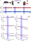
Blue light stimulation of baroreceptors elicits depressor responses. (A) AAVrg applied to the aortic arch to direct the expression of ChR2 and/or tdTom within baroreceptors (n = 6 per group). (B) Representative traces of arterial pressure and heart rate following the optogenetic stimulation of baroreceptors cell bodies at 1, 5, and 10 Hz (473 nm, 10 mW output; 20 ms pulse width; for 1 min). (C) Time-course changes in systolic blood pressure (SBP) and heart rate (HR) in control (tdTom) and experimental (ChR2) groups. *p < 0.05, **p < 0.01, ***p < 0.001, ****p < 0.0001; Two-way ANOVA followed by Tukey’s post hoc test. n = 6 per group.

References

    1. Alcaino C., Farrugia G., Beyder A. (2017). Mechanosensitive piezo channels in the gastrointestinal tract. Curr. Top Membr. 79 219–244. 10.1016/bs.ctm.2016.11.003 - DOI - PMC - PubMed
    1. Andresen M. C., Kunze D. L. (1994). Nucleus tractus solitarius–gateway to neural circulatory control. Annu. Rev. Physiol. 56 93–116. 10.1146/annurev.ph.56.030194.000521 - DOI - PubMed
    1. Andresen M. C., Paton J. F. R. (2011). ““The Nucleus of the Solitary Tract: Processing Information from Viscerosensory Afferents,”,” in Central regulation of autonomic functions, eds Llewellyn-Smith I. J., Verberne A. J. M. (Oxford: Oxford University Press; ), 23–46. 10.1093/acprof:oso/9780195306637.003.0002 - DOI
    1. Aumonier F. J. (1972). Histological observations on the distribution of baroreceptors in the carotid and aortic regions of the rabbit, cat and dog. Acta Anatomica. 82 1–16. 10.1159/000143797 - DOI - PubMed
    1. Bai L., Mesgarzadeh S., Ramesh K. S., Huey E. L., Liu Y., Gray L. A., et al. (2019). Genetic identification of vagal sensory neurons that control feeding. Cell 179 1129.e–1143.e. 10.1016/j.cell.2019.10.031 - DOI - PMC - PubMed

LinkOut - more resources