Can Seismocardiogram Fiducial Points Be Used for the Routine Estimation of Cardiac Time Intervals in Cardiac Patients?
- PMID: 35399285
- PMCID: PMC8986454
- DOI: 10.3389/fphys.2022.825918
Can Seismocardiogram Fiducial Points Be Used for the Routine Estimation of Cardiac Time Intervals in Cardiac Patients?
Abstract
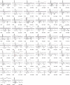
The indexes of cardiac mechanics can be derived from the cardiac time intervals, CTIs, i.e., the timings among the opening and closure of the aortic and mitral valves and the Q wave in the ECG. Traditionally, CTIs are estimated by ultrasound (US) techniques, but they may also be more easily assessed by the identification of specific fiducial points (FPs) inside the waveform of the seismocardiogram (SCG), i.e., the measure of the thorax micro-accelerations produced by the heart motion. While the correspondence of the FPs with the valve movements has been verified in healthy subjects, less information is available on whether this methodology may be routinely employed in the clinical practice for the monitoring of cardiac patients, in which an SCG waveform distortion is expected because of the heart dysfunction. In this study we checked the SCG shape in 90 patients with myocardial infarction (MI), heart failure (HF), or transplanted heart (TX), referred to our hospital for rehabilitation after an acute event or after surgery. The SCG shapes were classified as traditional (T) or non-traditional (NT) on whether the FPs were visible or not on the basis of nomenclature previously proposed in literature. The T shape was present in 62% of the patients, with a higher ∓ prevalence in MI (79%). No relationship was found between T prevalence and ejection fraction (EF). In 20 patients with T shape, we checked the FPs correspondence with the real valve movements by concomitant SCG and US measures. When compared with reference values in healthy subjects available in the literature, we observed that the Echo vs. FP differences are significantly more dispersed in the patients than in the healthy population with higher differences for the estimation of the mitral valve closure (-17 vs. 4 ms on average). Our results indicate that not every cardiac patient has an SCG waveform suitable for the CTI estimation, thus before starting an SCG-based CTI monitoring a preliminary check by a simultaneous SCG-US measure is advisable to verify the applicability of the methodology.
Keywords: cardiac mechanics; heart failure; heart transplant; myocardial infarction; telemedicine; telerehabilitation.
Copyright © 2022 Işilay Zeybek, Racca, Pezzano, Tavanelli and Di Rienzo.
Conflict of interest statement
The authors declare that the research was conducted in the absence of any commercial or financial relationships that could be construed as a potential conflict of interest.
Figures


References
-
- Biering-Sørensen T., Mogelvang R., Søgaard P., Pedersen S. H., Galatius S., Jørgensen P. G., et al. (2013). Prognostic value of cardiac time intervals by tissue Doppler imaging M-mode in patients with acute ST-segment-elevation myocardial infarction treated with primary percutaneous coronary intervention. Circ. Cardiovasc. Imaging 6 457–465. 10.1161/CIRCIMAGING.112.000230 - DOI - PubMed
-
- Crow R. S., Hannan P., Jacobs D., Hedquist L., Salerno D. M. (1994). Relationship between Seismocardiogram and Echocardiogram for Events in the Cardiac Cycle. Am. J. Noninvas. Cardiol. 8 39–46.
LinkOut - more resources
Full Text Sources
Research Materials
Miscellaneous

